Abstract
A 2-year-old boxer dog from southern Ontario was evaluated because of acute onset lethargy. Exploratory laparotomy revealed a hemorrhagic, destructive, liver mass. Histology, immunohistochemistry, and polymerase chain reaction confirmed Echinococcus multilocularis as the cause of the hepatic mass. This constitutes the first description of endemic E. multilocularis in Ontario.
Résumé
Hydatidose alvéolaire hépatique (Echinococcus multilocularis) chez un chien Boxer du Sud de l’Ontario. Un chien Boxer âgé de 2 ans du Sud de l’Ontario a été évalué en raison d’un début soudain d’une léthargie. Une laparatomie exploratoire a révélé une masse hépatique hémorragique et destructrice. L’histologie, l’immunohistochimie et l’amplification en chaîne par la polymérase ont confirmé Echinococcus multilocularis comme la cause de la masse hépatique. Il s’agit de la première description d’E. multilocularis endémique en Ontario.
(Traduit par Isabelle Vallières)
In June 2012, a 2-year-old 22-kg neutered male boxer dog was presented to a small animal clinic in southern Ontario with a 12-hour history of inappetance and lethargy. The dog was noted to have been standing in the backyard, reluctant to sit, lie down, or walk.
Case description
Physical examination of the dog revealed mild tachycardia (132 beats/min), moderate tachypnea (40 breaths/min), and tacky mucous membranes. The cranial abdomen was tense and painful on palpation. Right lateral abdominal radiography revealed loss of serosal detail throughout the abdomen. A complete blood cell count performed in-house (Vet ABC Animal Blood Analyzer; Heska, Loveland, Colorado, USA) revealed a mildly increased hemoglobin concentration [190 g/L; reference interval (RI): 119 to 180 g/L] and hematocrit (0.58 L/L; RI: 0.37 to 0.55 L/L). There were decreased numbers of lymphocytes (0.8 × 10⁹/L; RI: 1.2 to 4.5 × 10⁹/L) and monocytes (0.2 × 10⁹/L; RI: 0.3 to 1 × 10⁹/L). All other parameters were within the normal range. Biochemistry (Abaxis VetScan VS2; Abaxis North America, Union City, California, USA) indicated a mild increase in alanine transaminase activity (172 U/L; RI: 10 to 118 U/L).
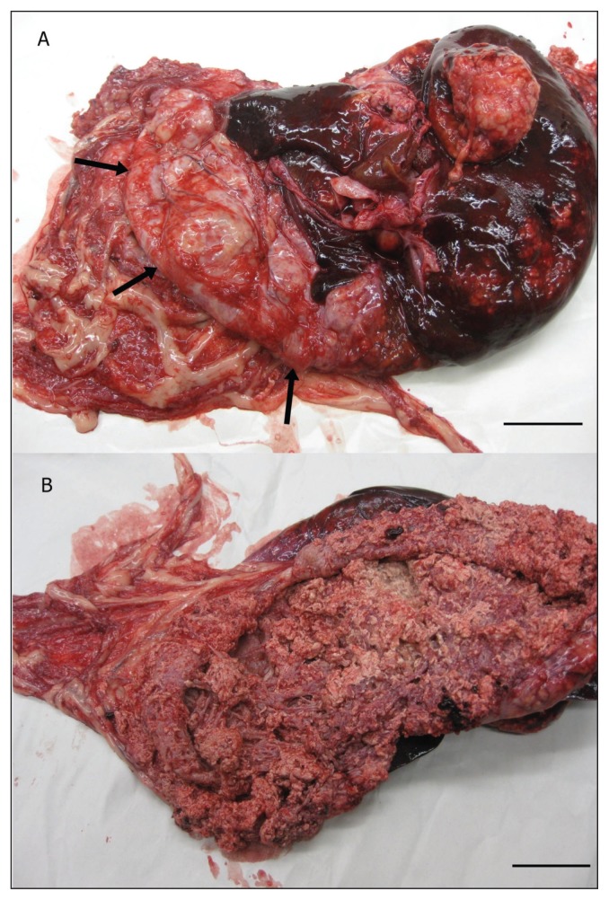
Exploratory laparotomy revealed hemoabdomen containing clots of blood and fibrin. At the time of the laparotomy, blood was actively filling the abdomen; exploration identified the liver as the source of hemorrhage. The liver contained generalized, multifocal, tan nodules, 1 to 5 mm in size. In addition, a 10-cm long space–occupying lesion was located centrally within the liver that had ruptured, leaving a cavity with uncontrollable hemorrhage (Figure 1 A,B). Excisional biopsies of the lesion were obtained for histopathology. Due to the poor prognosis, the dog was euthanized.
Figure 1.
Photograph of liver. A — Multifocal, tan-colored, multinodular masses in the liver. Arrows denote edge of liver lesion. Bar = 2.5 cm. B — Liver masses opened. The masses were cystic and had extensively infiltrated liver tissue. Bar = 2.5 cm.
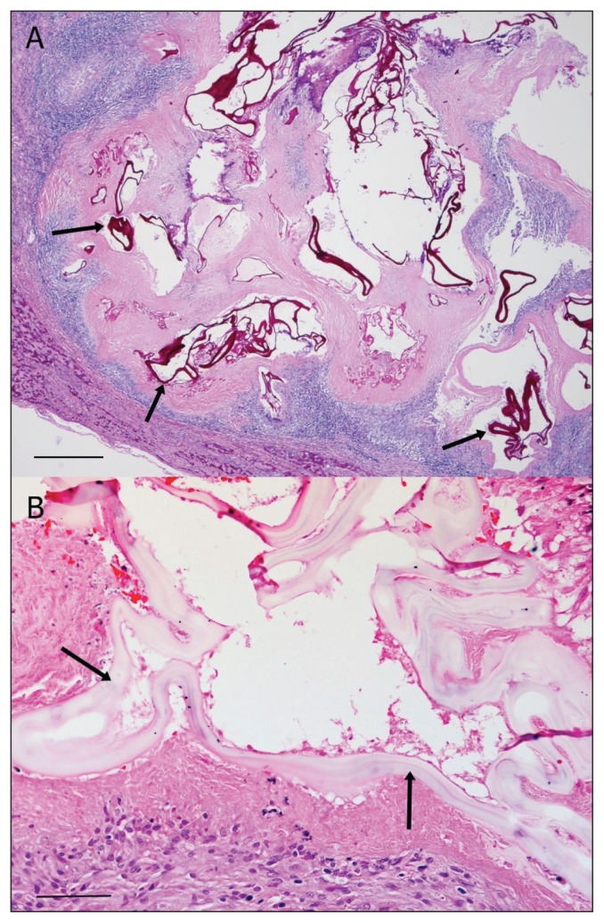
Histopathology of the intraoperative biopsies demonstrated the presence of multi-loculated cystic structures containing eosinophilic, fragmented, hyaline membranes that stained with Periodic acid-Schiff (PAS), necrotic debris, mineralized granular material, and prominent chronic eosinophilic and granulomatous inflammation (Figure 2 A,B). No protoscolices were observed in the tissue sections examined. These findings are consistent with the metacestode stage of Echinococcus multilocularis. As a result, formalin-fixed tissue was sent for polymerase chain reaction (PCR) testing at the Institut für Parasitologie, University of Bern, Switzerland. Sequence data from a PCR-generated fragment of the mitochondrial 12S rRNA gene (1), and restriction fragment length polymorphism analysis of PCR-generated fragments from the mitochondrial 12S rRNA and NADH dehydrogenase 1 genes (2) confirmed the identity of the metacestode as E. multilocularis. The final diagnosis was hepatic alveolar echinococcosis.
Figure 2.
Photomicrographs of a section of cystic tissue from the mass shown in Figure 1B. A — Note degenerating cysts in the liver containing Periodic acid-Schiff (PAS)-positive hyaline membranes (arrows) surrounded by eosinophilic granulomatous inflammation and necrosis. Stain = PAS, Bar = 250 μm. B — Note degenerating cysts in the liver that are lined by a hyaline membrane (“laminated layer”; arrows). Stain = hematoxylin and eosin, Bar = 50 μm.
Discussion
Echinococcus multilocularis is a cestode capable of causing potentially fatal zoonotic disease (3). The life cycle of this tapeworm typically involves red foxes, coyotes, and other wild canids as definitive hosts, and rodents as intermediate hosts (3). In some areas, domestic dogs, and to a lesser extent cats, may also act as definitive hosts, i.e., following consumption of infected rodents (4).
Humans, and occasionally dogs, may acquire infection with the metacestode stage of E. multilocularis via ingestion of infective eggs. Thereafter, alveolar hydatid cysts typically develop within the liver. Both humans and dogs are considered aberrant hosts as they play no role in parasite transmission (3). However, hepatic alveolar echinococcosis behaves like a malignant tumor and can have a prolonged incubation period prior to onset of clinical symptoms; in humans this is typically 5 to 15 y (3).
In North America, E. multilocularis occurs in 2 geographic regions (5). The first encompasses the west coast of Alaska and extends north and east to occupy most of the Canadian Arctic. The second region comprises the southern parts of Alberta, Saskatchewan, and Manitoba and 13 adjacent USA states (5). Echinococcus multilocularis was previously not known to occur in Ontario. However, the dog described here had only lived in southern Ontario and Manitoulin Island, Ontario, and therefore must have become infected in one of these 2 locations. Specifically, the dog was obtained at 8 wk of age from a breeder in southern Ontario, and then lived the rest of its life on a farm in the same region. However, on multiple occasions throughout the latter period, for a total of a few weeks, the dog visited Manitoulin Island. At both locations, the dog lived in a rural setting and had the freedom to roam on the properties, with potential exposure to foxes, coyotes, and rodents.
Hepatic alveolar echinococcosis in dogs is an extremely rare diagnosis in North America. To the best of the authors’ knowledge, only 1 case has previously been diagnosed: in British Columbia (6). However, in parts of central Europe this is an emerging issue, in both dogs and humans, associated with increasing fox populations especially in inner urban areas (5). Why the current case developed hepatic alveolar echinococcosis is unclear. However, data from Europe indicate that this clinical presentation occurs in dogs either as a result of ingestion of a large number of eggs from the environment, or as a result of an intestinal infection with the adult tapeworm (i.e., subsequent to ingestion of an infected rodent) and release of eggs into the intestinal lumen (7). Since the owners of the dog described here indicated that “the dog would eat just about anything,” either route of infection is a possibility for this case. However, either route would be novel in Ontario. Furthermore, cases of hepatic alveolar echinococcosis typically are only reported in highly endemic areas.
Data from Europe indicate that some dogs with hepatic alveolar echinococcosis may also have patent intestinal infections, and therefore may constitute a zoonotic threat. In light of the size of the hepatic lesion in the case described here, if the dog developed an intestinal infection, this most likely occurred within the first 2 to 6 mo of its life. As a result, 13 humans that came in contact with this dog at 2 to 6 mo of age were tested for antibody to E. multilocularis using 3 different screening enzyme-linked immunosorbent assays (ELISAs) [EgHF-ELISA, Em2-ELISA, and Em18-ELISA (8)]. Every serum sample was then additionally tested with an E. multilocularis-specific Western blot (9). On the basis of blood samples collected in September 2012, none of the in-contact people tested positive.
Interestingly, the case of hepatic alveolar hydatid disease in the aforementioned dog in British Columbia occurred in an animal that had never left that province. Furthermore, the parasite was shown to be genetically most closely related to European rather than North American strains (10). The origin of the case described here is unclear, as only formalin-fixed tissues were available and genotyping PCR analysis, as described in Jenkins et al (10), was not successful.
Lastly, it should be noted that some of the hematology findings in this dog are inconsistent with the findings at surgery. However, the discrepancy is likely due to the duration of time between blood collection and surgery, i.e., blood collection occurred prior to significant hemorrhage into the abdomen.
In summary, it is important that veterinary practitioners recognize that dogs can act as intermediate and definitive hosts for E. multilocularis, and that dogs with intestinal infections pose a serious zoonotic threat. Thus, in endemic areas, it is recommended that dogs and cats that hunt or scavenge on rodents should be dewormed on a monthly basis with a cestocide such as praziquantel (5). However, until the source of the infection in the dog described here is determined it would be premature to comment on whether preventative treatment of dogs with praziquantel is required in parts of Ontario; research is required to determine the distribution of E. multilocularis in Ontario.
Acknowledgments
We thank Dr. Dorothee Bienzle for comment on clinical pathology data, and the veterinarians and staff at the small animal clinic for their handling of this case. CVJ
Footnotes
Reprints will not be available from the authors.
Use of this article is limited to a single copy for personal study. Anyone interested in obtaining reprints should contact the CVMA office (hbroughton@cvma-acmv.org) for additional copies or permission to use this material elsewhere.
References
- 1.Dinkel A, von Nickisch-Rosenegk M, Bilger B, Merli M, Lucius R, Romig T. Detection of Echinococcus multilocularis in the definitive host: Coprodiagnosis by PCR as an alternative to necropsy. J Clin Microbiol. 1998;36:1871–1876. doi: 10.1128/jcm.36.7.1871-1876.1998. [DOI] [PMC free article] [PubMed] [Google Scholar]
- 2.Trachsel D, Deplazes P, Mathis A. Identification of taeniid eggs in the faeces from carnivores based on multiplex PCR using targets in mitochondrial DNA. Parasitology. 2007;134:911–920. doi: 10.1017/S0031182007002235. [DOI] [PubMed] [Google Scholar]
- 3.Deplazes P, Eckert J. Veterinary aspects of alveolar echinococcosis — A zoonosis of public health significance. Vet Parasitol. 2001;98:65–87. doi: 10.1016/s0304-4017(01)00424-1. [DOI] [PubMed] [Google Scholar]
- 4.Eckert J, Deplazes P. Biological, epidemiological, and clinical aspects of echinococcosis, a zoonosis of increasing concern. Clin Microbiol Rev. 2004;17:107–135. doi: 10.1128/CMR.17.1.107-135.2004. [DOI] [PMC free article] [PubMed] [Google Scholar]
- 5.Moro P, Schantz PM. Echinococcosis: A review. Int J Infect Dis. 2009;13:125–133. doi: 10.1016/j.ijid.2008.03.037. [DOI] [PubMed] [Google Scholar]
- 6.Peregrine AS, Jenkins EJ, Barnes B, et al. Alveolar hydatid disease (Echinococcus multilocularis) in the liver of a Canadian dog in British Columbia, a newly endemic region. Can Vet J. 2012;53:870–874. [PMC free article] [PubMed] [Google Scholar]
- 7.Haller M, Deplazes P, Guscetti F, Sardinas JC, Reichler I, Eckert J. Surgical and chemotherapeutic treatment of alveolar echinococcosis in a dog. J Am Anim Hosp Assoc. 1998;34:309–314. doi: 10.5326/15473317-34-4-309. [DOI] [PubMed] [Google Scholar]
- 8.Gottstein B, Jacquier P, Bresson-Hadni S, Eckert J. Improved primary immunodiagnosis of alveolar echinococcosis in humans by an enzyme-linked immunosorbent assay using the Em2plus-antigen. J Clin Microbiol. 1993;31:373–376. doi: 10.1128/jcm.31.2.373-376.1993. [DOI] [PMC free article] [PubMed] [Google Scholar]
- 9.Mueller N, Frei E, Nunez S, Gottstein B. Improved serodiagnosis of alveolar echinococcosis of humans using an in vitro-produced Echinococcus multilocularis antigen. Parasitology. 2007;134:879–888. doi: 10.1017/S0031182006002083. [DOI] [PubMed] [Google Scholar]
- 10.Jenkins EJ, Peregrine AS, Hill JE, et al. Detection of European strain of Echinococcus multilocularis in North America. Emerg Infect Dis. 2012;18:1010–1012. doi: 10.3201/eid1806.111420. [DOI] [PMC free article] [PubMed] [Google Scholar]


