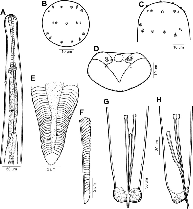
Figure 3.
Male of Philometra piscaria n. sp. from Epinephelus coioides. A: Anterior end of body, lateral view. B, C: Cephalic end, apical and subdorsoventral views. D: Caudal end, apical view. E, F: Distal end of gubernaculum, dorsal and lateral views. G, H: Posterior end of body, ventral and lateral views.

