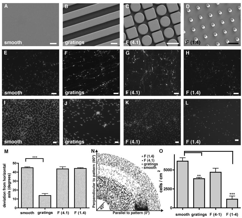
Figure 1. Tuj1+ induced neurons cultured on different topographies.
(A–D) Images of substrate topographies with micron-sized features fabricated by micro-imprinting. Scale bar 5 μm. (E) Neurites of tuj1+ iNs on smooth substrates displayed random orientation. Scale bar 100 μm. (F) Neurites of tuj1+ iNs on 5 μm gratings showed alignment with the underlying substrate features. Scale bar 100 μm. (G, H) Neurites of tuj1+ iNs on F (1.4) and F (4.1) substrates displayed random orientation, similar to smooth substrates. Scale bar 100 μm. (I–L) DAPI stain of cell nuclei attached to the different topographies. Scale bar 50 μm. (M) Quantification of iN’s directionality; 45° represents completely random orientation. A global one-way ANOVA of cell alignment revealed a significant effect of substrate gratings on iN alignment (p < 0.0001). (N) Visual representation of the iN’s angle in respect to the pattern axis. X-axis represents the axis parallel to the pattern, whereas y-axis represents iN alignment perpendicular to the pattern. (O) Quantification of cell attachment to the different topographies. Variation of cell attachment to the different substrates was observed by a global one-way ANOVA with a significant reduction of cell attachment to F (1.4) and grating substrates (p < 0.001).

