What is Aplysia
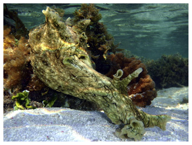
A genus of gastropod molluscs well-known as ‘model organisms’ in neuroscience, particularly work on the cellular biology of learning and memory (for his contributions Eric Kandel shared the 2000 Nobel prize for Physiology or Medicine). Their latin name comes from L’Aplysia — “that which one cannot wash”. Aplysia (Figure 1) has one of the earliest mentions in the literature of any animal, the first authentic description being by Pliny in his Historia Naturalis, c. 60 A.D. They are also commonly called sea hares because their posterior chemosensory tentacles, the rhinophores, stick up like ears.
Figure 1.

Aplysia dactylomela on a coral reef (Heron Island, Australia; photo: L.L. Moroz). This is one of the most abundant and cosmopolitan species of sea slugs. Charles Darwin, in January 1831, made a vivid description of this species at his very first stop on St. Jago (Cape Verde Islands) during the Voyage of the Beagle.
Where do they live?
Most species live in subtropical and tropical tide zones with a diversity of seaweeds. Thirty-seven Aplysia species are recognized; they have a widespread distribution, one species (A. punctata) even reaching the Arctic Circle. They vary in size from just a couple of centimeters up to 60–70 cm for the monstrous A. gigantea from Australia. The Californian A. vicaria can weigh up to 15.9 kg, the size of a medium dog. All Aplysia are benthic animals, grazing on the sea beds or corals and eating algae or other plants. Seven species, for example A. brasiliana, are good swimmers and can cross large distances by traveling up to 1 km in one swim episode.
Many Aplysia, when disturbed, secrete a purple ink, long considered toxic for humans; Darwin wrote that it causes “a sharp, stinging sensation”. It is actually harmless to people, though it can stain our clothes or hands on contact. The ink contains chemical deterrents that Aplysia uses to defend itself from opportunistic predators by stimulating their olfactory systems with consequent sensory disruption. Because of this, and the distasteful algal toxins accumulated from their diet, there are few natural predators for Aplysia.
What is so special about Aplysia
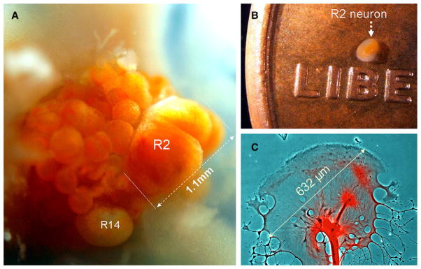
Their giant neurons, which are easy to work with and include the largest found in nature (Figure 2A). These neurons are the largest somatic cells in the animal kingdom (only eggs are larger) with beautiful natural orange, yellow, milk, reddish, or even in some species, greenish colorations. The remarkable features of these neurons were first recognized around 70 years ago by Angelique Arvanitaki, a graduate student working on a Mediterranean species (A. depilans). Aplysia is now a paramount model species for studies of how neurons and neural circuits control behaviors. It is also the best studied mollusc, with ongoing efforts to complete sequencing of its genome.
Figure 2.

Aplysia neurons (A,B) and growth cones (C) are some of the largest in the animal kingdom.
(A) A photograph of the freshly dissected right abdominal semi-ganglion from A. californica with the positions of two neurons marked (R2 and R14). This R2 cell is the largest neuron ever photographed reaching 1.1 mm in diameter. When this cell was isolated, yielding a record 1.965 μg of total RNA (while typical mammalian neurons contain less than 50 pg of RNA). These cells can also provide from 150 to 250 ng of DNA depending upon cell size. (B) R2 neurons can be seen by the naked eye on a penny. (C) Giant growth cone of a single motoneuron in cell culture (2nd day) stained with anti-β-tubulin antisera (red). Photographs by Leonid L. Moroz (A,C) and Sami Jezzini (B).
Aplysia’s central nervous system is composed of nine ganglia with ~10,000 neurons. Many neurons are accessible from the ganglionic surface, while unique colors and locations mean that they can be easily recognized from one preparation to the next — ‘personal’ names have been given to most of the cells. Aplysia neurons are truly colossal — one entire Drosophila brain or a dozen whole worms (Caenorhabitis elegans) can be packed in a single neuron like R2 (Figure 2A,B). Even the growth cones are gargantuan, they are the largest known in the animal kingdom and may reach up to 632 μm in diameter (Figure 2C).
Why are Aplysia neurons so large
They are giant because of their extraordinary polyploidy; they are probably the most polyploid somatic cells known, with up to 600,000 copies of a haploid genome in a single R2 neuron. How and why they achieve such a gigantic size is unclear. Their size correlates with the areas they innervate; a single large neuron can be a supply station for a highly extended tree of neuronal processes. Such a neuron can act as a single ‘true’ integrative center, controlling multiple behaviors. They may exploit a distinct strategy from other animal lineages, where complex tasks are controlled by a thousand times as many neurons. The larger diameters of neuronal processes can support an increase in the velocity of electrical signals. As a result, it enhances the speed of a behavioral response – an adaptation known in other species such as giant axons in squids, crustaceans or the Mauthner cells in teleost fishes.
How smart are Aplysia
As grazers with a relatively small number of neurons, Aplysia cannot compete in ‘intelligence’ with cephalopods, those ‘primates of the sea’. But this is exactly what neuroscientists want — an ideal organism with an array of simple stereotypic and learned behaviors that can be explained in terms of the molecular and integrative biology of individual neurons and synapses.
Aplysia can develop both non-associative and associative forms of long-term memory, following all fundamental learning paradigms (habituation, sensitization, classical and operant conditioning). Aplysia can remember a repetitively trained gill-withdrawal response for more than two to three weeks — impressive given its short life cycle (about one year). Importantly, the cellular and molecular mechanisms of long-term plasticity in Aplysia have many parallels in humans, suggesting profound evolutionary conservation of elementary events underlying learning and memory. Initial transcriptome analyses indicate that Aplysia has a complexity of synaptic and intracellular signaling pathways comparable to that of vertebrates. This fact also suggests the presence of diverse ancestral molecular toolkits supporting both elementary and higher neuronal functions as well as plasticity.
How do Aplysia reproduce and develop
Aplysia can lay eggs at a remarkable rate of 41,000 per minute, and a single egg string can contain as many as 83 million eggs. As they can lay eggs for several months, a few individuals can produce over a billion eggs within a month. They are hermaphrodites, but never self-fertilize in nature. In A. californica, for example, copulation frequently involves several individuals, who sometimes form a ring with up to 22–30 mating individuals, where an individual animal acts as both male and female.
Embryonic development, from spiral cleavage through blastula, gastrula and trochophore as well as the first larval stages (early ‘veligers’), occurs within the egg capsules. Veligers swim upwards using cilia and have a long planktotrophic life, feeding on microscopic algae and bacteria for up to several weeks until they detect settlement cues from the alga they will eat as adults. Veligers then undergo a rapid metamorphosis, dramatically changing both their physical organization and behavior, with loss of larval characteristics and formation of juveniles that resemble mature animals in all major features except reproductive organs.
Sexual maturity in A. californica is generally completed within three to four months; life expectancy is about 150–380 days under various conditions. Animals fed limited rations lived much longer and showed a lower initial mortality rate than those with unlimited food, suggesting that ‘caloric restriction’ prolongs lifespan, as shown in other species such as the worm C. elegans.
Why are Aplysia good for experimental biologists
The most obvious advantage of Aplysia is the large neurons that facilitate physiological, biochemical and genomic studies at the level of single cells and their compartments. Single-cell transcriptome and epigenomic tests can be done from individually identified Aplysia neurons, and less than 0.1% of such a cell is sufficient for direct microchemical analysis of dozens and even hundreds of metabolites and peptides using the tools of modern analytical chemistry. Thus, the coordinated activity of an entire genome within a single neuron can be linked to the operation of a given neural circuit and a learned behavior. The relative numerical simplicity of the Aplysia nervous system provides, potentially, an opportunity to understand the animal’s entire behavior in terms of the operation of distinct neural circuits. With the Aplysia genome to be finished soon, there are unprecedented opportunities for system biology approaches, addressing questions about how complex cell and nervous system features are integrated at all levels of biological organization.
Further, Aplysia is a representative of the largest superclade of bilaterian animals (Lophotrochozoa), which includes annelids, flat worms and about ten less known phyla. Aplysia research, therefore, can be very informative for a broad spectrum of questions in evolution and development, particularly for understanding the organization of animal body plans. Many genes from a common bilaterian ancestor (urbilateria) seem to be lost in representatives of the Ecdysozoa lineages, for example in nematodes and flies, which have extremely short lifecycles and particularly derived genomes. Thus, Aplysia, as a model organism, fills an important niche and together with other established experimental preparations undoubtedly will tell us more about the principles of the organization of the nervous system, animal development and evolution.
Where can I find out more?
- Carefoot TH. Aplysia: Its biology & ecology. Oceanogr Mar Biol Annu Rev. 1987;25:167–284. [Google Scholar]
- Eales NB. Aplysia. Liverpool Marine Biology Committee. Memoirs. 1921;24:1–91. [Google Scholar]
- Eales NB. Revision of the world species of Aplysia (Gastropoda, Opisthobrachia) Bull Bri Mus Nat Hist Zool. 1960;5:268–404. [Google Scholar]
- Gillette R. On the significance of neuronal gigantism in gastropods. Biol Bull. 1991;180:234–240. doi: 10.2307/1542393. [DOI] [PubMed] [Google Scholar]
- Kandel ER. Cellular Basis of Behavior. San Francisco: W.H. Freeman and Company; 1976. [Google Scholar]
- Kandel ER. Behavioral Biology of Aplysia. San Francisco: W.H. Freeman and Company; 1979. [Google Scholar]
- Kandel ER. The molecular biology of memory storage: a dialogue between genes and synapses. Science. 2001;294:1030–1038. doi: 10.1126/science.1067020. [DOI] [PubMed] [Google Scholar]
- Lovell P, Moroz LL. The largest growth cones in the animal kingdom and dynamics of neuronal growth in cell culture of Aplysia. Int Comp Biol. 2006;46:847–870. doi: 10.1093/icb/icl042. [DOI] [PubMed] [Google Scholar]
- Moroz LL, Ju J, Russo JJ, Puthanveetti S, Kohn A, Medina M, Walsh PJ, Birren B, Lander ES, Kandel ER. Sequencing the Aplysia Genome: a model for single cell, real-time and comparative genomics. National Human Genome Research Institute; 2004. online at http://www.genome.gov/Pages/Research/Sequencing/SeqProposals/AplysiaSeq.pdf. [Google Scholar]
- Moroz LL, Edwards JR, Puthanveettil SV, Kohn AB, Ha T, Heyland A, Knudsen B, Sahni A, Yu F, Liu L, et al. Neuronal transcriptome of Aplysia: Neuronal compartments and circuitry. Cell. 2006;127:1453–1467. doi: 10.1016/j.cell.2006.09.052. [DOI] [PMC free article] [PubMed] [Google Scholar]


