Abstract
Melkersson-Rosenthal syndrome (MRS) is a rare granulomatous inflammatory disease characterised by the triad of orofacial oedema, facial nerve palsy and furrowed tongue. We describe the case of a 29-year-old patient suffering from an oligosymptomatic form of the disease with orofacial oedema, cobblestone pattern on the buccal mucosa and swelling of the tongue, accompanied by intermittent fatigue, influenza-like symptoms, intermittent tinnitus and acute hearing loss. An increase of several autoimmune-associated antibodies was also detected. Treatment with prednisolone, azathioprine or methotrexate failed to adequately control all symptoms in the long term. In the absence of a specific and well-established therapy for MRS, treatment with adalimumab was administered. Under adalimumab, total remission of all symptoms was achieved, indicating that tumour necrosis factor-α blockers are a promising therapeutic option for patients with Melkersson-Rosenthal syndrome.
Background
To date, there is no well-established specific therapy for Melkersson-Rosenthal syndrome (MRS) and the literature is limited to isolated case reports. Owing to the lack of evidence-based therapy guidelines and the unpredictability of individual response to different therapeutic approaches, treatment of MRS can be protracted and complex, especially when the syndrome presents in an oligosymptomatic form. Multidisciplinary collaboration (eg, dentistry, rheumatology, internal medicine) is therefore essential for effective treatment. While the aetiology of MRS is unknown, this case provides additional evidence that it must be considered an autoimmune disease, and the success of off-label treatment with adalimumab in this case suggests that tumour necrosis factor-α (TNF α) blockers may be a useful and extremely effective treatment option.
Case presentation
MRS is a rare neuromucocutaneous disease (Orpha number 2483) characterised by non-caseating granulomatous inflammation and the triad of orofacial oedema, facial nerve palsy and furrowed tongue. The complete triad occurs only in a minority of 8–25% of patients suffering from MRS,1 whereas monosymptomatic or oligosymptomatic variants are more frequent. In this context, cheilitis granulomatosa with recurrent swelling of the lips is the most common monosymptomatic form of MRS, and the first symptom in approximately 75% of patients.2 3 MRS usually appears in the second decade of life with asymmetrical swelling involving one or both lips, often in association with orofacial swelling affecting the oral mucosa, gingiva, cheeks and the eyelids.4 In many cases, minor symptoms like migrainoid headaches, hyperacusis, tinnitus or trigeminus pain are also reported.5 6 With an estimated prevalence rate of 0.08%,7 MRS is a rare disorder, and large case series including therapies are limited. Nevertheless, successful treatment with intralesional corticosteroids,8 antibiotics,9 thalidomide10 or TNFα inhibitors11 12 has been reported. MRS is a recurrent intermittent disease with a low probability of complete remission, and while its aetiology is still unknown, there is speculation on an involvement of Mycobacterium tuberculosis, viral infection or genetic predisposition. However, the association of MRS with autoimmune diseases seems more probable.13 14
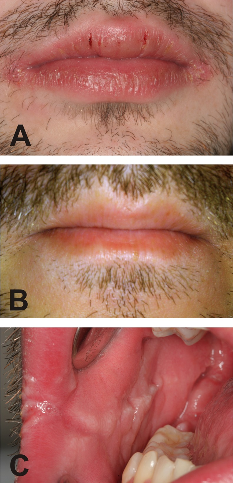
A 29-year-old man consulted our hospital in March 2005 a few weeks after hepatitis A+B immunisation. He presented with moderate inflammation of the oral mucosa and gingiva, with moderate gum proliferation. Worth mentioning in the medical history was acute hearing loss (figure 1) together with a minor decrease of lymphocytes (22%), a minor increase of IgG (16.8 g/L) and intermittent tingling and itching in the throat–ear section (without recognisable cause) 1 year previously. In April 2005, cheilitis vulgaris with pronounced labial lesions and bleeding tendency (figure 2A) occurred together with lingua geographica, facial swelling and intermittent swelling of the submandibular lymph nodes. Buccal swap and blood analyses were negative for fungal and viral infection, respectively, but a moderate shift within the leucocytes (neutrophilic granulocytes 78.1%, lymphocytes 13.4%) indicated the presence of systemic disease. Ultrasound and radiological examination gave no indication for sarcoidosis or Crohn's disease. Together with a loss of taste sensitivity for salty and sweet during June 2006, a cobblestone pattern on the buccal mucosa (figure 2C) and swelling of the tongue also developed, leading to the diagnosis of cheilitis granulomatosa MRS. Furthermore, intermittent fatigue and influenza-like symptoms occurred and the patient suffered from intermittent tinnitus together with soreness, tingling and itching in the throat–ear section. A biopsy of the oral mucosa exhibited non-caseating granulomatous inflammation and confirmed the suspicion of MRS. Under 60 mg/day prednisolone, the symptoms vanished entirely after a few days. After slow dose reduction and termination, the patient was free from symptoms for about 4 weeks before symptoms returned. Corticosteroid treatment was once again successfully initiated with 60 mg/day prednisolone, followed by a slow dose reduction to 20 mg/day, during which the patient remained free of symptoms. In October 2006, the antinuclear antibody (ANA) titre (1:320), thyroid autoantibodies (509 U/mL) and IgG cardiolipine antibodies (31.7 U/mL) increased and the screening test for lupus antibodies was positive. Neutrophil granulocytes (89%), IgG (21.4%), glutamate pyruvate transaminase (GPT) (59 U/L) and iron (199 µg/dL) were elevated and lymphocytes (7%) decreased. Treatment with 2×50 mg azathioprine was started, but the therapy had to be terminated after a few weeks due to severe side effects. Prednisolone therapy, initially continuing with 20 mg/day and subsequently reduced to 12.5 mg/day in combination with 15 mg/day methotrexate, was able to contain symptoms at a low level, with moderate labial and buccal swelling, cobblestone pattern on the buccal mucosa and minor lesions of the upper lip, but with reduced general condition and intermittent fatigue and tinnitus. In March 2007, facial and labial swelling suddenly increased, accompanied by attacks of acute hearing loss which were treated additionally with pentoxyfylline. Methotrexate therapy was terminated, and after another initial dose of 60 mg/day, prednisolone dosage was tapered to 20 mg/day. Thereafter, acute phases with increased facial, labial, buccal and gingival swelling and hearing loss alternated with phases of minor symptoms. It was not possible to reduce the prednisolone dose consistently (dose on demand from 3.75 to 60 mg/day) or to repress the symptoms completely, and the patient reported a creeping deterioration of his health status. At this point in time, conventional treatment options had thus proved unsuccessful and were exhausted.
Figure 1.

Tone audiogram of the right (R) and left (L) ear following acute hearing loss.
Figure 2.

Labial swelling and labial lesions with bleeding tendency (A); oral area 4.3 years after successful adalimumab therapy (B); buccal mucosa with beginning cobblestone pattern (C).
Treatment
After a review of the related literature and substantial discussion with the patient, anti-TNFα therapy was considered. The patient was provided with clear information regarding possible complications. In November 2007, the anti-TNFα antibody adalimumab was given off-label in an initial dose of 80 mg subcutaneously, followed by subsequent doses of 40 mg every 3 weeks, in addition to 20 mg/day prednisolone. Adalimumab was well tolerated, and after a few weeks the symptoms disappeared entirely and the patient reported an improved sense of well-being. Adalimumab administration was subsequently reduced to 40 mg every 4 weeks and prednisolone was carefully tapered out to zero by March 2009 without any relapse. During treatment with adalimumab, levels of lymphocytes, granulocytes, IgG and iron shifted to normal.
Outcome and follow-up
Adalimumab therapy was terminated in July 2009 with total remission of all symptoms (figure 2B). Follow-up examinations were subsequently carried out at irregular intervals. The patient has remained symptom free to this day (November 2013), without any recurrence of labial swelling, orofacial oedema or tinnitus/acute hearing loss.
Discussion
Both MRS and cheilitis granulomatosa are characterised by a chronic relapsing, remitting course of painless orofacial swelling. The aetiology remains unknown and treatment can be complicated, especially since the response to therapeutics is often unpredictable. While therapy usually focuses on treatment of the oedema, in the present case, systemic manifestations including intermittent acute hearing loss, tinnitus, fatigue and influenza-like symptoms more severely impaired the quality of life than the cosmetic deformity itself. The detection of lupus antibodies, the decrease of lymphocytes and the increase of granulocytes, IgG, ANA, thyroid autoantibodies and IgG cardiolipine antibodies together with the absence of fungal, bacterial or viral pathogens indicates the presence of autoimmune disease. Unfortunately, conventional immunomodulatory treatment was not entirely successful, failing to effectively prevent symptoms such as flares of tinnitus/acute hearing loss. We therefore took the decision—consensual with the patient—to initiate therapy with a TNFα blocker. Adalimumab was selected, as it allows safe and easy self-administration by the patient. Adalimumab is an IgG1 monoclonal antibody of human origin with a terminal half-life of approximately 2 weeks.15 It blocks the interaction of TNFα with the p55 and p75 receptors and leads to lysis of the cells expressing TNFα.15 Adalimumab is approved for the treatment of several autoimmune diseases (eg, in the EU: rheumatoid arthritis, Crohn's disease, psoriasis). As the increase of TNFα production is believed to play an important role in the mucosal damage of MRS,12 16 we concur with Riuz Villaverde11 in regarding anti-TNFα as an interesting and effective option in the treatment of MRS. This view is supported by other case reports describing successful therapy of MRS with adalimumab11 16 or infliximab.12 However, evidence-based treatment guidelines for MRS promising therapeutic success do not currently exist. Moreover, they are difficult to propose, since few case reports/studies are available in the literature and conventional large-scale placebo-controlled double-blind studies are impracticable due to the small number of patients affected by MRS. Nevertheless, our experience in this case suggests treatment with adalimumab to be a highly effective option when other therapeutic approaches have failed. We conclude that adalimumab appears to be able to cure MRS to the extent that all attendant symptoms were brought into lasting remission. Anti-TNFα therapy can therefore be considered a valid and effective therapy which may be administered to patients with MRS after careful consideration of individual risks and benefits.
Learning points.
Symptoms of Melkersson-Rosenthal syndrome (MRS) may include not only characteristic orofacial symptoms but also tinnitus and acute hearing loss.
This case provides further evidence that MRS is an autoimmune disease.
Interdisciplinary collaboration is required for successful therapy.
Antitumour necrosis factor-α therapy seems highly effective.
Acknowledgments
The authors thank Professor Peter Eickholz (J W von Goethe University, Frankfurt/Main, Germany) for his critical reading of the manuscript and valuable contribution to the diagnosis and follow-up care of the patient, and Janet Collins (Crohn Colitis Centre Rhein-Main, Frankfurt/Main, Germany) for language support and proofreading the manuscript.
Footnotes
Contributors: AP was involved in data collection and drafting of the manuscript. BS was involved in photography and revision of periodontology section of the manuscript. MN was involved in providing laboratory data and revision of immunological section of the manuscript. JS supervised manuscript preparation. All authors revised the manuscript for important intellectual content and approved the final version for publication.
Competing interests: None.
Patient consent: Obtained.
Provenance and peer review: Not commissioned; externally peer reviewed.
References
- 1.Sciubba JJ, Said-Al-Naief N. Orofacial granulomatosis: presentation, pathology and management of 13 cases. J Oral Pathol Med 2003;32:576–85 [DOI] [PubMed] [Google Scholar]
- 2.González-García C, Aguayo-Leiva I, Pian H, et al. Intralymphatic granulomas as a pathogenic factor in cheilitis granulomatosa/Melkersson-Rosenthal syndrome: report of a case with immunohistochemical and molecular studies. Am J Dermatopathol 2011;33:594–8 [DOI] [PubMed] [Google Scholar]
- 3.van der Waal RI, Schulten EA, van de Scheur MR, et al. Cheilitis granulomatosa. J Eur Acad Dermatol Venereol 2001;15:519–23 [DOI] [PubMed] [Google Scholar]
- 4.Worsaae N, Christensen KC, Schiodt M, et al. Melkersson-Rosenthal syndrome and cheilitis granulomatosa. A clinicopathological study of thirty-three patients with special reference to their oral lesions. Oral Surg Oral Med Oral Pathol 1982;54:404–13 [DOI] [PubMed] [Google Scholar]
- 5.Hornstein OP. Glossitis granulomatosa—an unusual subtype of Melkersson-Rosenthal syndrome. Mund Kiefer Gesichtschir 1998;2:14–9 [DOI] [PubMed] [Google Scholar]
- 6.Ziemssen F, Rohrbach JM, Scherwitz C, et al. Plastic reconstructive correction of persistent orofacial swelling and swelling of the eyelids in Melkersson-Rosenthal syndrome. Klin Monatsbl Augenheilkd 2003;220:352–6 [DOI] [PubMed] [Google Scholar]
- 7. 2006 Orphanet. Melkerson-Rosenthal syndrome. http://www.orpha.net/consor/cgi-bin/OC_Exp.php?lng=de&Expert=2483 (accessed 11 Oct 2013) [Google Scholar]
- 8.Lynde CB, Bruce AJ, Orvidas LJ, et al. Cheilitis granulomatosa treated with intralesional corticosteroids and anti-inflammatory agents. J Am Acad Dermatol 2011;65:e101. [DOI] [PubMed] [Google Scholar]
- 9.Pigozzi B, Fortina AB, Peserico A. Successful treatment of Melkersson-Rosenthal syndrome with lymecycline. Eur J Dermatol 2004;14:166–7 [PubMed] [Google Scholar]
- 10.Medeiros M, Jr, Araujo MI, Guimaraes NS, et al. Therapeutic response to thalidomide in Melkersson-Rosenthal syndrome: a case report. Ann Allergy Asthma Immunol 2002;88:421–4 [DOI] [PubMed] [Google Scholar]
- 11.Ruiz Villaverde R, Sánchez Cano D. Successful treatment of granulomatous cheilitis with adalimumab. Int J Dermatol 2012;51:118–20 [DOI] [PubMed] [Google Scholar]
- 12.Barry O, Barry J, Langan S, et al. Treatment of granulomatous cheilitis with infliximab. Arch Dermatol 2005;141:1080–2 [DOI] [PubMed] [Google Scholar]
- 13.Scagliusi P, Sisto M, Lisi S, et al. Hashimoto's thyroiditis in Melkersson-Rosenthal syndrome patient: casual association or related diseases? Panminerva Med 2008;50:255–7 [PubMed] [Google Scholar]
- 14.Giovannetti A, Mazzetta F, Cavani A, et al. Skewed T-cell receptor variable β repertoire and massive T-cell activation in idiopathic orofacial granulomatosis. Int J Immunopathol Pharmacol 2012;25:503–11 [DOI] [PubMed] [Google Scholar]
- 15.Rau R. Adalimumab (a fully human anti-tumour necrosis factor alpha monoclonal antibody) in the treatment of active rheumatoid arthritis: the initial results of five trials. Ann Rheum Dis 2002;61(Suppl 2):ii70–3 [DOI] [PMC free article] [PubMed] [Google Scholar]
- 16.Kakimoto C, Sparks C, White AA. Melkersson-Rosenthal syndrome: a form of pseudoangioedema. Ann Allergy Asthma Immunol 2007;99:185–9 [DOI] [PubMed] [Google Scholar]


