Abstract
The most important complication of intestinal malrotation is midgut volvulus because it may lead to intestinal ischaemia and necrosis. A 29-year-old male patient was admitted to the emergency department with abdominal pain. Ultrasonography (US), colour Doppler ultrasonography (CDUS), CT and barium studies were carried out. On US and CDUS, twisting of intestinal segments around the superior mesenteric artery (SMA) and superior mesenteric vein (SMV) and alteration of the SMA–SMV relationship were detected. CT demonstrated that the small intestine was making a rotation around the SMA and SMV, which amounted to more than 360°. The upper gastrointestinal barium series revealed a corkscrew appearance of the duodenum and proximal jejunum, which is a pathognomonic finding of midgut volvulus. Prior knowledge of characteristic imaging findings of midgut volvulus is essential in order to reach proper diagnosis and establish proper treatment before the development of intestinal ischaemia and necrosis.
Background
The inability of the small intestine to fulfil its physiological procedures of rotation and fixation during fetal development is called intestinal malrotation.1 The most important complication of malrotation is midgut volvulus because it may lead to intestinal ischaemia and necrosis.1 Midgut volvulus is simply the twisting of the mesentery of the small intestine around the mesenteric arterial axis. This happens due to adhesion of the mesentery to a narrow zone.2 Midgut volvulus may be primary or secondary to aetiological factors such as parasitic infections and diabetic autonomous neuropathy.2 Primary midgut volvulus is usually encountered in the newborn and infant eras. It is, however, an aetiological factor of intestinal obstruction during adulthood. This article presents an adult case which was referred for the evaluation of abdominal pain and findings of intestinal obstruction. The diagnosis of midgut volvulus was surgically proved. In this article, colour Doppler ultrasonography (CDUS), CT and barium study images are being presented as well.
Case presentation
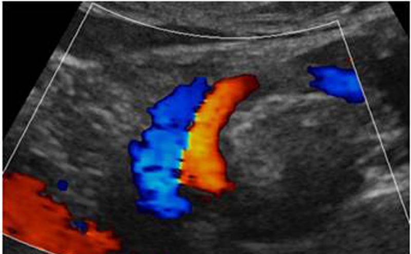
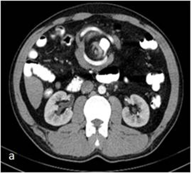
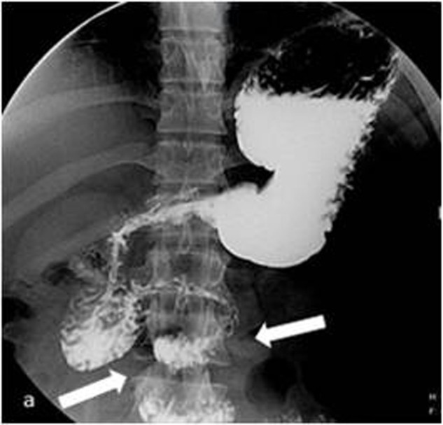
A 29-year-old male patient was admitted to the emergency department with abdominal pain and findings of intestinal obstruction. Clinical history revealed mild abdominal pain of a duration of 1 month, which had intensified during the past few days. His medical history was unremarkable. Physical examination revealed abdominal distention and generalised tenderness. An upright X-ray abdominal radiograph demonstrated air–fluid levels. On ultrasound (US), twisting of intestinal segments around the superior mesenteric artery (SMA) and superior mesenteric vein (SMV) was detected. On CDUS, alteration of the SMA–SMV relationship was noted and the SMV was displaced laterally to the left of the SMA. CDUS demonstrated a rotation of the SMV around SMA (figure 1). Flow was detected in the mesenteric vessels on CDUS. These findings led to the consideration of midgut volvulus. The patient was treated by nasogastric decompression for 2 days. The patients’ symptoms decreased gradually, and he passed gas and stool. Two days after admission, contrast-enhanced CT was performed. Arterial-phase CT demonstrated that the small intestine was making a rotation around the SMA and SMV, which amounted to more than 360° (figure 2). In addition, the upper gastrointestinal barium series revealed a corkscrew appearance of the duodenum and proximal jejunum, which is a pathognomonic finding of midgut volvulus (figure 3). Based on these clinical and radiological findings midgut volvulus was diagnosed in the patient and he underwent surgery.
Figure 1.

At colour Doppler ultrasonography, superior mesenteric artery and superior mesenteric vein are seen in rotation (whirlpool sign).
Figure 2.

At CT, the superior mesenteric vessels and the small intestines are seen to make a rotation together. This is called the whirlpool sign.
Figure 3.

The typical corkscrew appearance as seen at the barium series. This appearance is created by the duodenum and the proximal jejunum.
Treatment
In surgery, extended right hemicolectomy was performed.
Outcome and follow-up
The patient was discharged with full recovery following surgery.
Discussion
Intestinal malrotation is characterised by malfixation of the narrow mesentery and midgut to the posterior abdominal wall, and is usually encountered in infants and children. The most serious complication of malrotation is volvulus. Eighty per cent of midgut volvulus cases present during the first month of life. Only in a very small portion of cases may a diagnosis be made during adulthood,2 3 as it was in our case. In these patients, the clinical presentation of midgut volvulus is usually non-specific. Midgut volvulus may result in intestinal obstruction and even necrosis due to torsion of mesenteric vessels.2–4 For this reason, it is of paramount importance that diagnosis is made before the development of ischaemia and necrosis.
Conventional radiography is frequently non-specific. Air–fluid levels and dilated intestinal segments may be seen. In case of intestinal necrosis, air may be seen in the intestinal wall. In advanced cases, air may be present even in the portal vein.2 Plain radiography has low diagnostic value in the diagnosis of midgut volvulus.
It has been reported that CDUS is a valuable tool in the diagnosis of midgut volvulus. The intestinal segments and dilated SMV twist around the SMA in a clockwise pattern. This is called the whirlpool sign. The twisted mesenteric vessels which constitute the whirlpool sign, and even the SMV inversion to the left of the SMA, are clearly visible on CDUS as it was in our case.2 3
The whirlpool sign seen on abdominal CT refers to the spiral shape of the mesenteric vessels. However, this sign is not specific, and may be encountered in various other pathological conditions such as splenic torsion,5 but on the other hand, the presence of mesenteric vessels at the centrum of the abdominal whirlpool is virtually diagnostic of midgut volvulus. The intestinal segments and their feeding vessels may accompany the mesenteric vessels. CT is superior to other modalities in that it also demonstrates dilation of the small intestine, wall thickening, intramural gas, gas at the portal vein, ischaemia and necrosis.2 3
It has been reported that upper gastrointestinal barium series determine midgut volvulus with 100% accuracy.1 Barium studies may detect the abnormal position of the duodenojejunal junction. In the presence of malrotation the duodenojejunal junction is placed more inferiorly. If volvulus accompanies malrotation, a corkscrew sign may be present. The corkscrew sign refers to a spiral configuration of the fourth portion of the duodenum and the proximal jejunum.6 This sign was present in our case, too.
Alteration of the SMA–SMV relationship is one of the most frequently encountered findings in midgut volvulus and this finding may aid correct diagnosis, but it should be kept in mind that a normal SMA–SMV relationship does not necessary exclude the diagnosis of midgut volvulus. A study revealed that the SMA–SMV relationship was normal in 6 of the 14 midgut volvulus cases scanned retrospectively.7
Learning points.
Midgut volvulus is a pathological condition which is usually diagnosed in the newborn and treated surgically. Primary midgut volvulus cases diagnosed at adult ages constitute a rarity.
Cases with non-specific abdominal pain together with intestinal obstruction must remind us of the possibility of a midgut volvulus.
Prior knowledge of characteristic imaging findings is essential in order to reach proper diagnosis and establish proper treatment before the development of intestinal ischaemia and necrosis.
Footnotes
Competing interests: None.
Patient consent: Obtained.
Provenance and peer review: Not commissioned; externally peer reviewed.
References
- 1.Duran C, Öztürk E, Uraz S, et al. Midgut volvulus: value of multidetector computed tomography in diagnosis. Turk J Gastroenterol 2008;19:189–92 [PubMed] [Google Scholar]
- 2.Papadimitriou G, Marinis A, Papakonstantino A. Primary midgut volvulus in adults: report of two cases and review of the literature. J Gastrointest Surg 2011;15:1889–92 [DOI] [PubMed] [Google Scholar]
- 3.Bozlar U, Ugurel MS, Ustunsoz B, et al. CT angiographic demonstration of a mesenteric vessel “Whirlpool” in intestinal malrotation and midgut volvulus: a case report. Korean J Radiol 2008;9:466–9 [DOI] [PMC free article] [PubMed] [Google Scholar]
- 4.Önder H, Özmen CA, Gümüş M, et al. Erişkin Hastada İntestinal Malrotasyondan Kaynaklanan Midgut Volvulus. Sakaryamj 2012;2:102–4 [Google Scholar]
- 5.Yılmaz C, Esen OS, Colak A, et al. Torsion of a wandering spleen associated with portal vein thrombosis. J Ultrasound Med 2005;24:379–82 [DOI] [PubMed] [Google Scholar]
- 6.Ortiz-Neira CL. The corkscrew sign: midgut volvulus. Radiology 2007;242:315–16 [DOI] [PubMed] [Google Scholar]
- 7.Yang B, Chen WH, Zhang XF, et al. Adult midgut malrotation: multi-detector computed tomography (MDCT) findings of 14 cases. Jpn J Radiol 2013;31:328–35 [DOI] [PubMed] [Google Scholar]


