

Description
A 43-year-old man presented with recurrent pus discharge from his left upper leg for the past 3.5 years. He has had a gunshot injury to the left hip leading to fracture dislocation of the left hip. Initially, he was treated by skeletal traction using Steinmman pin through the upper left tibia. This was followed by pin tract infection, which persisted. The patient took various broad-spectrum antibiotics (eg, amoxiclav, erythromycin, cefuroxime, amikacin, ciprofloxacin, etc), off and on for about 2 years. Now, he had a pus discharging sinus on the anterolateral aspect of the upper leg with surrounding discolouration of the skin (figure 1). Wound swab culture grew Pseudomonas aurues. Radiograph of the upper leg showed a ring sequestrum in the upper tibia, surrounded by dense sclerosis (figures 2 and 3). Saucerisation of the cavity was done with complete removal of sequestra and all granulation tissues (figure 4). The wound healed in 10 days time, without any discharge or symptoms.
Figure 1.

Pus discharging sinus on the anterolateral aspect of the upper leg.
Figure 2.
Anteroposterior view of the leg showing chronic osteomyelitis of the upper tibia.
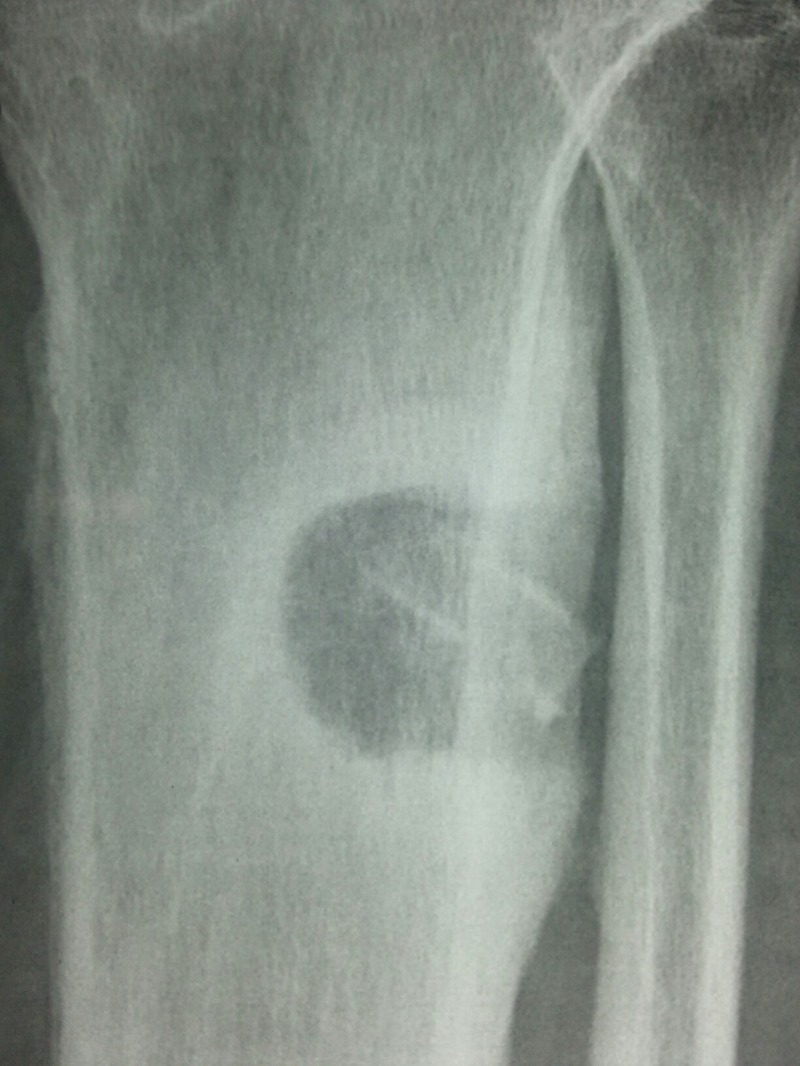
Figure 3.

Lateral view of the leg showing ring sequestrum with surrounding dense sclerosis in the upper tibia.
Figure 4.

Excised ring sequestrum with granulation tissue.
Metallic pin insertion for providing skeletal traction and external fixation, in major fractures of the lower limb is common.1 Minor pin tract infection occurs in 5–10 % of patients, and these usually respond to antibiotic therapy and local wound care. However, in 4 % of these patients, chronic pin tract osteomyelitis may develop,2 which is seen on the radiograph as a ring sequestrum.3 Staphylococcal osteomyelitis is reported more common in short-term fixator use, while Gram-negative rod infections are common with long-term use.4 The treatment of chronic pin tract infection includes removal of pins, appropriate antibiotic therapy and saucerisation of the cavity with removal of sequestra.2 5
Learning points.
Metallic pins in the bone must be inserted cautiously and kept with due care, as the damage to bone and superadded infection may lead to chronic osteomyelitis.
Treatment of chronic pin tract infection is usually surgical.
Footnotes
Competing interests: None.
Patient consent: Obtained.
Provenance and peer review: Not commissioned; externally peer reviewed.
References
- 1.Sisk TD. General principles and techniques of external skeletal fixation. Clinical Orthop Rel Res 1983;180:96–100 [PubMed] [Google Scholar]
- 2.Green SA. Complications of external skeletal fixation. Clin Orthop Rel Res 1983;180:109–16 [PubMed] [Google Scholar]
- 3.Nguyen VD, London J, Cone RO. Ring sequestrum: radiographic characteristics of skeletal fixation pin-tract osteomyelitis. Radiology 1986;158:129–31 [DOI] [PubMed] [Google Scholar]
- 4.Green SA, Ripley MJ. Chronic osteomyelitis in pin-tracts. J Bone Joint Surg 1984;66:1092–8 [PubMed] [Google Scholar]
- 5.Selegson D, Harman K. Negative experiences with pins-in-plaster for femoral fractures. Clin Orthop Rel Res 1979;138:243–145 [PubMed] [Google Scholar]