Abstract
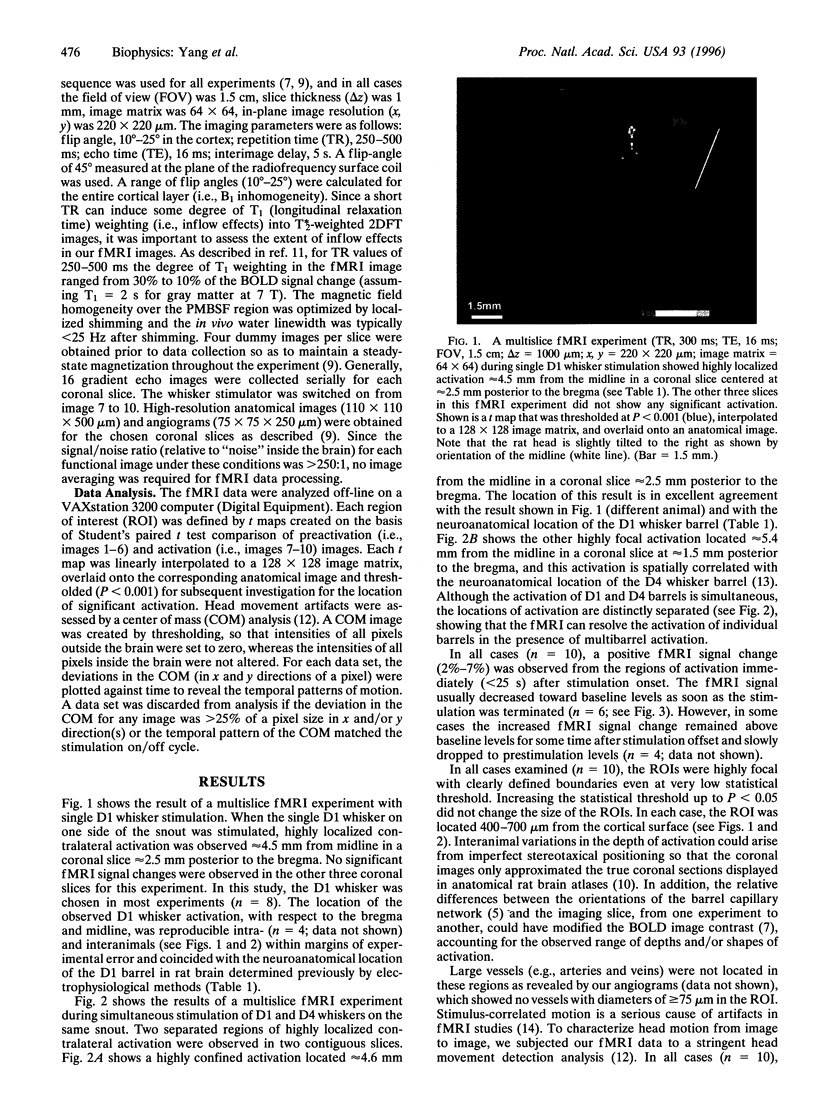
The previously established cortical representation of rat whiskers in layer IV of the cortex contains distinct cylindrical columns of cellular aggregates, which are termed barrels and correlate in a one-to-one relation to whiskers on the contralateral rat face. In the present study, functional magnetic resonance imaging (fMRI) of the rat brain was used to map whisker barrel activation during mechanical up-down movement (+/- 2.5 mm amplitude at 8 Hz) of single/multiple whisker(s). Multislice gradient echo fMRI experiments were performed at 7 T with in-plane image resolution of 220 x 220 microns, slice thickness of 1 mm, and echo time of 16 ms. Highly significant (P < 0.001) and localized contralateral regions of activation were observed upon stimulation of single/multiple whisker(s). In all experiments (n = 10), the locations of activation relative to bregma and midline were highly correlated with the neuroanatomical position of the corresponding whisker barrels, and the results were reproducible intra- and interanimal. Our results indicate that fMRI based on blood oxygenation level-dependent image contrast has the sensitivity to depict activation of a single whisker barrel in the rat brain. This noninvasive technique will supplement existing methods in the study of rat barrel cortex and should be particularly useful for the long-term investigations of central nervous system in the same animal.
Full text
PDF


Images in this article
Selected References
These references are in PubMed. This may not be the complete list of references from this article.
- Cox S. B., Woolsey T. A., Rovainen C. M. Localized dynamic changes in cortical blood flow with whisker stimulation corresponds to matched vascular and neuronal architecture of rat barrels. J Cereb Blood Flow Metab. 1993 Nov;13(6):899–913. doi: 10.1038/jcbfm.1993.113. [DOI] [PubMed] [Google Scholar]
- Hajnal J. V., Myers R., Oatridge A., Schwieso J. E., Young I. R., Bydder G. M. Artifacts due to stimulus correlated motion in functional imaging of the brain. Magn Reson Med. 1994 Mar;31(3):283–291. doi: 10.1002/mrm.1910310307. [DOI] [PubMed] [Google Scholar]
- Hyder F., Behar K. L., Martin M. A., Blamire A. M., Shulman R. G. Dynamic magnetic resonance imaging of the rat brain during forepaw stimulation. J Cereb Blood Flow Metab. 1994 Jul;14(4):649–655. doi: 10.1038/jcbfm.1994.81. [DOI] [PubMed] [Google Scholar]
- Kossut M., Hand P. J., Greenberg J., Hand C. L. Single vibrissal cortical column in SI cortex of rat and its alterations in neonatal and adult vibrissa-deafferented animals: a quantitative 2DG study. J Neurophysiol. 1988 Aug;60(2):829–852. doi: 10.1152/jn.1988.60.2.829. [DOI] [PubMed] [Google Scholar]
- Lindauer U., Villringer A., Dirnagl U. Characterization of CBF response to somatosensory stimulation: model and influence of anesthetics. Am J Physiol. 1993 Apr;264(4 Pt 2):H1223–H1228. doi: 10.1152/ajpheart.1993.264.4.H1223. [DOI] [PubMed] [Google Scholar]
- Masino S. A., Kwon M. C., Dory Y., Frostig R. D. Characterization of functional organization within rat barrel cortex using intrinsic signal optical imaging through a thinned skull. Proc Natl Acad Sci U S A. 1993 Nov 1;90(21):9998–10002. doi: 10.1073/pnas.90.21.9998. [DOI] [PMC free article] [PubMed] [Google Scholar]
- McCarthy G., Blamire A. M., Rothman D. L., Gruetter R., Shulman R. G. Echo-planar magnetic resonance imaging studies of frontal cortex activation during word generation in humans. Proc Natl Acad Sci U S A. 1993 Jun 1;90(11):4952–4956. doi: 10.1073/pnas.90.11.4952. [DOI] [PMC free article] [PubMed] [Google Scholar]
- Ogawa S., Lee T. M. Magnetic resonance imaging of blood vessels at high fields: in vivo and in vitro measurements and image simulation. Magn Reson Med. 1990 Oct;16(1):9–18. doi: 10.1002/mrm.1910160103. [DOI] [PubMed] [Google Scholar]
- Righini A., Pierpaoli C., Barnett A. S., Waks E., Alger J. R. Blue blood or black blood: R1 effects in gradient-echo echo-planar functional neuroimaging. Magn Reson Imaging. 1995;13(3):369–378. doi: 10.1016/0730-725x(94)00136-q. [DOI] [PubMed] [Google Scholar]
- Shulman R. G., Blamire A. M., Rothman D. L., McCarthy G. Nuclear magnetic resonance imaging and spectroscopy of human brain function. Proc Natl Acad Sci U S A. 1993 Apr 15;90(8):3127–3133. doi: 10.1073/pnas.90.8.3127. [DOI] [PMC free article] [PubMed] [Google Scholar]
- Simons D. J. Response properties of vibrissa units in rat SI somatosensory neocortex. J Neurophysiol. 1978 May;41(3):798–820. doi: 10.1152/jn.1978.41.3.798. [DOI] [PubMed] [Google Scholar]
- Waite P. M., Taylor P. K. Removal of whiskers in young rats causes functional changes in cerebral cortex. Nature. 1978 Aug 10;274(5671):600–602. doi: 10.1038/274600a0. [DOI] [PubMed] [Google Scholar]
- Welker C., Woolsey T. A. Structure of layer IV in the somatosensory neocortex of the rat: description and comparison with the mouse. J Comp Neurol. 1974 Dec 15;158(4):437–453. doi: 10.1002/cne.901580405. [DOI] [PubMed] [Google Scholar]
- Woolsey T. A., Van der Loos H. The structural organization of layer IV in the somatosensory region (SI) of mouse cerebral cortex. The description of a cortical field composed of discrete cytoarchitectonic units. Brain Res. 1970 Jan 20;17(2):205–242. doi: 10.1016/0006-8993(70)90079-x. [DOI] [PubMed] [Google Scholar]


