Abstract
Next generation nanomedicine will rely on innovative nanomaterials capable of unprecedented performance. Which ones are the most promising candidates for a medicinal chemist?
The expectations are high for the next generation of nanomedicines: a personalized and efficient therapy with lower side effects. In tissue engineering, nanomaterial-based scaffolds will offer a biodegradable support for cell growth and infiltration to be naturally replaced with time by new biological tissue. In drug delivery, smart nanodevices will target the disease site; there, an external trigger will prompt the controlled release of multiple agents for sensing, high-resolution imaging, and therapy. How can we conceive such a level of advanced performance without using innovative components? What are the nanomaterials of the future, and which ones are the most appealing for a medicinal chemist?
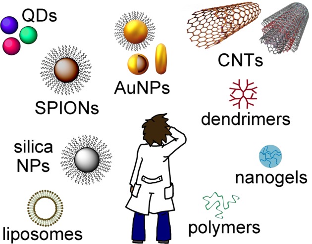
The nanomaterial landscape is vast (Figure 1). The medicinal chemist that looks close-by for familiar chemistry will see all of the well-characterized polymers, lipids, peptides and proteins, sugars, and surfactants that can be engineered into novel nanoformulations. Liposomes, dendrimers, and nanogels have been used for both controlled drug delivery and cell growth scaffolds. They are present in many nanomedicines that made it to the clinic, and most likely, they will appear to some extent also in the nanomedicines of the future.1 However, if we look a bit further and stretch our eyes toward the horizon, we will see how the landscape changes as we encounter the less-explored nanomaterials. Nanoparticles (NPs), quantum dots (QDs), and carbon nanotubes (CNTs), in one form or another, are all there. Which way does a medicinal chemist have to go for the best ingredients for the next generation nanomedicines?
Figure 1.

The medicinal chemist looks at the vast landscape of nanomaterials.
Many chemists would make their bet on mesoporous silica NPs. These are among the best-characterized NPs in vivo and indeed offer a number of advantages. Their synthesis can be fine-tuned to a variety of shapes and sizes (down to a few nanometers). The use of silane mixtures in their preparation allows for convenient incorporation of functional groups of choice (e.g., amino, carboxylic, thiol, etc.) for incorporation of therapeutic or imaging agents. Their porosity allows for high drug loading (up to 35 wt %).2 Remarkably, the “Cornell dots” are the first silica-based multimodal (optical/PET) diagnostic NPs recently approved for human clinical trials; their PEG coating and small size (<10 nm) allow for good biodistribution in a melanoma model, and fluorescent dye encapsulation in the NP core gives them notable brightness.3,4 However, despite these very encouraging advances, concerns still exist on the nanomaterial landscape about the coating stability of mesoporous silica NPs, since it has been shown that uncoated silica NPs are hemolytic.5
Magnetic NPs offer different advantages. They are well-known in medicine as MRI agents; in addition, their ability to respond to external magnetic fields gives an opportunity to develop cutting edge applications in protein and cell manipulation.6 In particular, superparamagnetic iron oxide NPs (SPIONs) have attracted a lot of attention for drug delivery applications in theranostics (i.e., combined therapy and diagnosis). One of the promises of SPIONs is targeted delivery to the disease site following an external magnetic force. In fact, their directional movement is usually hampered by blood flow, and their sensitivity to magnets can be notably reduced by the presence of a polymer coating (e.g., dextran). However, this organic “shell” is essential to reduce NPs undesired interaction with proteins and their subsequent opsonization. Therefore, SPIONs design needs careful fine-tuning of the “shell” for optimal performance. To date, opportunities exist to improve SPIONs colloidal stability in biological fluids (i.e., loss of the polymer coating) and to control drug delivery, avoiding undesired burst release from the polymer component.7
Another class that is drawing a lot of attention is gold NPs (AuNPs). Besides spherical NPs, the literature is rich with nanorods, nanocages, nanostars, and gold shells used to coat other NPs (e.g., SPION cores). Gold has been known in medicine for a very long time, but the attentive reader will note that the behavior of nanosized gold objects is a different matter, due to the high surface area and unique physicochemical properties. The shape of AuNPs has a big impact on their properties: spheres absorb visible light, and rods, cages, and shells absorb light in the near-infrared (NIR) region, where the human body is mostly transparent. NIR absorption is very useful, since it is employed in photothermal therapy (i.e., for heat generation to damage diseased tissue) and in high-resolution photoacustic imaging (i.e., for the generation of ultrasound waves). AuNPs, modified with both a strong Raman scatterer and an antibody, enhance the Raman response (surface enhanced Raman scattering, SERS), whereas the antibody imparts antigenic specificity.8
There is an increasing number of studies on the matter, but the heterogeneity of gold NP formulations makes it difficult to generalize important aspects such as biosafety assessments.9 In addition, despite the vast number of studies on gold nanomaterials, the functionalization chemistry of gold NPs usually revolves around the use of either thiols or amines, somewhat limiting the choice of triggers for drug loading and release. Nevertheless, imaginative variations have been found, such as the photothermal release of DNA cargos upon laser irradiation.10
QDs are yet another class of which we hear more and more in nanomedicine, especially in applications of multimodal imaging. The battle against cancer needs weapons of increasing sophistication, including tools to locate micrometastasis with exquisite spatiotemporal resolution. To this end, we may rely on multimodal imaging, because it is only with the combination of different techniques that we can go beyond the limitations of each modality, especially for imaging of deep tissues.11 QDs could be useful components of sophisticated nanodevices due to their very small size (typically of only a few nanometers), remarkable brightness, photostability, and ample offering of emission light colors for optical detection. Nevertheless, major limitations are posed by their chemical nature, since they are typically composed of heavy metals (e.g., cadmium, lead), for which QDs stability and safe excretion from the human body is a must.12
In the field of theranostics, CNTs are excellent candidates, as they exhibit many properties relevant to these objectives. For instance, CNTs possess relatively strong NIR absorption, which can be used for both high-resolution imaging (e.g., photoacustic modality) and photothermal therapy.11 Although biocompatibility and safety of CNTs are still an open issue, it is important to note that CNTs comprise a highly heterogeneous class of materials, for which biocompatibility data cannot, and should not, be generalized. There is a growing body of work that shows that CNT fate in vivo is highly dependent on their purity, physical properties (i.e., length, diameter, etc.), and chemical nature (i.e., functionalization). Importantly, there is increasing evidence that biodegradation of CNTs can be achieved by appropriate chemical modification.13 Derivatization of CNTs offers a variety of options for the imaginative medicinal chemist, who can covalently attach the polymer of choice for favorable interactions with biological entities. In addition to their high external surface, their hollow nature might permit loading with drugs or other bioactive cargoes, for their safe delivery in a cellular environment bypassing biological barriers otherwise encountered by other vectors.14 For instance, data exist on the ability of certain tubes to act as “cell membrane needles” and avoid the endocytic pathway.15,16 Another unique property is their ability to boost electrical activity of multilayered neuronal networks and cultured cardiac myocytes: the mechanisms of this phenomenon are still unclear and obviously deserve further investigation.17,18 Clearly, mastering such properties would pave the way to innovative tissue engineering that, until a few years back, was simply unthinkable. In our point of view, their unique properties offer ample opportunity for unprecedented performance in the field of “smart” nanomedicines; however, their application in the field is still in its infancy.19
In conclusion, the landscape of nanomaterials for medicines of the next generation is rich with options, and innovative solutions will likely be found in the wise combination of different components. It is clear that the examples reported in this viewpoint are only a few, representative of a class of materials that is continuously expanding and that includes an exceedingly high number of examples and ideas. The medicinal chemists who venture into this field should not impose limits on their imagination; instead, they should reach out to and partner with physicists, biologists, and clinicians to find creative solutions to these complex, multidisciplinary problems. We believe that hybrid, multifunctional nanomaterials will be the key components of the next generation of nanomedicines, and the brave medicinal chemists shall venture into the field to make them a reality.
Part of the work described in this article was supported by the European Union FP7 ERC Advanced Grant Carbonanobridge (ERC-2008-AdG-227135), the University of Trieste, the Italian Ministry of Education MIUR (Cofin Prot. 20085M27SS and FIRB prot. RBAP11ETKA), Regione Friuli-Venezia Giulia (Nanocancer), and AIRC (AIRC 5 per mille, Rif. 12214 “Application of Advanced Nanotechnology in the Development of Innovative Cancer Diagnostics Tools”).
Views expressed in this editorial are those of the authors and not necessarily the views of the ACS.
The authors declare no competing financial interest.
References
- Duncan R.; Gaspar R. Nanomedicine(s) under the Microscope. Mol. Pharmaceutics 2011, 862101–2141. [DOI] [PubMed] [Google Scholar]
- Mamaeva V.; Sahlgren C.; Lindén M. Mesoporous silica nanoparticles in medicine—Recent advances. Adv. Drug Delivery Rev. 2012, 10.1016/j.addr.2012.07.018. [DOI] [PubMed] [Google Scholar]
- Ma K.; Sai H.; Wiesner U. Ultrasmall Sub-10 nm Near-Infrared Fluorescent Mesoporous Silica Nanoparticles. J. Am. Chem. Soc. 2012, 134, 13180–13183. [DOI] [PubMed] [Google Scholar]
- Benezra M.; Penate-Medina O.; Zanzonico P. B.; Schaer D.; Ow H.; Burns A.; DeStanchina E.; Longo V.; et al. Multimodal silica nanoparticles are effective cancer-targeted probes in a model of human melanoma. J. Clin. Invest. 2011, 121, 2768–2780. [DOI] [PMC free article] [PubMed] [Google Scholar]
- Lin Y.-S.; Haynes C. L. Impacts of Mesoporous Silica Nanoparticle Size, Pore Ordering, and Pore Integrity on Haemolytic Activity. J. Am. Chem. Soc. 2010, 132, 4834–4842. [DOI] [PubMed] [Google Scholar]
- Pan Y.; Du X.; Zhao F.; Xu B. Magnetic nanoparticles for the manipulation of proteins and cells. Chem. Soc. Rev. 2012, 41, 2912–2942. [DOI] [PubMed] [Google Scholar]
- Mahmoudi M.; Sant S.; Wang B.; Laurent S.; Sen T. Superparamagnetic iron oxide nanoparticles (SPIONs): Development, surface modification and applications in chemotherapy. Adv. Drug Delivery Rev. 2011, 63, 24–46. [DOI] [PubMed] [Google Scholar]
- Porter M. D.; Lipert R. J.; Siperko L. M.; Wang G.; Narayanana R. SERS as a bioassay platform: fundamentals, design, and applications. Chem. Soc. Rev. 2008, 37, 1001–1011. [DOI] [PubMed] [Google Scholar]
- Akhter S.; Ahmad M. Z.; Ahmad F. J.; Storm G.; Kok R. J. Gold nanoparticles in theranostic oncology: Current state-of-the-art. Expert Opin. Drug Delivery 2012, 9, 1225–1243. [DOI] [PubMed] [Google Scholar]
- Vigderman L.; Zubarev E. R. Therapeutic platforms based on gold nanoparticles and their covalent conjugates with drug molecules. Adv. Drug Delivery Rev. 2012, 10.1016/j.addr.2012.05.004. [DOI] [PubMed] [Google Scholar]
- Lee D. E.; Koo H.; Sun I.-C.; Ryu J. H.; Kim K.; Kwon I. C. Multifunctional nanoparticles for multimodal imaging and theragnosis. Chem. Soc. Rev. 2012, 41, 2656–2672. [DOI] [PubMed] [Google Scholar]
- Cassette E.; Helle M.; Bezdetnaya L.; Marchal F.; Dubertret B.; Pons T. Design of new quantum dot materials for deep tissue infrared imaging. Adv. Drug Delivery Rev. 2012, 10.1016/j.addr.2012.08.016. [DOI] [PubMed] [Google Scholar]
- Bianco A.; Kostarelos K.; Prato M. Making carbon nanotubes biocompatible and biodegradable. Chem. Commun. 2011, 47, 10182–10188. [DOI] [PubMed] [Google Scholar]
- Li J.; Yap S. Q.; Yoong S. L.; Nayak T. R.; Chandra G. W.; Ang W. H.; Panczyk T.; Ramaprabhu S.; Vashist S. K.; Sheu F. S.; Tan A.; Pastorin G. Carbon nanotube bottles for incorporation, release and enhanced cytotoxic effect of cisplatin. Carbon 2012, 50, 1625–1634. [DOI] [PMC free article] [PubMed] [Google Scholar]
- Fabbro C.; Ali-Boucetta H.; Da Ros T.; Kostarelos K.; Bianco A.; Prato M. Targeting carbon nanotubes against cancer. Chem. Commun. 2012, 48, 3911–3926. [DOI] [PubMed] [Google Scholar]
- Al-Jamal K. T.; Nunes A.; Methven L.; Ali-Boucetta H.; Li S.; Toma F. M.; Herrero M. A.; et al. Degree of Chemical Functionalization of Carbon Nanotubes Determines Tissue Distribution and Excretion Profile. Angew. Chem., Int. Ed. 2012, 51, 6389–6393. [DOI] [PubMed] [Google Scholar]
- Fabbro A.; Bosi S.; Ballerini L.; Prato M. Carbon Nanotubes: Artificial Nanomaterials to Engineer Single Neurons and Neuronal Networks. ACS Chem. Neurosci. 2012, 3, 611–618. [DOI] [PMC free article] [PubMed] [Google Scholar]
- Martinelli V.; Cellot G.; Toma F. M.; Long C. S.; Caldwell J. H.; Zentilin L.; Giacca M.; Turco A.; Prato M.; Ballerini L.; Mestroni L. Carbon Nanotubes Promote Growth and Spontaneous electrical Activity in Cultured Cardiac Myocytes. Nano Lett. 2012, 12, 1831–1838. [DOI] [PubMed] [Google Scholar]
- Kostarelos K.; Bianco A.; Prato M. Promises, facts and challenges for carbon nanotubes in imaging and therapeutics. Nat. Nanotechnol. 2009, 4, 627–633. [DOI] [PubMed] [Google Scholar]


