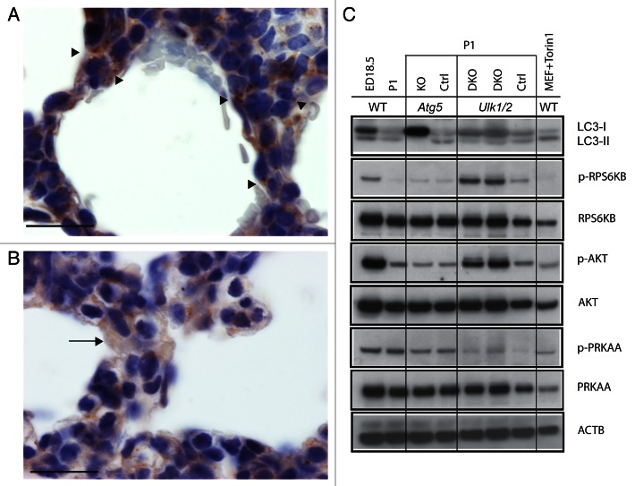
Figure 7. Analysis of autophagy in Ulk1/2 DKO and Atg5 KO neonatal lung tissue. Immunohistochemical staining of lung sections from (A) wild-type (WT) and (B) Ulk1/2 DKO mice at P1 with a LC3 antibody is shown after fixation, embedding in paraffin, and sectioning as described in Materials and Methods. Arrowheads in (A) show examples of alveolar epithelial cells with puncta. The arrow in (B) shows an example of an alveolar epithelial cell with diffuse, cytoplasmic LC3 staining. Scale bars: 20 μm. (C) Western blot analysis of protein lysates of lungs from wild-type mice at ED18.5 and P1, and from Atg5 KO mice and Ulk1/2 DKO mice and their respective littermate controls at P1 are shown. Blots were probed with antibodies specific for LC3, phospho-RPS6KB (Thr389), phospho-AKT (Ser473) and phospho-PRKAA/AMPKα (Thr172). Antibodies specific for total RPS6KB, AKT and PRKAA/AMPKα as well as ACTB were included as controls. Wild-type MEFs, which were serum starved and replated in medium with 10% FCS in the presence of Torin1 at 0.25 μM, were included as a positive control for autophagy.

An official website of the United States government
Here's how you know
Official websites use .gov
A
.gov website belongs to an official
government organization in the United States.
Secure .gov websites use HTTPS
A lock (
) or https:// means you've safely
connected to the .gov website. Share sensitive
information only on official, secure websites.
