

Abstract
Macroautophagy (hereafter autophagy) initiates at the phagophore assembly site (PAS), where most of the AuTophaGy-related (Atg) proteins are at least transiently localized. As the first protein complex targeted to the PAS, the Atg17-Atg31-Atg29 complex serves as the scaffold for other Atg proteins and plays a critical role for the organization of the PAS, and in autophagy initiation. We recently showed that this complex is constitutively formed and activated by the phosphorylation of Atg29 when autophagy is induced. Phosphorylation of Atg29 is required for its interaction with Atg11, another scaffold protein, and its function for promoting the proper assembly of the PAS. Single-particle electron microscopy analysis of the Atg17-Atg31-Atg29 complex reveals an elongated structure with Atg29 located at the opposing ends. This structural arrangement allows Atg29 to interact with Atg11, and is critical in the organization of the intact Atg1 complex.
Keywords: autophagy, PAS, scaffold, vacuole, yeast
Autophagy is a degradative pathway occurring in the lysosome/vacuole for the turnover of cytosol and organelles. Strict regulation of autophagy is crucial for cell metabolism, as insufficient or excessive autophagy will lead to an abnormal cell physiology. Therefore, induction as well as termination of autophagy has to be precisely controlled. When autophagy is induced, Atg proteins assemble at the PAS to promote the biogenesis of autophagosomes, the double-membrane vesicles that are involved in sequestration. The Atg1 protein complex plays a critical role in the recruitment of other Atg proteins to the PAS, and thus has a key role in autophagy induction. This complex is composed of at least the Atg1 protein kinase, its binding partners Atg13 and Atg11, and also the Atg17-Atg31-Atg29 (sub-)complex. The Atg17-Atg31-Atg29 complex is constitutively formed in both growing and starvation conditions, and is the first set of proteins targeted to the PAS under autophagy-inducing conditions, where it acts to recruit Atg13 and, thereby, Atg1, highlighting its significance in the induction of autophagy.
When autophagy is induced by either nitrogen starvation or treatment with rapamycin, Atg29 migrates as multiple bands during SDS-PAGE. This observation, coupled with a collapsing of the bands following phosphatase treatment, indicates that Atg29 is a phosphoprotein. In addition, the migration pattern suggests that Atg29 is phosphorylated at more than one site upon autophagy induction. Examination of the SDS-PAGE migration of different truncated forms of Atg29 indicates that most of the phosphorylated sites are located at the C terminus (amino acids 101–213). The presumably nonphosphorylated N terminus of Atg29 (Atg29[1–100]) constitutes a functional domain, as Atg29[1–100] is properly integrated into the Atg17-Atg31-Atg29 complex through binding to Atg31, and largely suppresses the autophagy defect of the atg29∆ strain.
The C terminus of Atg29 contains 23 serine or threonine residues, and constitutes a negative regulatory domain. Mutations of all of these sites to alanine (Atg29[23STA]) dramatically abolish the phosphorylation of Atg29, and affect its function; the mutant protein migrates as a single lower mass band during SDS-PAGE, and is unable to completely recue the autophagy defect of the atg29∆ mutant. However, the phosphorylation of Atg29 is not required for its association within the Atg17-Atg31-Atg29 complex. This is in agreement with the observation that the N-terminal half of the protein can bind Atg31, and the fact that the ternary complex is stable in both vegetative and starvation conditions, whereas phosphorylation is only detected (by SDS-PAGE) following autophagy induction. The C-terminal 17 amino acids of Atg29 (amino acids 197–213) are highly conserved in different yeast species (Fig. 1). This domain contains three serine residues (S197, S199, and S201) and an 11 amino acid nonphosphorylable peptide (amino acids 202–213). These 11 amino acids constitute an inhibitory peptide; the release of this inhibition is crucial for Atg29 function, and requires the phosphorylation of the C-terminal regulatory domain, especially the last 3 serine residues, which are extremely close to the inhibitory peptide. Computer-based protein sequence analysis suggested that Atg29 is an intrinsically disordered protein, with most of the disordered regions contained in the C-terminal regulatory domain. This property of the negative regulatory domain and the inhibitory peptide allow a rapid response to induce or terminate autophagy through phosphorylation or dephosphorylation.
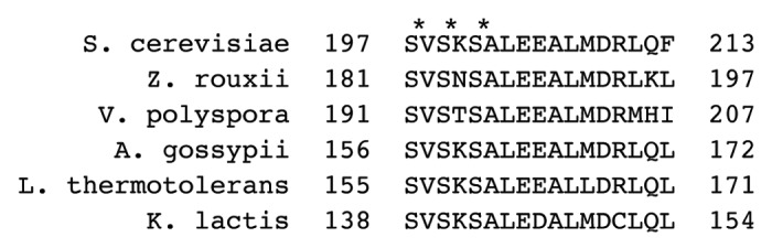
Figure 1. The C-terminal sequence of Atg29 is conserved. The C-terminal 17 amino acids of Atg29 from the indicated yeasts are indicated. The asterisks mark the last three serines (S197, S199, S201) in S. cerevisiae that appear to be particularly important in modulating the effect of the inhibitory peptide.
To better understand the function of Atg29 within the Atg17-Atg31-Atg29 complex, we performed negative stain single-particle EM analysis. In vitro purified Atg17, Atg31, and Atg29 form a stable 2:2:2 complex, and show an elongated shape resembling a stretched out letter “S.” In particular, the dimeric Atg17 forms the backbone, with Atg29 localized near the ends of this scaffold and Atg31 sandwiched in between; in conjunction with previous yeast two-hybrid studies and the recent crystal structure data, we can conclude that Atg31 bridges Atg17 with Atg29. The position of Atg29 within the Atg17-Atg31-Atg29 complex raised a possibility that Atg29 would bind to another protein; therefore we tried in vivo reconstitution of this complex using our multiple-knockout strain with Atg17-GFP as the reporter for the PAS targeting of the complex. In the absence of all other Atg proteins, the Atg17-Atg31-Atg29 complex is dispersed throughout the cytosol and is unable to target to the PAS, but localization is restored in the presence of Atg11. We found that the Atg17-Atg31-Atg29 complex associates with Atg11 through direct interaction between Atg29 and Atg11. Even though the phosphorylation of Atg29 is dispensable for its interaction with Atg31 and Atg17, it is crucial for interaction with Atg11, and the targeting of the Atg17-Atg31-Atg29 complex to the PAS. Atg11 is a scaffold protein required for selective autophagy, and deletion of ATG11 has no apparent effect on autophagy activity. However, atg11∆ shows a synthetic phenotype with an ATG29 deletion with regard to autophagy activity; the partial activity seen when Atg29 is absent from an otherwise wild-type strain is eliminated when Atg11 is also missing.
Atg1 binds Atg13 and Atg11, and the latter proteins directly bind Atg17 and Atg29, respectively. The Atg1-Atg11 interaction can be abolished by introducing 2 point mutations (L825H, W826S) in Atg1. Disturbance of this interaction has no obvious effect on the localization of Atg1 at the PAS, or on autophagy activity; we propose that this is due to the retention of Atg11 due to the Atg11-Atg29 interaction. In agreement with this hypothesis, deletion of ATG29 makes the Atg1-Atg11 interaction critical for both Atg1 puncta formation and autophagy activity.
Several subunits of the Atg1 complex are phosphoproteins, including Atg1, Atg13, Atg29, and Atg31. Previous studies found that the phosphorylation and dephosphorylation of Atg1 and Atg13 are related to autophagy activity. Here, we showed the role of Atg29 phosphorylation for autophagy induction. Future work is required to better understand the regulatory mechanism of the Atg1 complex, concerning its significant role in autophagy induction and termination.
Disclosure of Potential Conflicts of Interest
No potential conflicts of interest were disclosed.
Acknowledgments
This work was supported by NIH grant GM053396 to DJK, a University of Michigan Rackham Predoctoral Fellowship to MK, a Natural Sciences and Engineering Research Council of Canada Discovery Grant, a Michael Smith Foundation for Health Research Career Investigator Award, a Canadian Institutes of Health Research Operating Grant and New Investigator award to CKY, and a Natural Sciences and Engineering Research Council of Canada Postgraduate Scholarship award to LHC.
Footnotes
Previously published online: www.landesbioscience.com/journals/autophagy/article/26740