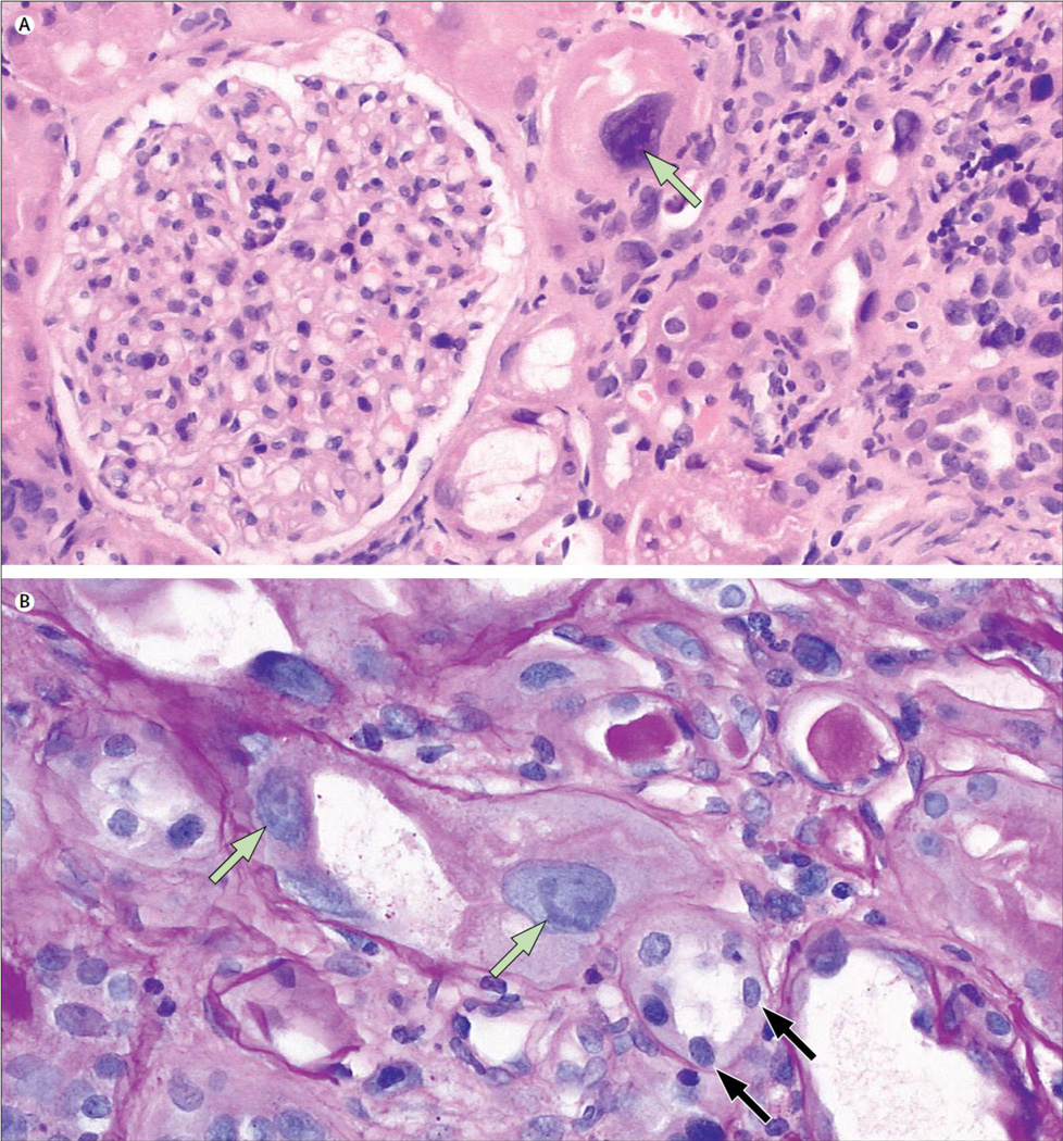
A 44-year-old white man was admitted to hospital for kidney biopsy, with progressive chronic kidney disease with mild proteinuria. Histological preparations showed bizarrely enlarged nuclei of proximal tubular epithelial cells, tubular atrophy, and interstitial fibrosis (figure).
Figure. Light micrographs of renal cells showing karyomegalic interstitial nephritis.
(A) Highly enlarged tubular epithelial cell nucleus (arrow) compared with glomerular nuclei (haemotoxylin and eosin stain, ×200). (B) Karyomegalic cells appear predominantly in proximal tubuli (green arrows). Distal tubular cells are mostly unaff ected (black arrows) (periodic acid-Schiff stain, ×400).
Clinically, karyomegalic interstitial nephritis is characterized by slow progressive renal failure in the absence of urinary sediment abnormalities, leading to end-stage renal disease in late adulthood. Because it is a systemic disease, enlarged nuclei can also be found in brain, lung, and liver tissue.


