Abstract
Human disease caused by highly pathogenic avian influenza (H5N1) is associated with fulminant viral pneumonia and mortality rates in excess of 60%. Cytokine dysregulation is thought to contribute to its pathogenesis. In comparison with human seasonal influenza (H1N1) viruses, clade 1, 2.1, and 2.2 H5N1 viruses induced higher levels of tumor necrosis factor-α in primary human macrophages. To understand viral genetic determinants responsible for this hyperinduction of cytokines, we constructed recombinant viruses containing different combinations of genes from high-cytokine (A/Vietnam/1203/04) and low-cytokine (A/WSN/33) phenotype H1N1 viruses and tested their cytokine-inducing phenotype in human macrophages. Our results suggest that the H5N1 polymerase gene segments, and to a lesser extent the NS gene segment, contribute to cytokine hyperinduction in human macrophages and that a putative H5 pandemic virus that may arise through genetic reassortment between H5N1 and one of the current seasonal influenza viruses may have a markedly altered cytokine phenotype.
Highly pathogenic avian influenza H5N1 viruses have affected poultry and other birds in >60 countries across 3 continents. They have been transmitted zoonotically to humans in 15 countries, causing human disease with an overall mortality rate of >60% and continuing to pose a pandemic threat. Since their first detection in 1996, these highly pathogenic avian influenza H5N1 viruses have continued to reassort and evolve, giving rise to a number of virus genotypes and clades [1]. Human disease has been caused by Z-genotype viruses of clade 1 in Thailand, Vietnam, and Cambodia, clade 2.1 in Indonesia, and clade 2.2 in the Middle East and Africa. More recently, clade 2.3.4 viruses of the V genotype have become dominant in China and North Vietnam, causing human disease in these countries. Although virus dissemination beyond the human respiratory tract does occur and sometimes contributes to disease severity, lung pathology remains the major cause of death. Studies in vitro and in vivo have indicated that high viral replication and cytokine dysregulation, and perhaps tissue tropism, are factors contributing to lung pathology [2]. The key target cells for the virus in the lung are the alveolar epithelial cells and macrophages [3]. We and others have shown elsewhere that primary human macrophages and alveolar epithelial cells infected with H5N1 viruses secrete markedly higher levels of proinflammatory cytokines and chemokines than do those infected with human H1N1 or H3N2 viruses [4-6] and that the interplay of such mediator interactions between macrophages and alveolar epithelial cells amplifies this inflammatory cascade [7]. So far, only a limited number of recent H5N1 viruses of clade 0 and clade 1 have been tested for their cytokine phenotype [4-6, 8, 9]. More importantly, the virus genetic determinants responsible for activating this cytokine hyperinduction remain obscure. Other studies have suggested that although the NS gene segment of the H5N1 virus may contribute modestly to the high-cytokine phenotype, it is not the major viral genetic determinant. In this study, we demonstrate that the H5N1 polymerases, rather than viral hemagglutinin (HA) and neuraminidase (NA), are the major viral genetic determinants of the cytokine dysregulation induced by H5N1 viruses in primary human macrophages.
METHODS
Viruses
The abbreviations, origins, and passage history of all the wild-type virus strains used in this study are shown in table 1. Virus infectivity was assessed by titration in Madin-Darby canine kidney (MDCK) cells. All procedures involving live H5N1 viruses and recombinant viruses were carried out in a biosafety level 3 facility.
Table 1. Wild-Type Influenza Viruses Used in This Study.
| Virus | Subtype | Abbreviation | H5N1 clade (genotype) | Country of origin | Passage history |
|---|---|---|---|---|---|
| A/Hong Kong/54/98 (H1N1) | H1N1 | 54/98 (H1N1) | … | Hong Kong | MDCK × 7 |
| A/Hong Kong/178394/04 (H1N1) | H1N1 | 178394/04 (H1N1) | … | Hong Kong | MDCK × 3 |
| A/Hong Kong/87856/05 (H1N1) | H1N1 | 87856/05 (H1N1) | … | Hong Kong | MDCK × 3 |
| A/Hong Kong/82965/05 (H1N1) | H1N1 | 829165 (H1N1) | … | Hong Kong | MDCK × 3 |
| A/goose/Hong Kong/437.6/99 (H5N1)a | H5N1 | 437.6/99 (H5N1) | 0 | Hong Kong | Egg × 4 |
| A/Hong Kong/483/97 (H5N1) | H5N1 | 483/97 (H5N1) | 0 | Hong Kong | MDCK × 8 |
| A/chicken/Indonesia/BL/03 (H5N1) | H5N1 | BL/03 (H5N1) | 2.1 (Z) | Indonesia | MDCK × 6 |
| A/chicken/Indonesia/2A/04 (H5N1) | H5N1 | 2A/04 (H5N1) | 2.1 (Z) | Indonesia | MDCK × 4 |
| A/chicken/Thailand/AIV-1/04 (H5N1) | H5N1 | AIV-1/04 (H5N1) | 1 (Z) | Thailand | Egg × 3; MDCK × 4 |
| A/chicken/Vietnam/33/04 (H5N1) | H5N1 | 33/04 (H5N1) | 1 (Z) | Vietnam | Egg × 3; MDCK × 3 |
| A/Vietnam/1203/04 (H5N1) | H5N1 | 1203/04 (H5N1) | 1 (Z) | Vietnam | MDCK × 4 |
| A/Vietnam/1194/04 (H5N1) | H5N1 | 1194/04 (H5N1) | 1 (Z) | Vietnam | MDCK × 6 |
| A/Thailand/MK2/04 (H5N1) | H5N1 | MK2/04 (H5N1) | 1 (Z) | Vietnam | MDCK × 4 |
| A/Vietnam/3046/04 (H5N1) | H5N1 | 3046/04 (H5N1) | 1 (Z) | Vietnam | MDCK × 4 |
| A/bar-headed goose/Qinghai/5/05 (H5N1) | H5N1 | QH5/05 (H5N1) | 2.2 (Z) | China | MDCK × 1 |
NOTE. MDCK, Madin-Darby canine kidney.
A/goose/Hong Kong/437.6/99 (H5N1) is from a A/goose/Guangdong/1/96 (H5N1)–like lineage.
Isolation of primary human macrophages and primary type I-like pneumocytes
Blood mononuclear cells from healthy donors (Hong Kong Red Cross Blood Transfusion Service) were separated by Ficoll-Paque centrifugation, and monocytes were purified by the adherence method and differentiated into macrophages, as described elsewhere [4]. Primary human type I-like pneumocytes were isolated from nontumor lung tissues using methods described elsewhere [5].
Infection of macrophages and pneumocytes
Primary macrophages and type I-like pneumocytes were seeded at 1 × 10⁵ cells per well in 24-well tissue-culture plates. The cells were infected at a multiplicity of infection of 2 unless otherwise indicated. After 30 min of virus adsorption, the virus inoculum was removed, and the cells were washed with phosphate-buffered saline and incubated in warm culture medium (serum-free medium for macrophages [GIBCO] and small airway growth medium for pneumocytes [Cambrex BioScience]) supplemented with 0.6 mg/L penicillin, 60 mg/L streptomycin, and 1 mg/L N-p-tosyl-l-phenylalanine chloromethyl ketone-treated trypsin (Sigma). Samples of culture supernatant were collected for virus titration and cytokine analysis. RNA was extracted from cells for analysis of cytokine gene expression. To confirm that the cells were all infected, they were fixed at 8 h after infection and analyzed by immunofluorescent staining specific for influenza virus nucleoprotein and matrix proteins (Dako Imagen; Dako Diagnostics).
Quantification of cytokine messenger RNA by real-time reverse-transcription polymerase chain reaction (PCR)
DNase-treated total RNA was isolated using the RNeasy Mini kit (Qiagen). The complementary DNA (cDNA) was synthesized from messenger RNA (mRNA) with poly(dT) primers and SuperScript II reverse transcriptase (Life Technologies). Transcript expression was determined using TaqMan Fast Universal Master Mix kit (Applied Biosystems) with specific primers. The fluorescence signals were measured by ABI Prism 7500 Sequence Detection System. The gene copy number of the target gene was normalized to the β-actin control and compared with the results of known amounts of plasmid encoding the target gene sequence. The results were presented as copy numbers of target genes per 1 × 10⁵ copies of β-actin control.
Quantitative analysis of tumor necrosis factor (TNF)-α by enzyme-linked immunosorbent assay in cell culture supernatants
The cell culture supernatants were collected at 6 h after infection and were irradiated with ultraviolet light (CL-100 ultraviolet crosslinker; UVP) for 15 min to inactivate the virus before the levels of TNF-α were measured by a specific TNF-α assay kit (R&D Systems), as described elsewhere [4].
Virus titration
The amount of virus in the supernatants of the influenza virus-infected macrophage cultures was titrated on MDCK cells, and the titers were reported as median tissue culture infective dose units per 100 μL (TCID50/100 μL).
Plasmid construction and reverse genetics
Recombinant viruses were generated by Reverse Genetics [10, 11]. The cDNAs of the A/Vietnam/1203/04 (H5N1) and A/WSN/33 (H1N1) strains were first synthesized by reverse transcription of viral RNA with an oligonucleotide (Uni-12) complementary to the conserved 3′ end of the viral RNA. The cDNA was then amplified by PCR with gene-specific primers. The reverse-transcription PCR products of viral RNA segments were cloned into the plasmid pHW2000. The newly introduced viral RNA in the recombinant viruses was sequenced for further confirmation. The mutant PB2 construct containing a mutation with amino acid at position 627 (1203[PB2-K627E]) was produced by PCR amplification with the primers possessing the mutation.
Statistical analysis
The statistical significance of differences between experimental groups was determined by using the unpaired, nonparametric Student’s t test. Correlation between experimental groups was determined by using the Spearman nonparametric test. For both tests, differences were considered significant at P < .05.
RESULTS
TNF-α induction in human macrophages infected by different clades of H5N1 viruses
Our previous studies showed that some H5N1 viruses isolated from 1997 through 2003 induced a high proinflammatory cytokine response in primary human macrophages and in infected humans [4, 7, 12]. To extend these findings to more recent clade 1, 2.1, and 2.2 viruses of the Z-genotype viruses isolated in 2004 and 2005, we compared viruses isolated from humans and poultry from 2004 through 2005 with the H5N1 virus isolated from humans in 1997 (H5N1/97). Human seasonal H1N1 influenza viruses and a virus representing the parental A/Gs/Gd/96-like H5N1 lineage (A/Gs/HK/437.6/99) were used for comparison. The viruses used are listed in table 1.
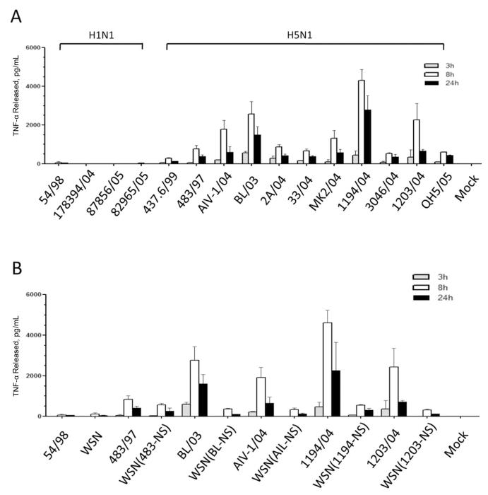
Primary human macrophages were infected at a multiplicity of infection of 2, and the level of TNF-α secreted in the supernatant was quantified at 3, 8, and 24 h after infection with use of an enzyme-linked immunosorbent assay (figure 1A). These H5N1 viruses induced varying levels of TNF-α, but they all induced higher levels of TNF-α than did H1N1 viruses (figure 1A). In particular, the H5N1 viruses isolated from 2 human subjects with fatal cases, 1194/04 and 1203/04, as well as the viruses isolated from chickens, BL/03 and AIV-1/04, induced [H11091]2-fold greater TNF-α levels than did H5N1 483/97. The levels of mRNA expression were consistent with results of the protein assays (data not shown). Our results suggest that, in comparison with a range of human H1N1 viruses, human and poultry Z-genotype viruses of clades 1, 2.1, and 2.2 isolated in 2004 and 2005 are all potent hyperinducers of cytokines such as TNF-α, possibly even more potent than H5N1 483/97.
Figure 1.
Tumor necrosis factor (TNF)-α secreted by primary human macrophages infected with H5N1 viruses of different clades and genotypes and H1N1 viruses (A) or recombinant H1N1 viruses containing NS gene segments of H5N1 viruses (B). Primary human macrophages were infected by H5N1 or H1N1 viruses at a multiplicity of infection of 2, and the amount of TNF-α released into the supernatant was quantified by enzyme-linked immunosorbent assay at 3 (gray), 8 (white), and 24 h (black) after infection. Error bars indicate standard deviations calculated from the results of 3 independent experiments that used macrophages from different donors. Mock, uninfected cells. A, Macrophages infected with H5N1 viruses of clades 0, 1, 2.1, and 2.2 and different H1N1 viruses. Virus abbreviations are shown in table 1. B, Macrophages similarly infected with recombinant H1N1 (A/WSN/33) viruses with NS gene segments of different H5N1 viruses and the relevant precursor wild-type controls. Virus abbreviations are shown in table 2.
Contribution by H5N1 NS proteins and polymerases but not surface proteins to high-cytokine phenotype
We have shown elsewhere that recombinant A/WSN/33 (WSN) influenza containing the NS gene segment of H5N1/97 virus is a more potent inducer of TNF-α than the WSN H1N1 parent, although such a recombinant virus did not fully regain the high-cytokine phenotype manifested by the H5N1 virus parent [4]. Because the internal genes (including the NS gene) of the contemporary Z-genotype H5N1 viruses have different evolutionary derivations compared with H5N1/97, we investigated whether the NS gene segment of contemporary Z-genotype viruses also contributes to the high-cytokine phenotype. Recombinant viruses carrying the NS gene segment of the H5N1 viruses A/Hong Kong/483/97, A/chicken/Indonesia/BL/03, A/chicken/Thailand/AIV-1/04, A/Vietnam/1194/04, or A/Vietnam/1203/04 were constructed in a background of WSN (H1N1) virus, which is a low cytokine inducer, and a plasmid-derived WSN wild-type virus was used as control. At 8 h after infection, the recombinant viruses produced higher concentrations of TNF-α than did the WSN control virus (figure 1B). However, as with the data reported elsewhere for the H5N1/97 virus [4], although each of the NS genes of Z-genotype H5N1 viruses increased the level of TNF-α induction compared with the parental WSN virus, the level of induction in each case was far below that induced with the wild-type H5N1 virus. We therefore concluded that the increase in TNF-α produced by Z-genotype H5N1 viruses is only partly attributable to the H5N1 NS gene segment.
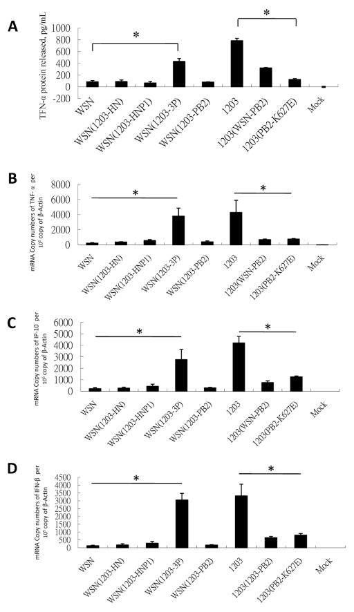
To determine the major viral genetic determinants of Z-genotype viruses that mediate the high-TNF-α phenotype, recombinant viruses were constructed that contained different combinations of a H5N1 virus A/Vietnam/1203/04 (1203) gene segments in a background of a H1N1 virus A/WSN/33 (WSN) (table 2). We investigated whether the surface proteins (HA or NA) or the polymerase genes and gene products are key factors that mediate the induction of high levels of TNF-α. The TNF-α levels induced in primary human macrophages infected by these recombinant viruses were tested (figure 2A and 2B), and the data are summarized in table 3. Recombinant virus carrying the H5N1-1203 surface proteins HA and NA in a WSN background, WSN(1203-HN), produced low levels of TNF-α in human macrophages, comparable to levels produced by the WSN control virus. Moreover, the WSN(1203-HNP1) virus, which carries HA, NA, and PB1 gene segments from Z-genotype H5N1, also did not induce high levels of TNF-α. We have independently confirmed these findings with use of a recombinant A/PR/8/34 (H1N1) virus (low-cytokine phenotype) containing 1203-HA, NA, and PB1 (data not shown).
Table 2. Recombinant Influenza Viruses Used in This Study.
| Recombinant virus | Virus used as background | Viral segments or point mutations substituted in the background |
|---|---|---|
| WSN | A/WSN/33 (H1N1) | … |
| WSN (483-NS) | A/WS N/33 (H1N1) | NS; A/Hong Kong/483/97 (H5N1) |
| WSN (BL-NS) | A/WSN/33 (H1N1) | NS; A/chicken/Indonesia/BL/03 (H5N1) |
| WSN (AIV-NS) | A/WSN/33 (H1N1) | NS; A/chicken/Thailand/AIV-1/04 (H5N1) |
| WSN (1194-NS) | A/WSN/33 (H1N1) | NS; A/Vietnam/1194/04 (H5N1) |
| WSN (1203-NS) | A/WSN/33 (H1N1) | NS; A/Vietnam/1203/04 (H5N1) |
| WSN (1203-HN) | A/WSN/33 (H1N1) | HA, NA; A/Vietnam/1203/04 (H5N1) |
| WSN (1203-HNP1) | A/WSN/33 (H1N1) | HA, NA, PB1; A/Vietnam/1203/04 (H5N1) |
| WSN (1203-3P) | A/WSN/33 (H1N1) | PA, PB1, PB2; A/Vietnam/1203/04 (H5N1) |
| WSN (1203-PB2) | A/WSN/33 (H1N1) | PB2; A/Vietnam/1203/04 (H5N1) |
| 1203 | A/Vietnam/1203/04 (H5N1) | … |
| 1203 (WSN-PB2) | A/Vietnam/1203/04 (H5N1) | PB2; A/WSN/33 (H1N1) |
| 1203 (WSN-3P) | A/Vietnam/1203/04 (H5N1) | PA, PB1, PB2; A/WSN/33 (H1N1) |
| 1203 (PB2-K627E) | A/Vietnam/1203/04 (H5N1) | PB2 residue at position 627 changed from lysine to glutamic acid |
| PR8 | A/PR/8/34 (H1N1) | … |
| PR8 (1203-HNP1) | A/PR/8/34 (H1N1) | HA, NA, PB1; A/Vietnam/1203/04 (H5N1) |
NOTE. HA, hemagglutinin; NA, neuraminidase.
Figure 2.
H5N1 polymerase genes contribute to hyperinduction of cytokine gene expression, but H5N1 surface proteins do not. Primary human macrophages were infected by recombinant viruses WSN or 1203 or the 1203 polymerase gene/surface protein-reassortant virus at a multiplicity of infection of 2. A, Tumor necrosis factor (TNF)-α released into the supernatant was quantified by enzyme-linked immunosorbent assay at 6 h after infection. The messenger RNA (mRNA) of the samples was collected at 3 h after infection. The copy numbers of TNF-α (B), interferon (IFN)-inducible protein (IP-10) (C), and IFN-β (D) mRNA were analyzed by Taqman real-time reverse-transcription polymerase chain reaction. Values were normalized by the copy numbers of β-actin gene and statistically analyzed by 2-tailed, paired t test. Mock, uninfected cells. *P < .05.
Table 3. Contribution of 1203/04 Gene Segments to Tumor Necrosis Factor (TNF)-α Induction in Human Macrophages.
| Recombinant virus | Virus segment | TNF-α induction, compared with 1203/04, % | |||||||
|---|---|---|---|---|---|---|---|---|---|
| HA | NA | PB1 | PB2 | PA | NS | NP | M | ||
| WSN | WSN | WSN | WSN | WSN | WSN | WSN | WSN | WSN | 11.0 |
| WSN (1203-HN) | H5N1 | H5N1 | WSN | WSN | WSN | WSN | WSN | WSN | 11.4 |
| WSN (1203-HNP1) | H5N1 | H5N1 | H5N1 | WSN | WSN | WSN | WSN | WSN | 8.4 |
| WSN (1203-3P) | WSN | WSN | H5N1 | H5N1 | H5N1 | WSN | WSN | WSN | 55.1 |
| WSN (1203-PB2) | WSN | WSN | WSN | H5N1 | WSN | WSN | WSN | WSN | 10.4 |
| 1203 | H5N1 | H5N1 | H5N1 | H5N1 | H5N1 | H5N1 | H5N1 | H5N1 | 100.0 |
| 1203 (WSN-PB2) | H5N1 | H5N1 | H5N1 | WSN | H5N1 | H5N1 | H5N1 | H5N1 | 40.9 |
| 1203(PB-2K627E) | H5N1 | H5N1 | H5N1 | H5N1a | H5N1 | H5N1 | H5N1 | H5N1 | 16.0 |
NOTE. HA, hemagglutinin; NA, neuraminidase.
Glutamic acid at position 627 of PB2.
WSN virus carrying the 3 polymerase gene segments of H5N1 (WSN[1203-3P]) was a strong inducer of TNF-α, comparable to the H5N1 virus itself (figure 2A and table 3). A reverse gene exchange (1203[WSN-3P]) virus was also a high TNF-α inducer (data not shown). H5N1 1203/04 PB2 in a WSN background (WSN[1203-PB2]) had low TNF-α induction (10.4% of that with the 1203 virus) (table 3), whereas a virus that carried the WSN PB2 gene segment in a H5N1 background (1203[WSN-PB2]) had moderate TNF-α induction (40.9% of that with the 1203 virus) (table 3). These findings were paralleled by TNF-α mRNA levels (figure 2B).
Other studies have shown that the lysine at position 627 in the H5N1 PB2 residue determines the high virulence of H5N1 viruses in the in vivo mouse model [13]. To understand whether cytokine induction was affected by this amino acid substitution, primary human macrophages were infected with recombinant 1203/04 virus with and without a single change at PB2 627 residue from lysine to glutamic acid (1203[PB2-K627E]). Surprisingly, the TNF-α protein level decreased >70%, suggesting that this amino acid residue in the H5N1 1203 PB2 gene is essential for cytokine hyperinduction within the context of the 1203 H5N1 gene constellation.
Because we showed elsewhere that H5N1/97 hyperinduces chemokines and interferons (IFNs) in addition to TNF-α in virus-infected human macrophages, we further examined IFN-induced protein (IP-10; chemokine [C-X-C motif] ligand 10) and IFN-β mRNA levels in human macrophages infected with the recombinant virus panel [4]. We found that the induction profile of these 2 cytokines showed a pattern similar to that seen with TNF-α (figure 2C and 2D). These results suggest that the polymerases of H5N1 control not only TNF-α induction but also the induction of other proinflammatory chemokines and IFN.
We have shown elsewhere that H5N1 viruses also manifest a high-cytokine phenotype when infecting primary human lung pneumocytes but do not directly induce TNF-α [5]. Here we tested whether this hyperinduction can be perturbed using H5N1 viruses with a mutation from lysine to glutamic acid at position 627 in the PB2 gene segment. Primary human type 1-like pneumocytes were infected with 1203(PB2-K627E), 1203/04, and WSN/33 viruses under the same conditions used for the human macrophage infection experiments. IP-10 and IFN-β transcription decreased significantly in 1203(PB2-K627E)-infected pneumocytes, compared with those infected with the 1203/04 control virus (figure 3). This showed that the hyper-induction of cytokine in human pneumocytes is also regulated by the lysine at PB2 627 in H5N1 virus.
Figure 3.
Lysine at position 627 of H5N1 PB2 affects cytokine gene expression in primary human pneumocytes. Primary human pneumocytes were infected by recombinant viruses WSN, 1203, or 1203(PB2-K627E) at a multiplicity of infection of 2. The messenger RNA (mRNA) of the samples was collected at 3, 6, and 12 h after infection. The copy numbers of interferon (IFN)-inducible protein (IP-10) (A) and IFN-β (B) mRNA were analyzed by Taqman real-time reverse-transcription polymerase chain reaction. Values were normalized by the copy numbers of β-actin gene and statistically analyzed by 2 tailed, paired t test. Mock, uninfected cells. *P < .05.
Replication of influenza viruses in human macrophages
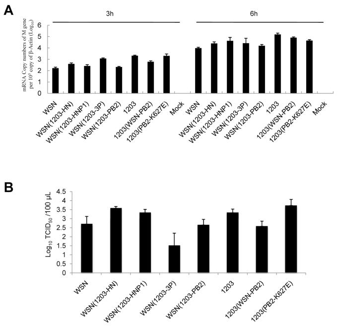
We next investigated whether the difference in cytokine induction is simply a manifestation of increased virus replication competence in primary human macrophages. Viral replication competence, as quantified by M gene virus load 3 h after infection, was correlated with TNF-α protein levels at 6-8 h after infection. When low- and high-cytokine phenotype viruses (ie, H5N1 and H1N1) were considered together, there was no correlation between M gene copy numbers and TNF-α levels (P = .34, by Spearman test). However, among the high-cytokine phenotype wild-type viruses (highly pathogenic avian influenza H5N1, excluding 437.6/99, which is a low-cytokine H5N1 precursor), there was a positive correlation between viral M gene copies and TNF-α levels (figure 1A) (P < .05, by Spearman test). There was a similar positive correlation between M gene levels and TNF-α levels within the WSN-H5N1 1203 recombinant viruses (figures 2A and 4A) (P < .05, by Spearman test). However, although the viral M gene copies and TCID50 titers of 1203(PB2-K627E) virus were similar to those of wildtype 1203 recombinant control virus (figure 4A and 4B), cytokine induction was significantly reduced (figure 2A), suggesting that glutamic acid at position 627 of H5N1 PB2 virus enhances cytokine induction by a mechanism other than alteration of virus replication competence.
Figure 4.
Viral messenger RNA (mRNA) transcription and infectious virus yield of the recombinant viruses. Primary human macrophages were infected by recombinant viruses WSN or 1203 or the 1203 polymerase gene/surface protein-reassortant virus at a multiplicity of infection of 2. A, Levels of matrix protein mRNA (M gene) at 3 and 6 h after infection were quantified by Taqman real-time reverse-transcription polymerase chain reaction. B, Primary human macrophages were infected by the recombinant viruses at an multiplicity of infection of 0.01. Median tissue culture infective dose (TCID50) of samples at 24 h after infection were determined by titration on Madin-Darby canine kidney cells. Mock, uninfected cells.
DISCUSSION
We and others have shown elsewhere that H5N1/97 viruses and those of the Z genotype associated with the more recent outbreaks of disease in poultry and humans have a high-cytokine phenotype in primary human macrophages [4, 6]. However, not all H5N1 viruses isolated during the past 14 years have the same phenotype. For example, viruses of the precursor A/goose/Guangdong/1/96 H5N1 virus genotype and those of genotype B, which do not share the same constellation of virus internal genes, also do not share this property of high cytokine induction [8]. In this study, we have shown that clade 1, 2.1, and 2.2 H5N1 viruses of Z genotype, isolated from 2004 through 2005 from humans or from birds, retain the high-cytokine phenotype in virus-infected primary human macrophages. The cytokine phenotype of the V-genotype viruses that predominated in southern China since 2005 [14] and in North Vietnam and Laos in 2006-2007 [15] have not yet been investigated. These V-genotype viruses differ from Z-genotype viruses only in their PA genes.
Because previous data have suggested that cytokine dysregulation is a contributory factor in H5N1-related disease pathogenesis, our results may reflect one aspect of the virulence of the Z-genotype viruses in humans [4, 5, 8, 12, 16]. We have demonstrated that the virus polymerase complex and, to a lesser extent, the NS gene segment of H5N1 are 2 viral genetic determinants that contribute to cytokine hyperinduction. Although the genetic background of the internal genes in H5N1/97 and Z-genotype viruses are different, we showed that NS genes of both viruses help contribute to the cytokine hyperinduction. The NS1 protein generated from the NS gene is known to play a role in the inhibition of type I IFN and IFN-induced proteins, such as double-stranded RNA-dependent protein kinase R and 2′5′-oligoadenylate synthetase/RNase L during infection [17-19]. In addition, it has been shown that the hyperinduction of TNF-α in H5N1-infected primary human macrophages is partly regulated by the activation of p38 kinase [20]. It remains to be investigated whether the NS gene of H5N1 viruses is responsible for activating p38 (either directly or through lack of inhibition), leading to the induction of TNF-α in primary human macrophages.
The PB2 gene and, to a lesser extent, the HA gene have been identified as contributing to the virulence of H5N1 viruses in mice [21]. Our in vitro study showed that the HA gene of H5N1 does not by itself regulate cytokine induction in human macrophages. However, it may well contribute to virus virulence in other ways, for example, by promoting virus dissemination beyond the respiratory tract.
It is of interest that the virulence of the 1203/04 virus in ferrets was determined by the polymerase complex and, to a lesser extent, the NS gene segment [22]. These results are concordant with our findings that the polymerase complex genes from H5N1 virus are needed to induce high levels of cytokine (WSN[1203-3P]). The mechanism by which the polymerase gene complex determines cytokine hyperinduction is still not known. Our findings suggest that the synergistic functionality of the homologous polymerase gene complex is important for the high-cytokine phenotype. WSN (H1N1) virus carrying the PB2, PB1, and PA gene segments of 1203 (H5N1) virus manifest a high cytokine activity. However, a 1203 (H5N1) virus carrying WSN (H1N1) PB2, PB1, and PA gene segments also manifested a high-cytokine phenotype, although the WSN polymerases in the homologous WSN background did not. Similarly, PB2-K627E substitution in 1203 affects the cytokine phenotype, although this substitution is not a sole determinant of cytokine induction. For example, WSN has PB2-K627, although it is a low-cytokine phenotype virus, and, conversely, some high-cytokine phenotype H5N1 viruses have PB2-E627. Similarly, avian A/quail/Hong Kong/G1/97-like H9N2 viruses, which induce high cytokine production in human macrophages, contain glutamic acid at the PB2 627 position. These findings suggest that PB2-K627 is not a sole determinant of the high-cytokine phenotype. Thus, the overall polymerase functionality and its context within the other viral genes all appear to be important in determining the ability of a virus to induce high cytokine levels in human cells.
Because viral transcription and replication are mainly regulated by the polymerases, the efficiency of virus replication may be related to the high-cytokine phenotype. Interestingly, within the high-cytokine phenotype H5N1 viruses and within the panel of recombinant viruses we generated, there was significant positive correlation between viral gene transcription (as assessed by virus M gene copy numbers 3 h after infection) (figure 4A) and TNF-α protein levels 6 h after infection (figures 1A and 2A) (P < .05, by Spearman test). However, when we considered all high- and low-cytokine phenotype wild-type H1N1 and H5N1 viruses studied here (figure 1A), there was no significant correlation between virus M gene copy numbers and TNF-α protein levels. Thus, we conclude that viral replication competence does not explain the marked differences in cytokine induction between low pathogenic H1N1 and highly pathogenic avian influenza H5N1 viruses. However, it does explain the variability in cytokine induction within the highly pathogenic avian influenza H5N1 virus group. The 1203(PB2-K627E) virus has similar virus replication competence in human macrophages when compared with the 1203 wild-type virus 1203(PB2-627K) but still has a low-cytokine phenotype.
The pandemics of 1957 and 1968 arose through reassortment, with the prevailing human seasonal influenza virus acquiring HA and PB1 genes (and the NA gene in 1957) from an avian influenza virus. Our results show that a human H1N1 virus with H5N1 HA, NA, and PB1 genes does not retain the high-cytokine phenotype. These results have also been repeated with a different H1N1 virus, A/PR/8/34. Thus, a pandemic virus that arises through reassortment between H5N1 and contemporary seasonal influenza viruses may have an unpredictable cytokine phenotype. However, we remain cognizant of the fact that the H5 HA retains virulence properties of its own, irrespective of cytokine induction.
Although antiviral drugs remain the mainstay of treatment for H5N1 disease in humans, an understanding of the pathogenesis of the disease process may lead to novel adjunctive therapies. This is particularly relevant in view of the poor overall survival in patients who begin treatment later in the course of the disease when the immunopathological cascades already initiated cannot be controlled by antiviral treatment alone [23].
Acknowledgments
The authors wish to thank Dr H. L. Yen and Dr L. L. M. Poon for advice and help with the reverse genetics experiments and May Yu, Carolina Leung, Ken Tong, and Wendy Yu for their technical support.
Financial support: National Institutes of Health (National Institute of Allergy and Infectious Diseases contract HHSN266200700005C), Research Grants Council of Hong Kong (Central Allocation grant HKU 1/05C), University Grants Committee of the Hong Kong Special Administrative Region, China (Area of Excellence grant; project AoE/M-12/06), and Research Fund for the Control of Infectious Diseases (grant 01030172).
Footnotes
Potential conflicts of interest: none reported.
References
- 1.Duan L, Bahl J, Smith GJ, et al. The development and genetic diversity of H5N1 influenza virus in China, 1996-2006. Virology. 2008;380:243–54. doi: 10.1016/j.virol.2008.07.038. [DOI] [PMC free article] [PubMed] [Google Scholar]
- 2.Peiris JSM, de Jong MD, Guan Y. Avian influenza virus (H5N1): a threat to human health. Clin Microbiol Rev. 2007;20:243–67. doi: 10.1128/CMR.00037-06. [DOI] [PMC free article] [PubMed] [Google Scholar]
- 3.Nicholls JM, Chan MC, Chan WY, et al. Tropism of avian influenza A (H5N1) in the upper and lower respiratory tract. Nat Med. 2007;13:147–9. doi: 10.1038/nm1529. [DOI] [PubMed] [Google Scholar]
- 4.Cheung CY, Poon LL, Lau AS, et al. Induction of proinflammatory cytokines in human macrophages by influenza A (H5N1) viruses: a mechanism for the unusual severity of human disease? Lancet. 2002;360:1831–7. doi: 10.1016/s0140-6736(02)11772-7. [DOI] [PubMed] [Google Scholar]
- 5.Chan MC, Cheung CY, Chui WH, et al. Proinflammatory cytokine responses induced by influenza A (H5N1) viruses in primary human alveolar and bronchial epithelial cells. Respir Res. 2005;6:135. doi: 10.1186/1465-9921-6-135. [DOI] [PMC free article] [PubMed] [Google Scholar]
- 6.Perrone LA, Plowden JK, García-Sastre A, Katz JM, Tumpey TM. H5N1 and 1918 pandemic influenza virus infection results in early and excessive infiltration of macrophages and neutrophils in the lungs of mice. PLoS Pathog. 2008;4:e1000115. doi: 10.1371/journal.ppat.1000115. [DOI] [PMC free article] [PubMed] [Google Scholar]
- 7.Lee SM, Cheung CY, Nicholls JM, et al. Hyperinduction of cyclooxygenase-2-mediated proinflammatory cascade: a mechanism for the pathogenesis of avian influenza H5N1 infection. J Infect Dis. 2008;198:525–35. doi: 10.1086/590499. [DOI] [PubMed] [Google Scholar]
- 8.Guan Y, Poon LL, Cheung CY, et al. H5N1 influenza: a protean pandemic threat. Proc Natl Acad Sci USA. 2004;101:8156–61. doi: 10.1073/pnas.0402443101. [DOI] [PMC free article] [PubMed] [Google Scholar]
- 9.Hui KP, Lee SM, Cheung CY, et al. Induction of proinflammatory cytokines in primary human macrophages by influenza A virus (H5N1) is selectively regulated by IFN regulatory factor 3 and p38 MAPK. J Immunol. 2009;182:1088–98. doi: 10.4049/jimmunol.182.2.1088. [DOI] [PubMed] [Google Scholar]
- 10.Neumann G, Kawaoka Y. Reverse genetics of influenza virus. Virology. 2001;287:243–50. doi: 10.1006/viro.2001.1008. [DOI] [PubMed] [Google Scholar]
- 11.Hoffmann E, Webster RG. Unidirectional RNA polymerase I-polymerase II transcription system for the generation of influenza A virus from 8 plasmids. J Gen Virol. 2000;81:2843–7. doi: 10.1099/0022-1317-81-12-2843. [DOI] [PubMed] [Google Scholar]
- 12.Peiris JS, Yu WC, Leung CW, et al. Re-emergence of fatal human influenza A subtype H5N1 disease. Lancet. 2004;363:617–9. doi: 10.1016/S0140-6736(04)15595-5. [DOI] [PMC free article] [PubMed] [Google Scholar]
- 13.Hatta M, Gao P, Halfmann P, et al. Molecular basis for high virulence of Hong Kong H5N1 influenza A viruses. Science. 2001; 293:1840–2. doi: 10.1126/science.1062882. [DOI] [PubMed] [Google Scholar]
- 14.Smith GJ, Fan XH, Wang J, et al. Emergence and predominance of an H5N1 influenza variant in China. Proc Natl Acad Sci U S A. 2006;103:16936–41. doi: 10.1073/pnas.0608157103. [DOI] [PMC free article] [PubMed] [Google Scholar]
- 15.Dung Nguyen T, Vinh Nguyen T, Vijaykrishna D, et al. Multiple sublineages of influenza A virus (H5N1), Vietnam, 2005-2007. Emerg Infect Dis. 2008;14:632–6. doi: 10.3201/eid1404.071343. [DOI] [PMC free article] [PubMed] [Google Scholar]
- 16.de Jong MD, Simmons CP, Thanh TT, et al. Fatal outcome of human influenza A (H5N1) is associated with high viral load and hypercytokinemia. Nat Med. 2006;12:1203–7. doi: 10.1038/nm1477. [DOI] [PMC free article] [PubMed] [Google Scholar]
- 17.Garcia-Sastre A. Inhibition of interferon-mediated antiviral responses by influenza A viruses and other negative-strand RNA viruses. Virology. 2001;279:375–84. doi: 10.1006/viro.2000.0756. [DOI] [PubMed] [Google Scholar]
- 18.Krug RM, Yuan W, Noah DL, Latham AG. Intracellular warfare between human influenza viruses and human cells: the roles of the viral NS1 protein. Virology. 2003;309:181–9. doi: 10.1016/s0042-6822(03)00119-3. [DOI] [PubMed] [Google Scholar]
- 19.Hale BG, Randall RE, Ortín J, Jackson D. The multifunctional NS1 protein of influenza A viruses. J Gen Virol. 2008;89:2359–76. doi: 10.1099/vir.0.2008/004606-0. [DOI] [PubMed] [Google Scholar]
- 20.Lee DC, Cheung CY, Law AH, et al. p38 mitogen-activated protein kinase-dependent hyperinduction of tumor necrosis factor alpha expression in response to avian influenza virus H5N1. J Virol. 2005;79:10147–54. doi: 10.1128/JVI.79.16.10147-10154.2005. [DOI] [PMC free article] [PubMed] [Google Scholar]
- 21.Hatta M, Gao P, Halfmann P, Kawaoka Y. Molecular basis for high virulence of Hong Kong H5N1 influenza A viruses. Science. 2001;293:1840–2. doi: 10.1126/science.1062882. [DOI] [PubMed] [Google Scholar]
- 22.Salomon R, Franks J, Govorkova EA, et al. The polymerase complex genes contribute to the high virulence of the human H5N1 influenza virus isolate A/Vietnam/1203/04. J Exp Med. 2006;203:689–97. doi: 10.1084/jem.20051938. [DOI] [PMC free article] [PubMed] [Google Scholar]
- 23.Writing Committee of the Second World Health Organization Consultation on Clinical Aspects of Human Infection with Avian Influenza A (H5N1) Virus. Abdel-Ghafar AN, Chotpitayasunondh T, et al. Update on avian influenza A (H5N1) virus infection in humans. N Engl J Med. 2008;358:261–73. doi: 10.1056/NEJMra0707279. [DOI] [PubMed] [Google Scholar]


