FIG 2 .
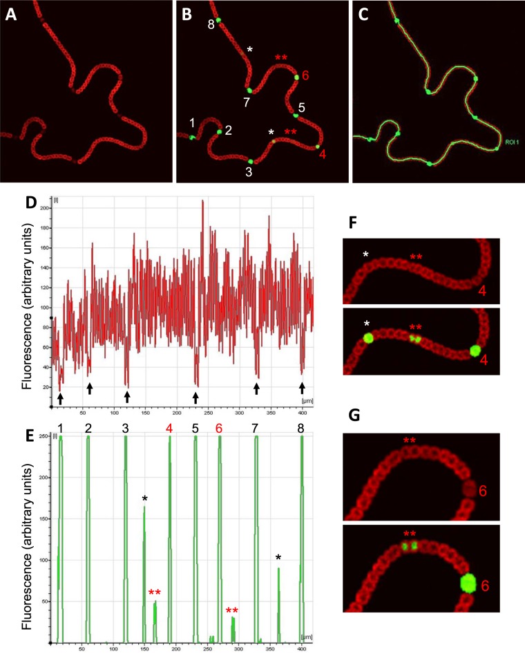
Expression of PnsiR1-gfp along nitrogen-fixing filaments. Confocal fluorescence images of a filament growing on top of nitrogen-free medium are shown for the red (A) and red plus green (B) channels, together with the region of interest (ROI) drawn to quantify signals along the filament (C). Quantification of the signals is shown for the red (D) and green (E) channels. Black arrows in panel D indicate the positions of mature heterocysts. Two regions of the filament are enlarged and enhanced for better observation of green fluorescence in panels F and G. Mature heterocysts (numbered 1, 2, 3, 5, 7, and 8) are indicated in white (B) or black (E). Immature heterocysts (numbered 4 and 6) are indicated in red. Prospective heterocysts are indicated by single asterisks. Double red asterisks indicate pairs of cells showing distinct green fluorescence above background.

