Abstract
As our population ages, there is a greater need for a suitable supply of engineered tissues to address a range of debilitating ailments. Stem cell based therapies are envisioned to meet this emerging need. Despite significant progress in controlling stem cell differentiation, it is still difficult to engineer human tissue constructs for transplantation. Recent advances in micro- and nanofabrication techniques have enabled the design of more biomimetic biomaterials that may be used to direct the fate of stem cells. These biomaterials could have a significant impact on the next generation of stem cell based therapies. Here, we highlight the recent progress made by micro- and nanoengineering techniques in the biomaterials field in the context of directing stem cell differentiation. Particular attention is given to the effect of surface topography, chemistry, mechanics and micro- and nanopatterns on the differentiation of embryonic, mesenchymal and neural stem cells.
Keywords: micro- and nanotopography, microwells, microarrays, embryonic and adult stem cells, stem cell therapy
1. Introduction
With the increasing number of patients suffering from damaged or diseased organs and the shortage of organ donors, the need for methods to construct human tissues outside the body has risen. To address this issue, the interdisciplinary field of tissue engineering has emerged in the past few years to generate biological tissue constructs that maintain or enhance normal tissue function [1,2].
One of the current challenges in the development of tissue engineered constructs is the lack of a renewable cell source. Embryonic, induced pluripotent and adult stem cells are promising cell sources in therapeutic and regenerative medicine. Due to their ability to self-renew and differentiate into various cell types, these cells could potentially be cultured and harvested for regeneration of damaged, injured and aged tissue [3,4]. Embryonic stem cells (ESC) are pluripotent with the ability to differentiate into cells of all three germ layers, ectoderm, endoderm, and mesoderm, whereas adult stem cells (ASC) are multipotent with the capacity to differentiate into a limited number of cell types [5]. For instance, mesenchymal stem cells (MSCs) which reside in the bone marrow, can differentiate into bone (osteoblasts) [6], muscle (myoblasts) [7], fat (adipocytes) [8] and cartilage (chrondocytes) [5] cells, while neural stem cells (NSCs) either give rise to support cells in the nervous system of vertebrates (astrocytes and oligodendrocytes) or neurons [9].
In vivo, differentiation and self-renewal of stem cells is dominated by signals from their surrounding microenvironment [10]. This microenvironment or “niche” is composed of other cell types as well as numerous chemical, mechanical and topographical cues at the micro- and nanoscale, which are believed to serve as signaling mechanisms to control the cell behavior [11]. For instance, extracellular matrix (ECM) molecules such as collagen [12] as well as the basement membrane of the tissue matrix [13] contain micro- and nanoscale features. Tissue stiffness is also known to vary depending on the organ type, disease state and aging process [14,15,16]. In tissue culture, stem cell differentiation has traditionally been controlled by the addition of soluble factors to the growth media [17]. However, despite much research, most stem cell differentiation protocols yield heterogeneous cell types [18,19]. Therefore, it is desirable to use more biomimetic in vitro culture conditions to regulate stem cell differentiation and self-renewal.
Recent advances in micro- and nanofabrication technology have paved the way to create substrates with precise micro- and nanocues, variable stiffness and chemical composition to better mimic the in vivo microenvironment [2,20,21]. By employing approaches such as self-assembled monolayers (SAMs), microcontact printing, e-beam, photo- and soft lithography, tissue engineers aim to incorporate topographical, mechanical and chemical cues into biomaterials to control stem cell fate decisions [2,21,22]. This review highlights recent progress made by using micro- and nanoengineered biomaterials to direct the fate of stem cells, with particular emphasis on ESCs, MSCs and NSCs.
2. Biomaterials with Micro- and Nanoscale Features for Directing Stem Cell Fate
2.1. Stem Cell Niche in Vivo
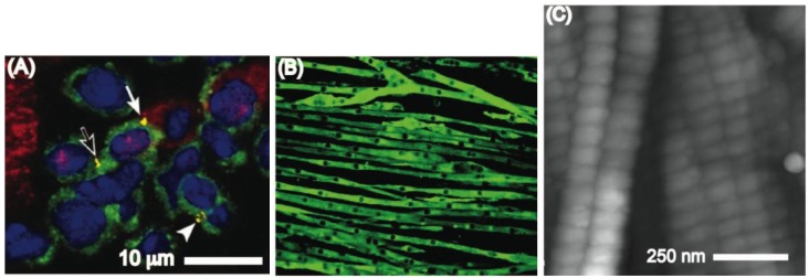
In the body the cellular microenvironment is comprised of other cells, matrix, and soluble factors that regulate the resulting cell behavior [22,23]. Direct cell-cell contact is an important regulator of cellular processes as well as tissue architecture. For instance, cell-cell contacts regulate the cardiac stem cell microenvironment (Figure 1A) and direct mature cardiomyocytes to form fibrous microstructures (Figure 1A) [24]. Furthermore, during myogenesis, myoblasts assemble into microscale tubes (Figure 1B) [25]. Nanotopographies in the basement membrane also affect cells [26]. These topographies are mainly composed of networks of nanoscale pores, ridges, and fibers made by ECM molecules such as collagen, fibronectin and laminin [26]. In addition, hydroxyapatite crystals and cell adhesive proteins such as osteopontin, osteocalcin and fibronectin can bind to collagen fibers (Figure 1C) [26,27] resulting in discrete nanopatterns of cell adhesive and mineral patches [26]. In summary, cells encounter and respond to topography in the in vivo environment at length scales ranging from the nano- to microscale [26]. It is therefore important to incorporate features at such length scales into the development of biomaterial-based platforms suitable for stem cell therapies.
Figure 1.

(A) Cluster of cardiac stem cells (green) and lineage committed cells (red). The yellow regions stain for connexin 43 (Published with permission from PNAS [24]); (B) A fluorescent image in which myoblast actin filaments are stained green and the myoblasts nuclei are shown as dark elongated spots. Each myoblast tube is measured to be approximately 12.5 μm in diameter (Published with permission from Am Physiol Soc [25]); (C) Atomic force microscopy images of the D-band patterns on collagen I (Published with permission from Royal society Publishing [27]).
2.2. Stem Cell Interactions with Microtopographies
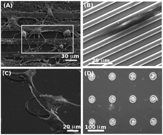
With the advances in photo- and soft lithographic techniques, there has been a growing interest towards fabrication of micro- and nanotopographies to address fundamental questions related to cell-substrate interactions. An excellent example is the alignment of cells along microgrooves, a phenomenon known as contact guidance [28,29,30,31]. Microstructures also influence basic cellular processes such as adhesion [32,33,34,35,36,37], migration [38,39,40], proliferation [41,42] and differentiation [43,44]. Recently, there has been significant interest towards utilizing microscale topographies in controlling stem cell behavior. Most of these studies have examined the effect of microgrooved topographies on alignment, morphology and differentiation of stem cells. In a study by Mallapragada et al. [45], adult rat hippocampal progenitor cells (AHPCs) exhibited an elongated morphology along microgrooved laminin coated polystyrene (PS) substrates (Figure 2A). On these substrates, the elongated morphology of the cells remained intact after seeding cortical astrocytes with the AHPCs, and the differentiation of AHPCs towards an early neural phenotype (III β-tubulin) was enhanced after co-culturing. Likewise, mouse mesenchymal stem cells (mMSCs) were shown to exhibit an elongated morphology when cultured on microgrooves [46] (Figure 2B). A number of studies have investigated the effect of microgroove widths on differentiation of MSCs [44,47]. It was shown that the differentiation of MSCs into neural-like cells was less pronounced on the 4 μm wide microgrooves compared to narrower 1 and 2 μm microgrooves. In addition, MSCs on 1 μm and 2 μm grooves showed an upregulation of the expression neurogenic markers such as microtubule associate protein 2 (MAP2) and neural nuclei (NeuN) [44]. In another study, Kurpinski et al. [47] applied a uniaxial strain to an elastomeric PDMS substrate containing parallel microgrooves on its surface. The uniaxial mechanical stimuli resulted in stem cell alignment along the microgrooves and an increase in cell proliferation. It was also evident that there was an increased expression of calponin 1, a gene-marker of smooth muscle cell contractility after 2 and 4 days of culture under induced mechanical strain.
Figure 2.
(A) Co-culture of adult rat hippocampal progenitor cells (AHPCs) with astrocytes on microgrooved PS substrate. The square illustrates how the cells align in the same direction as the microgrooves (Published with permission from Elsevier [45]); (B) Elongation of mouse mesenchymal stem cells (mMSCs) inside microgrooves on a silicon substrate (Published with permission from Elsevier [46]); (C) Attachment of rMSCs on ring shaped PMMA microstructures (Published with permissions from Elsevier [48]); (D) Formation of embryoid bodies (EBs) using an array of PEG microwells (Published with permission from Elsevier [53]).
The shape of the microtopographies has also been shown to be important in stem cell behavior [48,49]. For example Engel et al., [48] developed ring and square shaped poly(methyl methacrylate) (PMMA) patterns to control the attachment of rat MSCs (rMSCs) (Figure 2C). They showed that the attachment of rMSCs was most favorable to ring shaped microstructures compared to other geometries, while cell proliferation and differentiation were the same on the microstructured and flat surfaces. In another study [49], hMSCs were cultured on concave and convex shaped poly(L-Lactic-Acid) (PLLA) microtopographies. More than 50% of cells expressed CD71 after 10 days on the concave and convex surfaces confirming that hMSCs maintained their proliferative ability. Additionally the authors observed enhanced cell spreading on concave surfaces compared to the convex ones.
Non-adhesive microscale structures also direct cell differentiation. For example, it is widely recognized that the differentiation of ESCs into various cell types could benefit from cell aggregates called embryoid bodies (EBs) [50]. Typically, EBs are generated in non-adhesive dishes to yield cell aggregates of various sizes. To generate more homogenous EBs, the hanging drop method is used, however this technique is cumbersome and difficult to scale-up [51]. Recently, microtopography has been employed to generate homogenously sized EBs by trapping cells inside microwells with different diameters [52]. For example, non-adhesive PEG microwells have been used to generate and retrieve EBs of controlled sizes [53]. Such microwells have also been shown to direct the differentiation of stem cells by modulating the size of EBs [43]. In particular, larger EBs (450 μm) resulted in more cardiac cells whereas smaller EBs (150 μm) generated more endothelial cells. This behavior was shown to be regulated by the differential expression of non-canonical Wnt pathway molecules, Wnt5a and Wnt11. Overall, the above-mentioned studies demonstrated the potential of microstructures for creating EBs with a homogenous size distribution and in directing the fate of ESCs.
2.3. Stem Cell Behavior on Micropatterned Surfaces
Micropatterned substrates have been used extensively to pattern cells on substrates and to control the resulting cell shape [8,54,55,56]. These studies have revealed that cell shape is an important regulator of apoptosis [54,56], proliferation and differentiation [55]. For instance, McBeath et al. [55] demonstrated that hMSCs that were spread on large protein patterns differentiated into osteogenic cells, while rounded cells on smaller patterns generated adipogenic cells. In another study, Kilian et al. [8] explored the differentiation of hMSCs into osteogenic and adipogenic cells on different micropatterns. It was shown that cell attachment on ellipsoid and star shaped fibronectin micropatterns enhanced the differentiation into bone cells compared to square shaped geometries.
Micropatterns have also been used to demonstrate a relationship between NSCs shape and differentiation. Solanki et al. [9] was able to control the fate of rat NSCs (rNSCs) by varying the geometry and dimensions (10–250 μm) of laminin patterns. They reported that grid patterns resulted in axon-like outgrowths from the cell body accompanied by neural differentiation, while square shaped islands resulted in an increase in the number of cells expressing astrocyte markers. Likewise, a more stellate-like cell morphology was observed by Ruiz et al. [57] on grid shaped micropatterns compared to a nonpatterned surface. The stellate morphology resulted in an enhanced expression of the neural marker β-TubIII. In summary, these reports [8,9,57] validate the feasibility of controlling the fate of both MSCs and NSCs by culturing cells on micropatterned surfaces.
2.4. Nanoscale Engineering Approaches for Controlling Stem Cell Fate
Early studies of cells on nanostructured surfaces have mainly focused on nanogrooves. These studies have demonstrated that nanoscale grooves can direct cell alignment and migration through contact guidance even on feature sizes that were only 30 nm deep [58,59,60,61]. Moreover, studies have shown that stem cell alignment on nanogrooves lead to a more pronounced differentiation profile. In the paper by Lee et al., polymeric nanogrooves (350 nm wide) were used to demonstrate a correlation between cell alignment and hESCs differentiation into the neuronal linage [62]. A similar relationship between the alignment of hMSCs and their neuronal differentiation was shown using 350 nm wide grooves by Yim et al. [63]. With the advances in the nanofabrication technology, other topographies have now become available for use in stem cell studies. For example, approaches such as polymer phase separation [64,65,66], metal anodization [67,68,69], dip-pen nanolithography [70], colloidal lithography [71,72,73], UV-assisted capillary force lithography [62,74,75] molecular beam epitaxy (MBE) [76,77,78], and glancing angle deposition [79,80] have been employed to fabricate sub-100 nm nanotubes, islands, and pyramids.
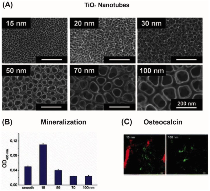
It has been shown that the nanostructured surfaces can direct MSCs into osteogenic cells [6,67,68,81,82,83]. Much of this focus has been on MSCs interaction with vertical TiO2 nanotubes fabricated by metal anodization [6,67,68,81,82]. Park et al. [67,68] performed extensive studies on the behavior of rMSCs on TiO2 nanotubes with tube diameters in the range of 15 to 100 nm (Figure 3A) [67,68]. A more pronounced cell response was observed on smaller nanotubes (15–30 nm), where cell adhesion, spreading, bone mineralization (Figure 3B) and bone marker expression (Figure 3C) were found to be enhanced compared to flat TiO2 [68,69,84]. Moreover, by examining the cytoskeletal structure of the cells, more focal contacts were observed on the smallest nanotubes [67], in agreement with the upregulated stem cell differentiation observed on the 15 and 30 nm nanotubes [85]. Other groups have observed similar behavior for hMSCs and rMSCs that were cultured on 70 and 100 nm TiO2 nanotubes [6,86]. For example, a higher alkaline phosphate activity was observed on 80 nm nanotubes followed by a larger mineralization of calcium and phosphate [86]. Furthermore, Oh et al. [6] found that the expression of bone proteins such as osteopontin and osteocalcin were significantly higher on 100 nm nanotubes. In conclusion, there is a strong indication that generation of nanotubes by metal anodization could enhance the performance of orthopedic titanium implants. This is either linked directly to mechanical stresses transmitted from the nanostructures to the cell nucleus [87,88,89] or indirectly by structural modulation of ECM proteins to expose cell adhesive domains [90,91,92] or a combination of both.
Figure 3.
(A) Scanning electron micrographs of the TiO2-nanotubes (15, 20, 30, 50, 70, 100 nm); (B) Plot of alkaline phosphatase activity versus nanotube diameter; (C) Osteocalcin (red) and F-actin (green) staining of cells seeded on 15 nm and 100 nm TiO2-nanotubes. The scale bar is 20 μm (Published with permission from ACS publications [67]).
As described previously, the nanoscale chemical and topographical cues in vivo have different shapes and sizes. By employing UV-assisted capillary force lithography it is possible to examine stem cell behavior on nanotopographies with various chemistries, shapes and sizes [23,62,75]. In a study by You et al. [75], the osteogenic differentiation of hMSCs on polyurethane acrylate nanogrooves and columns with different sizes were investigated. They noticed that the highest expression of osteogenic markers and alkaline phosphatase activity was on the 400 nm wide nanocolumns.
Moreover, the distribution of topographical cues in the stem cell microenvironment may also influence stem cell behavior. Dalby et al. [83] found that surfaces composed of nanopits with controlled disorder resulted in increased expression of osteogenic markers relative to surfaces consisting of either highly ordered or randomly displaced nanopits. In another study by Hunt and coworkers [70], dip-pen nanolithography was used to fabricate nanopatterns with different chemistries and spacings for analysis of stem cell behavior [70]. Specifically, they patterned thiolated molecules terminated with various chemistries (including carboxyl, amino, methyl and hydroxyl) onto gold surfaces. The chemically functionalized islands were 70 nm wide with inter-island spacing that ranged from 140 to 1000 nm. They cultured hMSCs on the fabricated surfaces and found that the adhesion and expression of several stem cell markers depended on the specific chemistry and the distance between the nanoscale islands [70]. This approach provided an efficient method to precisely control size, spacing and chemistry of nanofabricated patterns and could in theory be used to fabricate randomly ordered nanoscale islands. Thus, various nanoscale fabrication methods can be used to create nanostructured surfaces for directing stem cell differentiation. These approaches are implemented in 2D, therefore to further advance the use of these systems to regulate stem cell behavior, it is necessary to implement the nanosculpturing in a 3D environment to better replicate the in vivo environment of the stem cells.
3. The Role of Chemical Moieties and Substrate Stiffness on Stem Cell Fate
3.1. Chemically Functionalized Surfaces
The stem cell microenvironment consists of numerous molecular cues including proteins and polysaccharides. It is becoming increasingly clear that the biochemical cues in the cellular microenvironment to a large extent determine processes such as cell attachment, proliferation and differentiation [93]. By using SAMs to coat surfaces, it is possible to test stem cell behavior on a range of chemistries [94,95]. Wu and coworkers [95] used SAMs with different chain lengths and hydrophobic head groups to develop various surface hydrophobicities. They observed that an increase in surface hydrophobicity resulted in higher hESC proliferation and differentiation [95]. In the future, such SAM coated surfaces could potentially be used to control cell size and enhance the differentiation profile of hESCs in vitro.
SAMS conjugated to various ligands, such as peptides or proteins, have also been synthesized and used in stem cell studies [96,97,98,99]. For example, surfaces functionalized with RGD ligands increased osteogenic [100,101,102,103], chondrogenic [104] and neurogenic [105] differentiation of stem cells compared to non-functionalized substrates. Although many studies have used RGD functionalized surfaces, different polymer coatings have likewise been used to induce stem cell differentiation. In one approach, Joy et al. [106] coated surfaces with different polymer compositions and observed that the surface chemistry had a significant influence on the osteogenic and adipogenic differentiation of hMSCs. However, after functionalization with RGD ligands, no differences were found in the expression of these markers between surfaces coated with different polymers. The fact that immobilized adhesive ligands can override the effect of the underlying polymer coating can have important implications in designing polymer-based biomaterials. In conclusion, altering the surface chemistry influences the behavior of stem cells and their differentiation in a notable way.
3.2. Substrate Stiffness
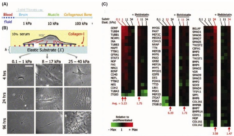
Mammalian cells can sense the elasticity of the substrates on which they are cultured [7]. This is caused by transmission of mechanical forces between substrate and cell, which generates contractile forces in the cell. These contractile forces in turn influence cell behaviors such as spreading [107,108], migration [109], proliferation [110] and apoptosis [111]. Pitelka and coworkers [112] provided early evidence in 1979 that substrate stiffness also affects differentiation. They found that mouse epithelial cells (mECs) differentiate better on softer collagen substrates compared to harder plastic tissue culture dishes. In another study, myoblasts were seeded on substrates with different stiffness [113] to show that actin/myosin striation, as it is seen in natural muscle, occurred only on the substrates with mechanical properties similar to that of a muscle [114]. More recently, Engler et al. [7] cultured hMSCs on a polyacrylamide gel homogeneously coated with collagen I ligands. The substrates had variable stiffness representing that of nerve (0.1–1 kPa), muscle (8–17 kPa) and bone tissue (25–40 kPa) and it was it was observed that the hMSCs differentiated along the neurogenic, myogenic and osteogenic lineage, respectively (Figure 4) [7]. Cooper-White and coworkers [113] further hypothesized that ECM proteins could influence hMSCs fate and therefore analyzed the combined effect of various ECM proteins (collagen I, collagen IV, laminin, and fibronectin). Their results revealed a significant interplay between ECM proteins and the underlying substrate elasticity affecting the myo- or osteogenic differentiation patterns. These studies suggest that both the elastic modulus of the substrate and the coated ECM proteins play a significant role in hMSC differentiation [7,113].
Figure 4.
(A) The elastic moduli of different solid tissues ranging from blood to collagenous bone; (B) The images show how different substrate stiffness values influence cell morphology. Scale bar is 20 μm; (C) Microarray profiling of differentiation marker expression on substrates with different stiffnesses. The microarray profiling showed that neurogenic markers were highest on 0.1–1 kPa gels, while myogenic markers were highest on 11 kPa gels and osteogenic markers were highest on 34 kPa gels. (Published with permission from Elsevier [7]).
In vivo, stem cells exist in 3D microenvironments, hence it is important to understand the effect of 3D matrix stiffness on stem cell differentiation. Over the past few years, many new techniques have emerged to fabricate 3D constructs with precise mechanical properties [93]. In particular, hydrogels have proven as a promising tool for the fabrication of 3D microenvironments [115,116,117,118,119,120,121]. In one study Pek et al. [116] used a thixotropic polyethylene glycol-silica (PEG-silica) to generate 3D gels with different stiffnesses [116]. Their findings showed that the highest expression of neural (ENO2), myogenic (MYOG) and osteogenic (Runx2, OC) markers occurred on gels corresponding to low (7 Pa), intermediate (25 Pa) and high (75 Pa) gel stiffness respectively, consistent with previous findings on 2D surfaces [7].
3.3. High-Throughput Screening of Stem Cell Differentiation on Biomaterials
Most of today's biomaterials are prepared and tested individually for various applications. This process is time-consuming and expensive. An emerging approach in the development of biomaterials has been the use of combinatorial high-throughput screening methods to lower the cost and the experimentation time. This approach can be applied to stem cell bioengineering by simultaneously examining numerous parameters on stem cell fate. Kohn and co-workers [122] developed one of the first high-throughput biomaterial library systems in 1997. By employing polyacrylates, they successfully generated a microarray with 112 different combinations and used the array to examine fibroblast proliferation.
Microarray printing technologies have been more widely used in the biomaterials field over the past few years to screen for various stem cell material-interactions [100,123,124]. For example, Flaim et al. [124] used a DNA spotter to develop an ECM matrix microarray for probing the differentiation of primary rat hepatocyte cells (rHCs) and mESCs towards an early hepatic phenotype, by using five different proteins (collagen I, collagen III, collagen IV, laminin and fibronectin) in 32 combinations. This platform was used to identify specific ECM mixtures, containing either collagen I or fibronectin, that directed mESCs into a hepatic fate.
The differentiation profile of hESCs have also been examined on high-throughput biomaterial platforms [123,125,126]. The growth of hESCs on arrays with 18 different laminin-derived peptides was investigated by Derda et al. [125]. Their results revealed that the RNIAEIIKDI laminin peptide resulted in undifferentiated cells, while LGTIPG peptide promoted differentiation. Thus, they demonstrated that high-throughput platforms can be used to quickly identify peptide sequences that can regulate stem cell fate. In another study by Anderson et al. [123], the growth and differentiation of hESCs into cytokeratin positive cells on a microarray containing 1728 polymer mixtures were examined. Later, they investigated additional parameters such as root mean square roughness (0 –100 nm), stiffness (0.002 –2.262 GPa) and wettability (30–110°). They found that surface wettability and the elastic modulus of the polymers modulated the colony formation frequency (CFF) of hESCs, while surface roughness did not have a significant effect [126]. Taken together, these studies show that high-throughput screening platforms could more rapidly identify important parameters in culture dishes for better control of stem cell fate.
4. Conclusions
Surface topography as well as micro- and nanoscale chemical patterns on biomaterials have proven to be efficient methods to direct stem cell behavior. In addition, substrate stiffness and chemical cues are important factors in controlling stem cell fate. Further advances in controlling stem cell fate could be achieved by combining the above mentioned parameters in a more scalable and combinatorial manner to address the complexity of the natural stem cell niche. Overall, it is becoming clearer that the advances in micro- and nanoengineering can be used to precisely control stem cell behavior through cell-substrate interactions with enormous potential implications in science and medicine.
Acknowledgments
AK acknowledges funding from the National Institutes of Health (HL092836, DE019024, EB008392, DE021468, AR05837, EB012597, HL099073), the National Science Foundation CAREER Award (DMR 0847287) and the office of Naval Research Young Investigator Award. ADP is grateful for the postdoctoral fellowship grant awarded by the Danish Council for Technology and Innovation, and the University of Aarhus for administration of the grant.
References
- 1.Griffith L.G., Naughton G. Tissue engineering-current challenges and expanding opportunities. Science. 2002;295:1009–1014. doi: 10.1126/science.1069210. [DOI] [PubMed] [Google Scholar]
- 2.Khademhosseini A., Langer R., Borenstein J., Vacanti J.P. Microscale technologies for tissue engineering and biology. Proc. Natl. Acad. Sci. USA. 2006;103:2480–2487. doi: 10.1073/pnas.0507681102. [DOI] [PMC free article] [PubMed] [Google Scholar]
- 3.Sorensen M.L. Stem Cell Applications in Diseases. Nova Biomedical Books; New York, NY, USA: 2008. [Google Scholar]
- 4.Zipori D. Biology of Stem Cells and the Molecular Basis of the Stem State (Stem Cell Biology and Regenerative Medicine) Humana Press; New York, NY, USA: 2009. [Google Scholar]
- 5.Zandstra P.W., Nagy A. Stem cell bioengineering. Annu. Rev. Biomed. Eng. 2001;3:275–305. doi: 10.1146/annurev.bioeng.3.1.275. [DOI] [PubMed] [Google Scholar]
- 6.Oh S., Brammer K.S., Li J.Y.S., Teng D., Engler A.J., Chien S., Jin S. Stem cell fate dictated solely by altered nanotube dimension. Proc. Natl. Acad. Sci. USA. 2009;106:2130–2135. doi: 10.1073/pnas.0813200106. [DOI] [PMC free article] [PubMed] [Google Scholar]
- 7.Engler A.J., Sen S., Sweeney H.L., Discher D.E. Matrix elasticity directs stem cell lineage specification. Cell. 2006;126:677–689. doi: 10.1016/j.cell.2006.06.044. [DOI] [PubMed] [Google Scholar]
- 8.Kilian K.A., Bugarija B., Lahn B.T., Mrksich M. Geometric cues for directing the differentiation of mesenchymal stem cells. Proc. Natl. Acad. Sci. USA. 2010;107:4872–7487. doi: 10.1073/pnas.0903269107. [DOI] [PMC free article] [PubMed] [Google Scholar]
- 9.Solanki A., Solanki A., Shah S., Memoli K.A., Park S.Y., Hong S., Lee K.-B. Controlling differentiation of neural stem cells using extracellular matrix protein patterns. Small. 2010;6:2509–2513. doi: 10.1002/smll.201001341. [DOI] [PMC free article] [PubMed] [Google Scholar]
- 10.Moore K.A., Lemischka I.R. Stem cells and their niches. Science. 2006;311:1880–1885. doi: 10.1126/science.1110542. [DOI] [PubMed] [Google Scholar]
- 11.Place E.S., Evans N.D., Stevens M.M. Complexity in biomaterials for tissue engineering. Nat. Mater. 2009;8:457–470. doi: 10.1038/nmat2441. [DOI] [PubMed] [Google Scholar]
- 12.Pamula E., de Cupere V., Dufrêne Y.F., Rouxhet P.G. Nanoscale organization of adsorbed collagen: Influence of substrate hydrophobicity and adsorption time. J. Colloid Interface Sci. 2004;271:80–91. doi: 10.1016/j.jcis.2003.11.012. [DOI] [PubMed] [Google Scholar]
- 13.Goodman S.L., Sims P.A., Albrecht R.M. Three-dimensional extracellular matrix textured biomaterials. Biomaterials. 1996;17:2087–2095. doi: 10.1016/0142-9612(96)00016-6. [DOI] [PubMed] [Google Scholar]
- 14.Gefen A., Gefen N., Zhu Q., Raghupathi R., Margulies S.S. Age-dependent changes in material properties of the brain and braincase of the rat. J. Neurotrauma. 2003;20:1163–1177. doi: 10.1089/089771503770802853. [DOI] [PubMed] [Google Scholar]
- 15.Engler A.J., Griffin M.A., Sen S., Bönnemann C.G., Sweeney H.L., Discher D.E. Myotubes differentiate optimally on substrates with tissue-like stiffness: Pathological implications for soft or stiff microenvironments. J. Cell Biol. 2004;166:877–887. doi: 10.1083/jcb.200405004. [DOI] [PMC free article] [PubMed] [Google Scholar]
- 16.Mitchell G.F., Parise H., Benjamin E.J., Larson M.G., Keyes M.J., Vita J.A., Vasan R.S., Levy D. Changes in arterial stiffness and wave reflection with advancing age in healthy men and women: The Framingham Heart Study. Hypertension. 2004;43:1239–1245. doi: 10.1161/01.HYP.0000128420.01881.aa. [DOI] [PubMed] [Google Scholar]
- 17.Pittenger M.F., Mackay A.M., Beck S.C., Jaiswal R.K., Douglas R., Mosca J.D., Moorman M.A., Simonetti D.W., Craig S., Marshak D.R. Multilineage potential of adult human mesenchymal stem cells. Science. 1999;284:143–147. doi: 10.1126/science.284.5411.143. [DOI] [PubMed] [Google Scholar]
- 18.Ding S., Schultz P.G. A role for chemistry in stem cell biology. Nat. Biotechnol. 2004;22:833–840. doi: 10.1038/nbt987. [DOI] [PubMed] [Google Scholar]
- 19.Hwang N.S., Varghese S., Elisseeff J. Controlled differentiation of stem cells. Adv. Drug Delivery Rev. 2008;60:199–214. doi: 10.1016/j.addr.2007.08.036. [DOI] [PMC free article] [PubMed] [Google Scholar]
- 20.Park T.H., Shuler M.L. Integration of cell culture and microfabrication technology. Biotechnol. Prog. 2003;19:243–253. doi: 10.1021/bp020143k. [DOI] [PubMed] [Google Scholar]
- 21.Bettinger C.J., Langer R., Borenstein J.T. Engineering substrate topography at the micro- and nanoscale to control cell function. Angew. Chem. Int. Ed. 2009;48:5406–5415. doi: 10.1002/anie.200805179. [DOI] [PMC free article] [PubMed] [Google Scholar]
- 22.Pollard T.D., Earnshaw W.C., Lippincott-Schwartz J. Cell Biology. 2nd ed. Elsevier Inc.; Philadelphia, PA, USA: 2008. [Google Scholar]
- 23.Gupta K., Kim D.H., Ellison D., Smith C., Kundu A., Tuan J., Suh K.Y., Levchenko A. Lab-on-a-chip devices as an emerging platform for stem cell biology. Lab chip. 2010;10:2019–2031. doi: 10.1039/c004689b. [DOI] [PubMed] [Google Scholar]
- 24.Urbanek K., Cesselli D., Rota M., Nascimbene A., de Angelis A., Hosoda T., Bearzi C., Boni A., Bolli R., Kajstura J., et al. Stem cell niches in the adult mouse heart. Proc. Natl. Acad. Sci. USA. 2006;103:9226–9231. doi: 10.1073/pnas.0600635103. [DOI] [PMC free article] [PubMed] [Google Scholar]
- 25.Stevenson E.J., . Koncarevic A., Giresi P.G., Jackman R.W., Kandarian S.C. Transcriptional profile of a myotube starvation model of atrophy. J. Appl. Physiol. 2005;98:1396–406. doi: 10.1152/japplphysiol.01055.2004. [DOI] [PubMed] [Google Scholar]
- 26.Stevens M.M., George J.H. Exploring and engineering the cell surface interface. Science. 2005;310:1135–1138. doi: 10.1126/science.1106587. [DOI] [PubMed] [Google Scholar]
- 27.Janko M., Zink A., Gigler A.M., Heckl W.M., Stark R.W. Nanostructure and mechanics of mummified type I collagen from the 5300-year-old Tyrolean Iceman. Proc. Biol. Sci. 2010;277:2301–2309. doi: 10.1098/rspb.2010.0377. [DOI] [PMC free article] [PubMed] [Google Scholar]
- 28.Flemming R.G., Murphy C.J., Abrams G.A., Goodman S.L., Nealey P.F. Effects of synthetic micro- and nano-structured surfaces on cell behavior. Biomaterials. 1999;20:573–588. doi: 10.1016/s0142-9612(98)00209-9. [DOI] [PubMed] [Google Scholar]
- 29.Walboomers X.F., Croes H.J., Ginsel L.A., Jansen J.A. Growth behavior of fibroblasts on microgrooved polystyrene. Biomaterials. 1998;19:1861–1868. doi: 10.1016/s0142-9612(98)00093-3. [DOI] [PubMed] [Google Scholar]
- 30.Walboomers X.F., Monaghan W., Curtis A.S., Jansen J.A. Attachment of fibroblasts on smooth and microgrooved polystyrene. J. Biomed. Mater. Res. 1999;46:212–220. doi: 10.1002/(sici)1097-4636(199908)46:2<212::aid-jbm10>3.0.co;2-y. [DOI] [PubMed] [Google Scholar]
- 31.Bettinger C.J., Orrick B., Misra A., Langer R., Borenstein J.T. Microfabrication of poly (glycerol-sebacate) for contact guidance applications. Biomaterials. 2006;27:2558–2565. doi: 10.1016/j.biomaterials.2005.11.029. [DOI] [PubMed] [Google Scholar]
- 32.Strobl J.S., Nikkhah M., Agah M. Actions of the anti-cancer drug suberoylanilide hydroxamic acid (SAHA) on human breast cancer cytoarchitecture in silicon microstructures. Biomaterials. 2010;31:7043–7050. doi: 10.1016/j.biomaterials.2010.05.023. [DOI] [PubMed] [Google Scholar]
- 33.Lu J., Rao M.P., MacDonald N.C., Khang D., Webster T.J. Improved endothelial cell adhesion and proliferation on patterned titanium surfaces with rationally designed, micrometer to nanometer features. Acta Biomater. 2008;4:192–201. doi: 10.1016/j.actbio.2007.07.008. [DOI] [PubMed] [Google Scholar]
- 34.Teixeira A.I., Abrams G.A., Bertics P.J., Murphy C.J., Nealey P.F. Epithelial contact guidance on well-defined micro- and nanostructured substrates. J. Cell Sci. 2003;116:1881–1892. doi: 10.1242/jcs.00383. [DOI] [PMC free article] [PubMed] [Google Scholar]
- 35.Turner A.M., Dowell N., Turner S.W., Kam L., Isaacson M., Turner J.N., Craighead H.G., Shain W. Attachment of astroglial cells to microfabricated pillar arrays of different geometries. J. Biomed. Mater. Res. 2000;51:430–441. doi: 10.1002/1097-4636(20000905)51:3<430::aid-jbm18>3.0.co;2-c. [DOI] [PubMed] [Google Scholar]
- 36.Nikkhah M., Strobl J.S., de Vita R., Agah M. The cytoskeletal organization of breast carcinoma and fibroblast cells inside three dimensional (3-D) isotropic silicon microstructures. Biomaterials. 2010;31:4552–4561. doi: 10.1016/j.biomaterials.2010.02.034. [DOI] [PubMed] [Google Scholar]
- 37.Nikkhah M., Strobl J.S., Peddi B., Agah M. Cytoskeletal role in differential adhesion patterns of normal fibroblasts and breast cancer cells inside silicon microenvironments. Biomed. Microdevices. 2009;11:585–595. doi: 10.1007/s10544-008-9268-2. [DOI] [PubMed] [Google Scholar]
- 38.Miyoshi H., Ju J., Lee S.M., Cho D.J., Ko J.S., Yamagata Y., Adachi T. Control of highly migratory cells by microstructured surface based on transient change in cell behavior. Biomaterials. 2010;31:8539–8545. doi: 10.1016/j.biomaterials.2010.07.076. [DOI] [PubMed] [Google Scholar]
- 39.Tzvetkova-Chevolleau T., Stéphanou A., Fuard D., Ohayon J., Schiavone P., Tracqui P. The motility of normal and cancer cells in response to the combined influence of the substrate rigidity and anisotropic microstructure. Biomaterials. 2008;29:1541–1551. doi: 10.1016/j.biomaterials.2007.12.016. [DOI] [PubMed] [Google Scholar]
- 40.Mai J.Y., Sun C., Li S., Zhang X. A microfabricated platform probing cytoskeleton dynamics using multidirectional topographical cues. Biomed. Microdevices. 2007;9:523–531. doi: 10.1007/s10544-007-9060-8. [DOI] [PubMed] [Google Scholar]
- 41.den Braber E.T., de Ruijter J.E., Smits H.T., Ginsel L.A., von Recum A.F., Jansen J.A. Quantitative analysis of cell proliferation and orientation on substrata with uniform parallel surface micro-grooves. Biomaterials. 1996;17:1093–1099. doi: 10.1016/0142-9612(96)85910-2. [DOI] [PubMed] [Google Scholar]
- 42.Matsuzaka K., Walboomers F., de Ruijter A., Jansen J.A. Effect of microgrooved poly-l-lactic (PLA) surfaces on proliferation, cytoskeletal organization, and mineralized matrix formation of rat bone marrow cells. Clin. Oral Implants Res. 2000;11:325–333. doi: 10.1034/j.1600-0501.2000.011004325.x. [DOI] [PubMed] [Google Scholar]
- 43.Hwang Y.S., Chung B.G., Ortmann D., Hattori N., Moeller H.C., Khademhosseini A. Microwell-mediated control of embryoid body size regulates embryonic stem cell fate via differential expression of WNT5a and WNT11. Proc. Natl. Acad. Sci. USA. 2009;106:16978–16983. doi: 10.1073/pnas.0905550106. [DOI] [PMC free article] [PubMed] [Google Scholar]
- 44.Kim S.J., Lee J.K., Kim J.W., Jung J.W., Seo K., Park S.B., Roh K.H., Lee S.R., Hong Y.H., Kim S.J., et al. Surface modification of polydimethylsiloxane (PDMS) induced proliferation and neural-like cells differentiation of umbilical cord blood-derived mesenchymal stem cells. J. Mater. Sci. Mater. Med. 2008;19:2953–2962. doi: 10.1007/s10856-008-3413-6. [DOI] [PubMed] [Google Scholar]
- 45.Recknor J.B., Sakaguchi D.S., Mallapragada S.K. Directed growth and selective differentiation of neural progenitor cells on micropatterned polymer substrates. Biomaterials. 2006;27:4098–4108. doi: 10.1016/j.biomaterials.2006.03.029. [DOI] [PubMed] [Google Scholar]
- 46.Zahor D., Radko A., Vago R., Gheber L. Organization of mesenchymal stem cells is controlled by micropatterned silicon substrates. Mater. Sci. Eng. C. 2007;27:117–121. [Google Scholar]
- 47.Kurpinski K., Chu J., Hashi C., Li S. Anisotropic mechanosensing by mesenchymal stem cells. Proc. Natl. Acad. Sci. USA. 2006;103:16095–100. doi: 10.1073/pnas.0604182103. [DOI] [PMC free article] [PubMed] [Google Scholar]
- 48.Engel E., Martínez E., Mills C.A., Funes M., Planell J.A., Samitier J. Mesenchymal stem cell differentiation on microstructured poly (methyl methacrylate) substrates. Ann. Anat. 2009;191:136–144. doi: 10.1016/j.aanat.2008.07.013. [DOI] [PubMed] [Google Scholar]
- 49.Lee I.C., Lee Y.T., Yu B.Y., Lai J.Y., Young T.H. The behavior of mesenchymal Stem cells on micropatterned PLLA membranes. J. Biomed. Mater. Res. A. 2009;91A:929–938. doi: 10.1002/jbm.a.32309. [DOI] [PubMed] [Google Scholar]
- 50.Itskovitz-Eldor J., Schuldiner M., Karsenti D., Eden A., Yanuka O., Amit M., Soreq H., Benvenisty N. Differentiation of human embryonic stem cells into embryoid bodies compromising the three embryonic germ layers. Mol. Med. 2000;6:88–95. [PMC free article] [PubMed] [Google Scholar]
- 51.Banerjee M., Bhonde R.R. Application of hanging drop technique for stem cell differentiation and cytotoxicity studies. Cytotechnology. 2006;51:1–5. doi: 10.1007/s10616-006-9001-z. [DOI] [PMC free article] [PubMed] [Google Scholar]
- 52.Khademhosseini A., Ferreira L., Blumling J., Yeh J., Karp J.M., Fukuda J., Langer R. Co-culture of human embryonic stem cells with murine embryonic fibroblasts on microwell-patterned substrates. Biomaterials. 2006;27:5968–5977. doi: 10.1016/j.biomaterials.2006.06.035. [DOI] [PubMed] [Google Scholar]
- 53.Karp J.M., Yeh J., Eng G., Fukuda J., Blumling J., Suh K.Y., Cheng J., Mahdavi A., Borenstein J., Langer R., Khademhosseini A. Controlling size, shape and homogeneity of embryoid bodies using poly(ethylene glycol) microwells. Lab Chip. 2007;7:786–794. doi: 10.1039/b705085m. [DOI] [PubMed] [Google Scholar]
- 54.Chen C.S., Mrksich M., Huang S., Whitesides G.M., Ingber D.E. Geometric control of cell life and death. Science. 1997;276:1425–1428. doi: 10.1126/science.276.5317.1425. [DOI] [PubMed] [Google Scholar]
- 55.McBeath R., Pirone D.M., Nelson C.M., Bhadriraju K., Chen C.S. Cell shape, cytoskeletal tension, and RhoA regulate stem cell lineage commitment. Dev. Cell. 2004;6:483–495. doi: 10.1016/s1534-5807(04)00075-9. [DOI] [PubMed] [Google Scholar]
- 56.Singhvi R., Kumar A., Lopez G.P., Stephanopoulos G.N., Wang D.I. Engineering cell shape and function. Science. 1994;264:696–698. doi: 10.1126/science.8171320. [DOI] [PubMed] [Google Scholar]
- 57.Ruiz A., Buzanska L., Gilliland D., Rauscher H., Sirghi L., Sobanski T., Zychowicz M., Ceriotti L., Bretagnol F., Coecke S., et al. Micro-stamped surfaces for the patterned growth of neural stem cells. Biomaterials. 2008;29:4766–4774. doi: 10.1016/j.biomaterials.2008.08.017. [DOI] [PubMed] [Google Scholar]
- 58.Clark P., Connolly P., Curtis A.S., Dow J.A., Wilkinson C.D. Cell guidance by ultrafine topography in vitro. J. Cell Sci. 1991;99:73–77. doi: 10.1242/jcs.99.1.73. [DOI] [PubMed] [Google Scholar]
- 59.Wojciak-Stothard B., Curtis A., Monaghan W., MacDonald K., Wilkinson C. Guidance and activation of murine macrophages by nanometric scale topography. Exp. Cell Res. 1996;223:426–435. doi: 10.1006/excr.1996.0098. [DOI] [PubMed] [Google Scholar]
- 60.Rajnicek A., Britland S., McCaig C. Contact guidance of CNS neurites on grooved quartz: Influence of groove dimensions, neuronal age and cell type. J. Cell Sci. 1997;110:2905–2913. doi: 10.1242/jcs.110.23.2905. [DOI] [PubMed] [Google Scholar]
- 61.Wilkinson C.D.W., Curtis A.S.G., Crossan J. Nanofabrication in cellular engineering. J. Vac. Sci. Technol. B. 1998;16:3132–3136. [Google Scholar]
- 62.Lee M.R., Kwon K.W., Jung H., Kim H.N., Suh K.Y., Kim K., Kim K.S. Direct differentiation of human embryonic stem cells into selective neurons on nanoscale ridge/groove pattern arrays. Biomaterials. 2010;31:4360–4366. doi: 10.1016/j.biomaterials.2010.02.012. [DOI] [PubMed] [Google Scholar]
- 63.Yim E.K., Pang S.W., Leong K.W. Synthetic nanostructures inducing differentiation of human mesenchymal stem cells into neuronal lineage. Exp. Cell Res. 2007;313:1820–1829. doi: 10.1016/j.yexcr.2007.02.031. [DOI] [PMC free article] [PubMed] [Google Scholar]
- 64.Dalby M.J., Riehleô M.O., Johnstone H., Affrossman S., Curtis A.S. In vitro reaction of endothelial cells to polymer demixed nanotopography. Biomaterials. 2002;23:2945–2954. doi: 10.1016/s0142-9612(01)00424-0. [DOI] [PubMed] [Google Scholar]
- 65.Affrossman S., Bertrand P., Hartshorne M., Kiff T., Leonard D., Pethrick R.A., Richards R.W. Surface segregation in blends of polystyrene and perfluorohexane double end capped polystyrene studied by static SIMS, ISS, and XPS. Macromolecules. 1996;29:5432–5437. [Google Scholar]
- 66.Dalby M.J., Riehle M.O., Johnstone H., Affrossman S., Curtis A.S.G. Investigating the limits of filopodial sensing: A brief report using SEM to image the interaction between 10 nm high nano-topography and fibroblast filopodia. Cell Biol. Int. 2004;28:229–236. doi: 10.1016/j.cellbi.2003.12.004. [DOI] [PubMed] [Google Scholar]
- 67.Park J., Bauer S., von der Mark K., Schmuki P. Nanosize and vitality: TiO2 nanotube diameter directs cell fate. Nano Lett. 2007;7:1686–1691. doi: 10.1021/nl070678d. [DOI] [PubMed] [Google Scholar]
- 68.Park J., Bauer S., Schmuki P., von der Mark K. Narrow window in nanoscale dependent activation of endothelial cell growth and differentiation on TiO2 nanotube surfaces. Nano Lett. 2009;9:3157–3164. doi: 10.1021/nl9013502. [DOI] [PubMed] [Google Scholar]
- 69.Park J., Bauer S., Schlegel K.A., Neukam F.W. TiO2 nanotube surfaces: 15 nm-an optimal length scale of surface topography for cell adhesion and differentiation. Small. 2009;5:666–671. doi: 10.1002/smll.200801476. [DOI] [PubMed] [Google Scholar]
- 70.Curran J.M., Stokes R., Irvine E., Graham D., Amro N.A., Sanedrin R.G., Jamil H., Hunt J.A. Introducing dip pen nanolithography as a tool for controlling stem cell behaviour: unlocking the potential of the next generation of smart materials in regenerative medicine. Lab Chip. 2010;10:1662–1670. doi: 10.1039/c004149a. [DOI] [PubMed] [Google Scholar]
- 71.Denis F.A., Hanarp P., Sutherland D.S., Dufrene Y.F. Fabrication of nanostructured polymer surfaces using colloidal lithography and spin-coating. Nano Lett. 2002;2:1419–1425. [Google Scholar]
- 72.Dolatshahi-Pirouz A., Jensen1 T., Vorup-Jensen T., Bech R., Chevallier J., Besenbacher F., Foss M., Sutherland D.S. Synthesis of functional nanomaterials via colloidal mask templating and glancing angle deposition (GLAD) Adv. Eng. Mater. 2010;12:899–905. [Google Scholar]
- 73.Dolatshahi-Pirouz A., Sutherland D.S., Foss M., Besenbacher F. Growth characteristics of inclined columns produced by Glancing Angle Deposition (GLAD) and colloidal lithography. Appl. Surf. Sci. 2011;257:2226–2230. [Google Scholar]
- 74.Jeong H.E., Kwak R., Khademhosseini A., Suh K.Y. UV-assisted capillary force lithography for engineering biomimetic multiscale hierarchical structures: From lotus leaf to gecko foot hairs. Nanoscale. 2009;1:331–338. doi: 10.1039/b9nr00106a. [DOI] [PubMed] [Google Scholar]
- 75.You M.H., Kwak M.K., Kim D.H., Kim K., Levchenko A., Kim D.Y., Suh K.Y. Synergistically enhanced osteogenic differentiation of human mesenchymal stem cells by culture on nanostructured surfaces with induction media. Biomacromolecules. 2010;11:1856–1862. doi: 10.1021/bm100374n. [DOI] [PMC free article] [PubMed] [Google Scholar]
- 76.Dolatshahi-Pirouz A., Jensen T.H.L., Kraft D.C., Foss M. Fibronectin adsorption, cell adhesion, and proliferation on nanostructured tantalum surfaces. ACS Nano. 2010;4:2874–2882. doi: 10.1021/nn9017872. [DOI] [PubMed] [Google Scholar]
- 77.Pennisi C.P., Sevcencuô C., Dolatshahi-Pirouz A., Foss M., Hansen J.L., Larsen A.N., Zachar V., Besenbacher F., Yoshida K. Responses of fibroblasts and glial cells to nanostructured platinum surfaces. Nanotechnology. 2009;20:385103. doi: 10.1088/0957-4484/20/38/385103. [DOI] [PubMed] [Google Scholar]
- 78.Mo Y., Savage D.E., Swartzentruber B.S., Lagally M.G. Kinetic pathway in Stranski-Krastanov growth of Ge on Si(001) Phys. Rev. Lett. 1990;65:1020–1023. doi: 10.1103/PhysRevLett.65.1020. [DOI] [PubMed] [Google Scholar]
- 79.Dolatshahi-Pirouz A., Pennisi C.P., Skeldal S., Foss M., Chevallier J., Zachar V., Andreasen P., Yoshida K., Besenbacher F. The influence of glancing angle deposited nano-rough platinum surfaces on the adsorption of fibrinogen and the proliferation of primary human fibroblasts. Nanotechnology. 2009;20:095101. doi: 10.1088/0957-4484/20/9/095101. [DOI] [PubMed] [Google Scholar]
- 80.Dolatshahi-Pirouz A., Hovgaard M.B., Rechendorff K., Chevallier J., Foss M., Besenbacher F. Scaling behavior of the surface roughness of platinum films grown by oblique angle deposition. Phys. Rev. B. 2008;77:115427. [Google Scholar]
- 81.Kim H.S., Myung Y., Cho Y.J., Jang D.M., Jung C.S., Park J., Ahn J.P. Three-dimensional structure of twinned and zigzagged one-dimensional nanostructures using electron tomography. Nano Lett. 2010;10:1682–1691. doi: 10.1021/nl1000168. [DOI] [PubMed] [Google Scholar]
- 82.Mooney E., Dockery P., Greiser U., Murphy M., Barron V. Carbon nanotubes and mesenchymal stem cells: Biocompatibility, proliferation and differentiation. Nano Lett. 2008;8:2137–2143. doi: 10.1021/nl073300o. [DOI] [PubMed] [Google Scholar]
- 83.Dalby M.J., Gadegaard N., Tare R., Andar A., Riehle M.O., Herzyk P., Wilkinson C.D., Oreffo R.O. The control of human mesenchymal cell differentiation using nanoscale symmetry and disorder. Nat. Mater. 2007;6:997–1003. doi: 10.1038/nmat2013. [DOI] [PubMed] [Google Scholar]
- 84.Agrawal A., Park J., Ryu D.Y., Hammond P.T., Russell T.P., McKinley G.H. Controlling the location and spatial extent of nanobubbles using hydrophobically nanopatterned surfaces. Nano Lett. 2005;5:1751–1756. doi: 10.1021/nl051103o. [DOI] [PubMed] [Google Scholar]
- 85.Burridge K., Chrzanowska-Wodnicka M. Focal adhesions, contractility, and signaling. Annu. Rev. Cell Dev. Biol. 1996;12:463–518. doi: 10.1146/annurev.cellbio.12.1.463. [DOI] [PubMed] [Google Scholar]
- 86.Popat K.C., Leoni L., Grimes C.A., Desai T.A. Influence of engineered titania nanotubular surfaces on bone cells. Biomaterials. 2007;28:3188–3197. doi: 10.1016/j.biomaterials.2007.03.020. [DOI] [PubMed] [Google Scholar]
- 87.Berger S.L. The complex language of chromatin regulation during transcription. Nature. 2007;447:407–412. doi: 10.1038/nature05915. [DOI] [PubMed] [Google Scholar]
- 88.Maniotis A.J., Bojanowski K., Ingber D.E. Mechanical continuity and reversible chromosome disassembly within intact genomes removed from living cells. J. Cell. Biochem. 1997;65:114–130. [PubMed] [Google Scholar]
- 89.Maniotis A.J., Chen C.S., Ingber D.E. Demonstration of mechanical connections between integrins cytoskeletal filaments, and nucleoplasm that stabilize nuclear structure. Proc. Natl. Acad. Sci. USA. 1997;94:849–854. doi: 10.1073/pnas.94.3.849. [DOI] [PMC free article] [PubMed] [Google Scholar]
- 90.Dolatshahi-Pirouz A., Rechendorff K., Hovgaard M.B., Foss M., Chevallier J., Besenbacher F. Bovine serum albumin adsorption on nano-rough platinum surfaces studied by QCM-D. Colloids Surf. B. 2008;66:53–59. doi: 10.1016/j.colsurfb.2008.05.010. [DOI] [PubMed] [Google Scholar]
- 91.Dolatshahi-Pirouz A., Skeldal S., Hovgaard M.B., Jensen T., Foss M., Chevallier J., Besenbacher F. Influence of nanoroughness and detailed surface morphology on structural properties and water-coupling capabilities of surface-bound fibrinogen films. J. Phys. Chem. C. 2009;113:4406–4412. [Google Scholar]
- 92.Roach P., Farrar D., Perry C.C. Surface tailoring for controlled protein adsorption: Effect of topography at the nanometer scale and chemistry. J. Am. Chem. Soc. 2006;128:3939–3945. doi: 10.1021/ja056278e. [DOI] [PubMed] [Google Scholar]
- 93.Ratner B.D. Biomaterials Science: An Introduction to Materials in Medicine. 2nd ed. Elsevier Academic Press; Amsterdam, The Netherlands: 2004. [Google Scholar]
- 94.Phillips J.E., Petrie T.A., Creighton F.P., García A.J. Human mesenchymal stem cell differentiation on self-assembled monolayers presenting different surface chemistries. Acta Biomater. 2010;6:12–20. doi: 10.1016/j.actbio.2009.07.023. [DOI] [PMC free article] [PubMed] [Google Scholar]
- 95.Valamehr B., Jonas S.J., Polleux J., Qiao R., Guo S., Gschweng E.H., Stiles B., Kam K., Luo T.-J.M., Witte O.N., et al. Hydrophobic surfaces for enhanced differentiation of embryonic stem cell-derived embryoid bodies. Proc. Natl. Acad. Sci. USA. 2008;105:14459–14464. doi: 10.1073/pnas.0807235105. [DOI] [PMC free article] [PubMed] [Google Scholar]
- 96.Keselowsky B.G., Collard D.M., Garcia A.J. Integrin binding specificity regulates biomaterial surface chemistry effects on cell differentiation. Proc. Natl. Acad. Sci. USA. 2005;102:5953–5957. doi: 10.1073/pnas.0407356102. [DOI] [PMC free article] [PubMed] [Google Scholar]
- 97.Shin H., Temenoff J.S., Bowden G.C., Zygourakis K., Farach-Carson M.C., Yaszemski M.J., Mikos A.G. Osteogenic differentiation of rat bone marrow stromal cells cultured on Arg-Gly-Asp modified hydrogels without dexamethasone and beta-glycerol phosphate. Biomaterials. 2005;26:3645–3654. doi: 10.1016/j.biomaterials.2004.09.050. [DOI] [PubMed] [Google Scholar]
- 98.Saha K., Irwin E.F., Kozhukh J., Schaffer D.V., Healy K.E. Biomimetic interfacial interpenetrating polymer networks control neural stem cell behavior. J. Biomed. Mater. Res. A. 2007;81A:240–249. doi: 10.1002/jbm.a.30986. [DOI] [PubMed] [Google Scholar]
- 99.Shin H., Zygourakis K., Farach-Carson M.C., Yaszemski M.J., Mikos A.G. Modulation of differentiation and mineralization of marrow stromal cells cultured on biomimetic hydrogels modified with Arg-Gly-Asp containing peptides. J. Biomed. Mater. Res. A. 2004;69A:535–543. doi: 10.1002/jbm.a.30027. [DOI] [PubMed] [Google Scholar]
- 100.Anderson J.M., Kushwaha M., Tambralli A., Bellis S.L., Camata R.P., Jun H.-W. Osteogenic differentiation of human mesenchymal stem cells directed by extracellular matrix-mimicking ligands in a biomimetic self-assembled peptide amphiphile nanomatrix. Biomacromolecules. 2009;10:2935–2944. doi: 10.1021/bm9007452. [DOI] [PMC free article] [PubMed] [Google Scholar]
- 101.Paletta J.R.J., Bockelmann S., Walz A., Theisen C., Wendorff J.H., Greiner A., Fuchs-Winkelmann S., Schofer M.D. RGD-functionalisation of PLLA nanofibers by surface coupling using plasma treatment: influence on stem cell differentiation. J. Mater. Sci. Mater. Med. 2010;21:1363–1369. doi: 10.1007/s10856-009-3947-2. [DOI] [PubMed] [Google Scholar]
- 102.Park J.S., Yang H.N., Jeon S.Y., Woo D.G., Na K., Park K.-H. Osteogenic differentiation of human mesenchymal stem cells using RGD-modified BMP-2 coated microspheres. Biomaterials. 2010;31:6239–6248. doi: 10.1016/j.biomaterials.2010.05.002. [DOI] [PubMed] [Google Scholar]
- 103.Hosseinkhani H., Hosseinkhani M., Tian F., Kobayashi H., Tabata Y. Osteogenic differentiation of mesenchymal stem cells in self-assembled peptide-amphiphile nanofibers. Biomaterials. 2006;27:4079–4086. doi: 10.1016/j.biomaterials.2006.03.030. [DOI] [PubMed] [Google Scholar]
- 104.Re'em T., Tsur-Gang O., Cohen S. The effect of immobilized RGD peptide in macroporous alginate scaffolds on TGF beta 1-induced chondrogenesis of human mesenchymal stem cells. Biomaterials. 2010;31:6746–6755. doi: 10.1016/j.biomaterials.2010.05.025. [DOI] [PubMed] [Google Scholar]
- 105.Ananthanarayanan B., Little L., Schaffer D.V., Healy K.E., Tirrell M. Neural stem cell adhesion and proliferation on phospholipid bilayers functionalized with RGD peptides. Biomaterials. 2010;31:8706–8715. doi: 10.1016/j.biomaterials.2010.07.104. [DOI] [PMC free article] [PubMed] [Google Scholar]
- 106.Joy A., Cohen D.M., Luk A., Anim-Danso E., Chen C., Kohn J. Control of surface chemistry, substrate stiffness, and cell function in a novel terpolymer methacrylate library. Langmuir. 2011;27:1891–1899. doi: 10.1021/la103722m. [DOI] [PMC free article] [PubMed] [Google Scholar]
- 107.Pelham R.J., Wang Y.L. Cell locomotion and focal adhesions are regulated by substrate flexibility. Proc. Natl. Acad. Sci. USA. 1997;94:13661–13665. doi: 10.1073/pnas.94.25.13661. [DOI] [PMC free article] [PubMed] [Google Scholar]
- 108.Discher D.E., Janmey P., Wang Y.L. Tissue cells feel and respond to the stiffness of their substrate. Science. 2005;310:1139–1143. doi: 10.1126/science.1116995. [DOI] [PubMed] [Google Scholar]
- 109.Guo W.H., Frey M.T., Burnham N.A., Wang Y.-L. Substrate rigidity regulates the formation and maintenance of tissues. Biophys. J. 2006;90:2213–2220. doi: 10.1529/biophysj.105.070144. [DOI] [PMC free article] [PubMed] [Google Scholar]
- 110.Peyton S.R., Raub C.B., Keschrumrus V.P., Putnam A.J. The use of poly(ethylene glycol) hydrogels to investigate the impact of ECM chemistry and mechanics on smooth muscle cells. Biomaterials. 2006;27:4881–4893. doi: 10.1016/j.biomaterials.2006.05.012. [DOI] [PubMed] [Google Scholar]
- 111.Wang H.B., Dembo M., Wang Y.L. Substrate flexibility regulates growth and apoptosis of normal but not transformed cells. Am. J. Physiol. Cell Physiol. 2000;279:C1345–C1350. doi: 10.1152/ajpcell.2000.279.5.C1345. [DOI] [PubMed] [Google Scholar]
- 112.Emerman J.T., Burwen S.J., Pitelka D.R. Substrate properties influencing ultrastructural differentiation of mammary epithelial-cells in culture. Tissue Cell. 1979;11:109–119. doi: 10.1016/0040-8166(79)90011-9. [DOI] [PubMed] [Google Scholar]
- 113.Rowlands A.S., George P.A., Cooper-White J.J. Directing osteogenic and myogenic differentiation of MSCs: interplay of stiffness and adhesive ligand presentation. Am. J. Physiol. Cell Physiol. 2008;295:C1037–C1044. doi: 10.1152/ajpcell.67.2008. [DOI] [PubMed] [Google Scholar]
- 114.Engler A.J., Griffin M.A., Sen S., Bönnemann C.G., Lee Sweeney H., Discher D.E. Myotubes differentiate optimally on substrates with tissue-like stiffness: Pathological implications for soft or stiff microenvironments. J. Cell Biol. 2004;166:877–887. doi: 10.1083/jcb.200405004. [DOI] [PMC free article] [PubMed] [Google Scholar]
- 115.Coutinho D.F., Sant S.V., Shin H., Oliveira J.T., Gomes M.E., Neves N.M. Modified Gellan Gum hydrogels with tunable physical and mechanical properties. Biomaterials. 2010;31:7494–7502. doi: 10.1016/j.biomaterials.2010.06.035. [DOI] [PMC free article] [PubMed] [Google Scholar]
- 116.Pek Y.S., Wan A.C.A., Ying J.Y. The effect of matrix stiffness on mesenchymal stem cell differentiation in a 3D thixotropic gel. Biomaterials. 2010;31:385–391. doi: 10.1016/j.biomaterials.2009.09.057. [DOI] [PubMed] [Google Scholar]
- 117.Saha K., Keung A.J., Irwin E.F., Li Y., Little L., Schaffer D.V., Healy K.E. Substrate modulus directs neural stem cell behavior. Biophys. J. 2008;95:4426–4438. doi: 10.1529/biophysj.108.132217. [DOI] [PMC free article] [PubMed] [Google Scholar]
- 118.Li Y.J., Chung E.H., Rodriguez R.T., Firpo M.T., Healy K.E. Hydrogels as artificial matrices for human embryonic stem cell self-renewal. J. Biomed. Mater. Res. A. 2006;79A:1–5. doi: 10.1002/jbm.a.30732. [DOI] [PubMed] [Google Scholar]
- 119.Khademhosseini A., Langer R. Microengineered hydrogels for tissue engineering. Biomaterials. 2007;28:5087–5092. doi: 10.1016/j.biomaterials.2007.07.021. [DOI] [PubMed] [Google Scholar]
- 120.Yeh J., Ling Y., Karp J.M., Gantz J., Chandawarkar A., Eng G., Blumling J., III., Langer R., Khademhosseini A. Micromolding of shape-controlled, harvestable cell-laden hydrogels. Biomaterials. 2006;27:5391–5398. doi: 10.1016/j.biomaterials.2006.06.005. [DOI] [PubMed] [Google Scholar]
- 121.Nichol J.W., Koshy S.T., Bae H., Hwang C.M., Yamanlar S., Khademhosseini A. Cell-laden microengineered gelatin methacrylate hydrogels. Biomaterials. 2010;31:5536–5544. doi: 10.1016/j.biomaterials.2010.03.064. [DOI] [PMC free article] [PubMed] [Google Scholar]
- 122.Brocchini S., James K., Tangpasuthadol V., Kohn J. A combinatorial approach for polymer design. J. Am. Chem. Soc. 1997;119:4553–4554. [Google Scholar]
- 123.Anderson D.G., Levenberg S., Langer R. Nanoliter-scale synthesis of arrayed biomaterials and application to human embryonic stem cells. Nat. Biotechnol. 2004;22:863–866. doi: 10.1038/nbt981. [DOI] [PubMed] [Google Scholar]
- 124.Flaim C.J., Chien S., Bhatia S.N. An extracellular matrix microarray for probing cellular differentiation. Nat. Methods. 2005;2:119–125. doi: 10.1038/nmeth736. [DOI] [PubMed] [Google Scholar]
- 125.Derda R., Li L., Orner B.P., Lewis R.L., Thomson J.A., Kiessling L.L. Defined substrates for human embryonic stem cell growth identified from surface arrays. ACS Chem. Biol. 2007;2:347–355. doi: 10.1021/cb700032u. [DOI] [PubMed] [Google Scholar]
- 126.Mei Y., Saha K., Bogatyrev S.R., Yang J., Hook A.L., Kalcioglu Z.I., Cho S.-W., Mitalipova M., Pyzocha N., Rojas F., et al. Combinatorial development of biomaterials for clonal growth of human pluripotent stem cells. Nat. Mater. 2010;9:768–778. doi: 10.1038/nmat2812. [DOI] [PMC free article] [PubMed] [Google Scholar]


