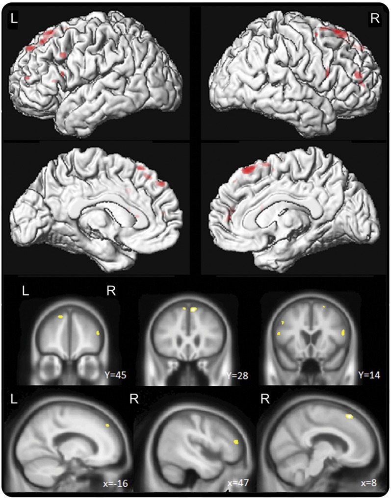
Figure. Voxel-based morphometric analysis comparing patients with and without NVOA.

Regions of gray matter loss in the patients with nonverbal oral apraxia (NVOA) compared with the matched patients without NVOA. Results are shown on lateral and medial 3-dimensional renderings of the brain, and on representative coronal and sagittal slices, uncorrected for multiple comparisons at p < 0.001.
