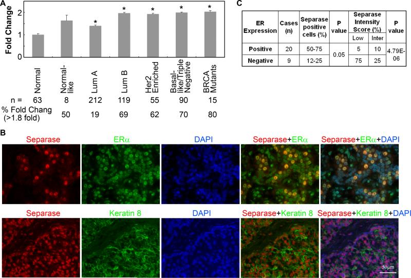
Figure 5. Expression of Separase transcript and protein in human breast cancer.
(A) Analysis of ESPL1 transcript that encodes Separase protein using the TCGA breast cancer data sets 32 showing overexpression of Separase mRNA in various tumor subtypes. Bottom panel shows the total number of tumors used in the analysis and % of tumors that express Separase transcripts >1.8 times compared to the normal breast tissues. (A) Representative tissue microarrays (TMA) of human breast cancers that reveal strong Separase expression and nuclear co-localization with ERα (top panel) in pre-dominantly cytokeratin-8/18 positive tumors (bottom panel). (B) Percentage of Separase positive cells and the corresponding intensity scores of Separase expression in ERα positive and negative tumors in the human TMA.

