SUMMARY
Microglia are the resident macrophages of the central nervous system and their functions have been extensively studied in various brain pathologies. The physiological roles of microglia in brain plasticity and function, however, remain unclear. To address this question, we generated CX3CR1CreER mice expressing tamoxifen-inducible Cre recombinase that allow for specific manipulation of gene function in microglia. Using CX3CR1CreER to drive diphtheria toxin receptor expression in microglia, we found that microglia could be specifically depleted from the brain upon diphtheria toxin administration. Mice depleted of microglia show deficits in multiple learning tasks and a significant reduction in motor learning-dependent synapse formation. Furthermore, Cre-dependent removal of brain-derived neurotrophic factor (BDNF) from microglia largely recapitulated the effects of microglia depletion. Microglial BDNF increases neuronal TrkB phosphorylation, a key mediator of synaptic plasticity. Together, our findings reveal important physiological functions of microglia in learning and memory by promoting learning-related synapse formation through BDNF signaling.
INTRODUCTION
Microglia are a population of resident myeloid cells that occupy all regions of the mammalian central nervous system (CNS). Recent studies have shown that microglia colonize the brain early during development (embryonic day 9.5) (Ginhoux et al., 2010). As development proceeds, microglia transition from an amoeboid to a highly ramified morphology with multiple fine processes that display a constant motility within neural tissues (Davalos et al., 2005; Swinnen et al., 2013). Although the role of microglia in CNS pathologies has been extensively studied, their contribution to normal CNS physiology remains unclear. Disruptions in the colony stimulating factor-1 (CSF-1) signaling pathway in mice cause a reduction in the number of microglia as well as defects in neuronal structure and function (Michaelson et al., 1996; Roumier et al., 2004). In humans, mutations in CSF-1 signaling have been associated with presenile-dementia (Paloneva et al., 2000). A variety of structural and functional deficiencies have also been associated with deletion or loss of function mutations in a number of genes expressed in microglia, including the fractalkine receptor CX3CR1, methyl CpG binding protein 2 (Mecp2), and homeobox protein HoxB8 (Chen et al., 2010; Derecki et al., 2012; Paolicelli et al., 2011). Together, these studies suggest that microglial dysfunction has a significant detrimental impact on the development and function of the CNS. However, because genes such as CSF-1, CX3CR1, Mecp2 and HoxB8 function in many myeloid populations, both microglia and peripheral myeloid cells are affected in these studies. As deficits in peripheral myeloid cells could have significant impacts on the CNS (Dantzer et al., 2008), caution is warranted in deducing the precise function of microglia in the brain from experiments using knockout mice that affect both peripheral and CNS myeloid cells.
Many lines of evidence indicate that experience-dependent synaptic structural plasticity is important for the CNS development as well as for learning and memory formation (Bailey and Kandel, 1993; Grutzendler et al., 2002; Yang et al., 2009a). For example, motor skill learning induces the formation of postsynaptic dendritic spines in the motor cortex and the survival of these spines strongly correlates with performance improvement after learning (Liston et al., 2013; Yang et al., 2009a). Recent studies have shown that microglial processes are often in close proximity to neuronal somata and dendritic spines, and that the dynamics of microglial processes are regulated by sensory experience and/or neuronal activity (Tremblay et al., 2010; Wake et al., 2009). These findings suggest that microglia may play a role in regulating experience-dependent synaptic plasticity. Recently, several studies have suggested that microglia are involved in synaptic pruning through the phagocytosis of synapses during early postnatal periods, and that this process can be disrupted by loss of the fractalkine receptor CX3CR1 (Paolicelli et al., 2011) or complement receptor 3 (CR3/CD11b) (Schafer et al., 2012). However, synaptic phagocytosis by microglia and synaptic pruning defects observed in CX3CR1 and CR3 null mice are absent during later postnatal development and adulthood (Paolicelli et al., 2011; Schafer et al., 2012). Furthermore, similar to other myeloid genes, CX3CR1 and CR3 have functions not only in microglia but also in peripheral myeloid populations, making it difficult to pinpoint the precise functions of microglia using CX3CR1 and CR3 knockout mice. Therefore, it remains unknown whether and how microglia are involved in experience-dependent changes of synaptic circuits, particularly in later post-natal and adult life. It is also unclear whether microglial dysfunction would contribute significantly to learning deficits as seen in neurological diseases.
To investigate the precise roles of microglia in the brain, we generated a mouse line that allows, for the first time, specific genetic manipulation of microglia in an inducible fashion. Here, we report that specific depletion of microglia leads to deficits in multiple learning tasks and learning-induced synaptic remodeling. Furthermore, genetic depletion of BDNF from microglia recapitulates many of the phenotypes generated by deletion of microglia, indicating that microglial BDNF is an important factor for synaptic remodeling associated with learning and memory.
RESULTS
Generation of CX3CR1CreER mice to manipulate gene expression in microglia
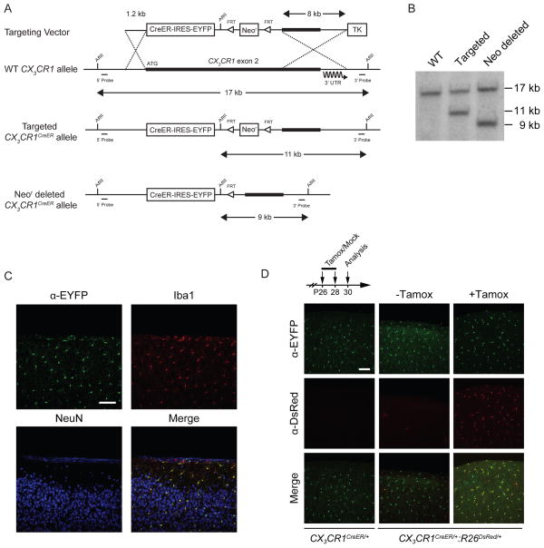
In order to manipulate microglial function, we generated CX3CR1CreER mice expressing tamoxifen-inducible Cre recombinase (CreER) in microglia under the control of the endogenous CX3CR1 promoter (Fig. 1A). The gene encoding CreER was followed by an IRES-EYFP element and the insertion site was chosen according to previous studies in which the CX3CR1 coding region was replaced with EGFP (Jung et al., 2000). Correct targeting of the CX3CR1 locus and subsequent FLP recombinase mediated excision of the neomycin resistance cassette was verified by Southern blot analysis (Fig. 1B). We performed immunostaining for the microglial marker Iba1 and neuronal marker NeuN in brain slices from CX3CR1CreER/+ heterozygous mice. As expected, we found that virtually all (98.8 ± 1.3%, 172 cells) EYFP+ cells were Iba1+ while no NeuN+ cells (401 cells) were EYFP+ (Fig. 1C), indicating that the CreER transcript is specifically expressed in microglia in the brain. To test the functionality of tamoxifen-inducible Cre recombinase, we crossed CX3CR1CreER mice to Rosa26-stop-DsRed reporter allele mice (R26DsRed) (Luche et al., 2007) to generate CX3CR1CreER/+:R26DsRed/+ animals. In the absence of tamoxifen, few (0.3 ± 0.01%) DsRed+ microglia were found in the brain of CX3CR1CreER/+:R26DsRed/+ mice (Fig. 1D). In contrast, five days after tamoxifen treatment, the majority (93.9 ± 0.5%) of CX3CR1-EYFP+ microglia in CX3CR1CreER/+:R26DsRed/+ mice were found to co-express DsRed (Figs. 1D and 2A). Thus, Cre-mediated recombination in microglia is highly efficient in CX3CR1CreER/+ mice.
Figure 1. Generation of mice carrying the CX3CR1CreER allele.
(A) Schematic of targeting strategy used for knock-in of CreER-IRES-YFP at the CX3CR1 locus. (B) Southern blot analysis of AflII digested genomic DNA from untargeted (WT), CX3CR1CreER targeted, or CX3CR1CreER mice after deletion of the neomycin resistance sequence. (C) Coronal sections of motor cortex from P45 CX3CR1CreER mice stained for EYFP, Iba1, and NeuN. (D) Coronal sections of motor cortex from mice of the indicated genotypes and treatments (scale bar = 100 μm).
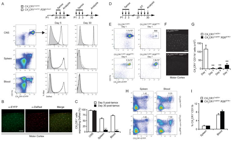
Figure 2. A strategy to restrict Cre-mediated manipulation of gene function including deletion of microglia.
(A) CX3CR1-EYFP+ CD11b+ populations in various tissues from mice of the indicated genotypes 5 or 30 days post-tamoxifen treatment. (B) Coronal sections of motor cortex from CX3CR1CreER/+:R26DsRed/+ mice stained for EYFP and DsRed 30 days after tamoxifen treatment. (C) Quantification of flow cytometry fluorescence-activated cell sorting (FACS)analysis showing the percentage of CX3CR1-EYFP+ cells co-expressing DsRed in multiple tissues at 5 or 30 days post-tamoxifen treatment. (D) Time-course of tamoxifen/DT administration and analysis. (E) FACS analysis of microglia in the brain of control and microglia-depleted mice at indicated time points after DT administration. Dot plots show total number of CX3CR1-EYFP+ CD11b+ cells gated on DAPI− CD3− CD19− CD45int. (F) Coronal sections of motor cortex from control or microglia-depleted mice stained for Iba1 one day after DT administration. (G) Number of CX3CR1-EYFP+ CD11b+ microglia in the brain after DT administration at various time-points. (H) FACS analysis showing percentage of CX3CR1-EYFP+ CD11b+ cells in the spleen and blood of mice after DT administration. (I) Quantification of data shown in (H). n=4 animals for each experimental condition. Data are represented as mean +/− SEM. ****p<0.0001, *p<0.05; Scale bar= 100 μm. See also Figures S1, S2, S3.
Cre-dependent manipulation of gene expression in microglia
Consistent with previous studies of CX3CR1 expression patterns (Jung et al., 2000), EYFP expression was also observed in multiple CD11b+ myeloid populations in peripheral tissues of CX3CR1CreER/+ mice (Fig. 2A). We found that the percentages of several myeloid populations in the brain, blood, and spleen were comparable between WT mice and CX3CR1CreER/+ mice at postnatal day 14 (P14) and P30, suggesting that the development and maturation of myeloid populations, including microglia in the brain, were not altered in heterozygous CX3CR1CreER/+ mice lacking one copy of the endogenous CX3CR1 gene (Fig. S1A–B). As expected, in CX3CR1CreER/+:R26DsRed/+ mice, Cre-mediated recombination occurred not only in CNS microglia but also in CX3CR1+ cell populations in the spleen (59.0 ± 3.1%), and blood (70.9 ± 4%) 5 days after tamoxifen administration (Fig. 2A, C).
In order to restrict Cre-mediated recombination exclusively to microglia, we took advantage of the fact that microglia and other CX3CR1+ cells have substantially different rates of turnover and are derived from different precursor populations. Microglia are a self-renewing population (Ajami et al., 2007) with a low turnover rate (Lawson et al., 1992), while monocytes and inflammatory macrophages exhibit rapid turnover (van Furth and Cohn, 1968) and are replenished through a CX3CR1− bone marrow precursor population (Fogg et al., 2006). Therefore, upon exposure to tamoxifen, we expect that microglia would undergo recombination that persists once tamoxifen has dissipated. Conversely, peripheral CX3CR1+ populations would initially undergo recombination, but subsequently be replaced by non-recombined cells from CX3CR1− progenitors in the absence of additional tamoxifen (Fig. S1C). Indeed, when CX3CR1CreER/+:R26DsRed/+ mice were pulsed with tamoxifen at P1–P3 and examined 30 days later, 93.0 ± 1.0% of EYFP+ microglia were found to be DsRed+ (Fig. 2A–C). In contrast, only 7.9 ± 1.9% of cells in the spleen, and 1.7 ± 0.5% of the cells within the blood were DsRed+ (Fig. 2A, C). These results demonstrate that CX3CR1CreER/+ mice, when utilized 30 days after tamoxifen administration, permit Cre-dependent manipulation of gene expression almost exclusively in microglia.
We next used a similar strategy to express the diphtheria toxin receptor (DTR) specifically in microglia and administered diphtheria toxin (DT) to deplete microglia in the CNS while leaving other CX3CR1+ populations intact. To accomplish this, CX3CR1CreER mice were crossed with mice harboring the Rosa26-stop-DTR (R26iDTR) allele (Buch et al., 2005), and CX3CR1CreER/+:R26iDTR/+ mice were given tamoxifen ~30 days prior to the administration of diphtheria toxin (DT) (Fig. 2D). We found that CX3CR1CreER/+:R26iDTR/+ mice had a marked (99.1± 0.8%) reduction of CNS microglia within one day after administration of DT (Fig. 2E–G). In contrast, the number of microglia was not affected in the brain of control mice carrying a single copy of the CX3CR1CreER allele only. Seven days after DT administration, the number of microglia remained significantly lower (84.8 ± 3.0% reduction) in CX3CR1CreER/+:R26iDTR/+ mice than that in control mice (Fig. 2E, G), suggesting limited repopulation of microglia within one week of depletion. Importantly, we observed no significant difference in the number of CX3CR1+ CD11b+ cells in the spleen or blood between CX3CR1CreER/+:R26iDTR/+ mice and control mice (Fig. 2H–I). These experiments demonstrate that CX3CR1CreER/+:R26iDTR/+ mice can be used to specifically and robustly delete microglia in the CNS.
Within the first week after microglial depletion, we found no difference in animal viability (n=14–16 per group), or weight change (Fig. S2A) between microglia-depleted and non-depleted control mice. To investigate whether microglial ablation may cause inflammatory responses in the CNS, we measured transcript and protein levels of the inflammatory cytokines TNFα, IL-1β, and IL-6 in brain tissues and observed no significant difference between the microglia-depleted and control mice (Fig. S2B–D). We also found no significant difference in the immunostaining of glial fibrillary acidic protein (GFAP) and glutamate transporter GLT-1 within 7 days after microglial depletion in either motor cortex or hippocampal CA1 region (Fig. S2E–J). In addition, we found no alterations in blood-brain-barrier permeability between microglia-depleted and control mice as measured by the amount of Evans Blue dye in the brain after intravenous injection (Fig. S2K). To determine if depletion of microglia altered overall densities of neurons and synapses, we stained sections of the motor cortex and hippocampal CA1 region for NeuN, cleaved caspase 3 (CC3), or the pre-synaptic marker synaptic vesicle protein 2 (SV2) and found no significant difference in these parameters between microglia-depleted and control mice (Fig. S3). Taken together, these findings suggest that depleting microglia leaves surrounding brain tissues minimally disturbed. Thus, CX3CR1CreER/+:R26iDTR mice provide an important tool to delete microglia and examine their function in the CNS.
Depletion of microglia reduces synaptic structural plasticity associated with learning
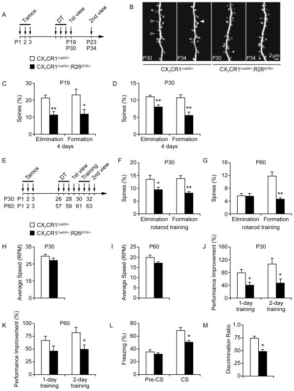
Recent studies have suggested that microglia participate in synapse elimination through a phagocytic engulfment mechanism involving the fractalkine receptor CX3CR1 (Paolicelli et al., 2011) or complement receptor 3 (CR3/CD11b) (Schafer et al., 2012). This process of synaptic phagocytosis seems to occur only during early, but not late postnatal periods (Paolicelli et al., 2011; Schafer et al., 2012). To determine the potential function of microglia in the mature brain, we first investigated whether microglia depletion might alter synaptic structural plasticity during late postnatal period (P19) or young adulthood (P30). Using transcranial two-photon microscopy (Yang et al., 2009a), we examined the effect of microglial depletion on the baseline remodeling of postsynaptic dendritic spines of layer V pyramidal neurons in the motor cortex of Thy1 YFP-H line mice crossed with CX3CR1CreER/+ or CX3CR1CreER/+:R26iDTR/+ mice (Fig. 3A–B). We found that at either P19 or P30, microglial depletion caused a significant decrease in both spine formation and elimination over 4 days (Fig. 3C–D). In addition, we observed that baseline spine remodeling was not altered in CX3CR1CreER/+ mice lacking a single copy of the endogenous CX3CR1 gene as CX3CR1CreER/+:Thy1 YFP-H and CX3CR1+/+:Thy1 YFP-H mice showed similar rates of spine turnover (Fig. S4A). Taken together, these findings suggest that microglia are involved in not only spine elimination but also spine formation during late postnatal development and young adulthood.
Figure 3. Microglia are important for learning-dependent spine remodeling and performance improvement.
(A) Timeline of tamoxifen/DT administration and in vivo imaging in CX3CR1-iDTR mice. (B) Transcranial two-photon imaging of dendritic spines in control and microglia-depleted mice. Filled and empty arrowheads indicate spines formed or eliminated between two views. Asterisk indicates filopodia. (C–D) Percentage of spines formed or eliminated within 4 days in the motor cortex was significantly reduced after microglia depletion in both P19 (C) and P30 animals (*p<0.05, **p<0.01, n=4–6). (E) Timeline of tamoxifen/DT administration, rotarod training, and in vivo imaging. (F) Motor learning-related spine remodeling was significantly reduced in P30 mice with microglia depletion (*p<0.05, **p<0.01, n=4–5). (G) Motor learning-related spine formation was significantly reduced in P60 mice with microglia depletion (**p<0.01, n=4–5). (H) Average speed reached during the first rotarod training session in P30 mice (n=6–7). (I) Average speed reached during the first rotarod training session in P60 mice (n=8). (J) Microglia-depleted mice showed impaired performance improvement compared to non-depleted control mice over one or two days of training (*p<0.05, n=6–7). (K) P60 microglia-depleted mice showed impaired performance improvement compared to non-depleted control mice over one or two days of training (*p<0.05, n=8). (L) Percentage of freezing in control or microglia-depleted mice before (pre-CS) and during (CS) presentation of the conditioned stimulus in the recall test (*p<0.05, n=8) (M) Discrimination ratio of time spent interacting with a novel object vs. a familiar object in a novel object recognition assay was significantly altered in microglia-depleted mice (*p<0.05, n=8). Data are represented as mean +/− SEM. See also Figure S4.
Previous studies have shown that motor skill learning causes an increase in dendritic spine remodeling in the motor cortex, and that the degree of new spine formation correlates with performance improvement after learning (Liston et al., 2013; Yang et al., 2009a). To investigate the role of microglia in this process, we examined whether depletion of microglia altered spine formation and elimination over 4 days in response to rotarod motor learning (Fig. 3E). We found a significant decrease in learning-dependent formation and elimination of dendritic spines in 1-month-old microglia-depleted mice as compared with age-matched controls (Fig. 3F). Furthermore, in 2-month-old adult mice, microglia-depletion caused a significant decrease in learning-dependent spine formation but not spine elimination (Fig. 3G). Notably, although the baseline performance of microglia-depleted animals remained unchanged (Fig. 3H–I), there was a significant decrease in performance improvement after motor learning in microglia-depleted mice as compared to control mice at P30 and P60 (Fig. 3J–K; Fig. S4B). These results demonstrate that microglia have an important role in learning-induced remodeling of excitatory synapses, as well as in animals’ performance improvement after motor learning.
To further understand the effect of microglia deletion on learning and memory, we compared microglia-depleted and control mice in two additional behavioral paradigms: auditory-cued fear conditioning (FC) and novel object recognition (NOR). We found that microglia-depleted animals exhibited significantly reduced freezing fear response to the auditory cue during the recall test as compared to non-depleted controls (Fig. 3L). Additionally, while the non-depleted control mice showed a preference towards the novel object during the NOR test, microglia-depleted mice showed no such preference (Fig. 3M). Thus, mice lacking microglia demonstrate deficits in multiple behavioral tasks, underscoring an important role of microglia in learning and memory that involves multiple brain regions.
Depletion of microglia alters synaptic protein levels and glutamatergic synaptic function
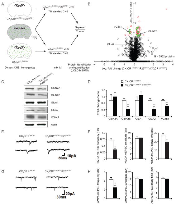
Our results indicate that microglial depletion causes defects in learning-induced dendritic spine remodeling and behavioral performance. To better understand synaptic alterations after microglial deletion, we examined the levels of various proteins in microglia-depleted brain, focusing on those involved in synaptic plasticity and function. We first performed a quantitative proteomic screen from whole-brain protein extracts by shotgun LCLC-MS/MS high-resolution mass spectrometric proteome analysis (Washburn et al., 2001). Specifically, we utilized 15N-labeled mouse brains as an internal standard and mixed 15N whole brain 1:1 with P30 microglia-depleted or control littermate brains to calculate relative protein profiles (Fig. 4A). A similar approach has been used previously for proteome-wide quantitative analysis of long-lived proteins and synaptosome changes during development (Savas et al., 2012). We quantified a total of 6562 proteins and found that the levels of 61 proteins were significantly altered as a result of microglia depletion (Fig. 4B and Table S1, ANOVA p value < 0.05 & Log2 fold change ≥20%). Importantly, 21/61 (34%) of the significantly altered proteins had known roles in synaptic plasticity/function, such as the post-synaptic Glutamate [NMDA] receptor subunit epsilon-2 (GluN2B) and the presynaptic vesicular glutamate transporter 1 (VGlut1). We additionally found the Glutamate [AMPA] receptor subunit 2 (GluA2) to be decreased, although it did not meet our significance criteria.
Figure 4. Biochemical and electrophysiological properties of synapses are altered in microglia-depleted brains.
(A) Quantitative proteomic scheme to identify CNS proteins altered after microglial depletion. Control (n=3) or microglia-depleted (n=3) brain homogenates were mixed 1:1 with 15N internal standard and were prepared together. Samples were then analyzed by LCLC-MS/MS shotgun proteomics. Green dots represent microglia. (B) Proteomic summary volcano plot (x-axis=log2 CX3CR1CreER/+/CX3CR1CreER/+:R26iDTR/+, y-axis=−log10 ANOVA p value). Black open circles: quantified proteins, red open circles: significantly altered proteins, green filled circles: significantly altered proteins with known synaptic functions. (C) Synaptosome fractions from control or microglia-depleted brains probed with indicated antibodies by Western blot. (D) Densitometric quantification of Western blots in (C) (*p<0.05, n=6). (E) Examples of NMDA mEPSCs in layer V pyramidal neurons from control and microglia-depleted mice. (F) Average NMDA mEPSC frequency, amplitude and decay time in control (n=17 cells) and microglia-depleted mice (n=17 cells). mEPSC frequency and decay time were significantly reduced in microglia-depleted mice (p<0.001). (G) Examples of AMPA mEPSCs in layer V pyramidal neurons from control and microglia-depleted mice. (H) Average mEPSC frequency, amplitude and decay time in control (n=9 cells) and microglia-depleted mice (n=8 cells). mEPSC frequency was significantly reduced in microglia depleted mice (p<0.05). Data are represented as mean +/− SEM. See also Table S1.
To further investigate the alteration of synaptic proteins after microglia depletion, we generated synaptosome fractions from microglia-depleted mice and compared them to those from the non-depleted controls. In line with the results of our proteomic screen, we found VGlut1 and GluA2 to be significantly decreased in synaptosomes from microglia-depleted brains. Although GluN2B protein was increased in the whole brain fraction as shown in our proteomic screen, its level was decreased in the synaptosome fraction, suggesting differential alteration of GluN2B in synaptosome versus non-synaptosome fractions after microglial depletion. The NMDAR subunit GluN2A and the AMPAR subunit GluA1 remained unaltered between microglia-depleted mice and controls (Fig 4C–D). These results support the data from our proteomic screen, and demonstrate that microglia-depleted brains display significant and specific changes of synaptic proteins at glutamatergic synapses.
To test whether changes in the abundance of the synaptic proteins GluN2B, VGlut1, and GluA2 after microglia depletion affect glutamatergic synaptic function, we performed whole-cell patch clamp recordings from layer V pyramidal neurons in the motor cortex of P30 mice one day after the depletion of microglia. We found that the frequencies of both NMDA and AMPA receptor-mediated miniature excitatory post-synaptic currents (mEPSCs) were significantly decreased as compared to controls (Fig. 4E–H), suggesting a decrease in spontaneous glutamate release in the absence of microglia. Furthermore, the decay time of NMDA mEPSCs but not AMPA mEPSCs was significantly reduced in microglia-depleted animals while the amplitude of spontaneous NMDA and AMPA mEPSCs remained unchanged (Fig. 4F, H). The decrease in the decay time of NMDA mEPSCs is consistent with the reduction of GluN2B subunits in microglia-depleted mice as GluN2A-dominant NMDA receptors have faster decay times (Chen et al., 1999). Taken together, these data indicate that microglia play a role in regulating the level of several synaptic proteins important for the function of glutamatergic excitatory synapses in the brain.
Removal of BDNF from microglia reduces learning-dependent synaptic structural plasticity
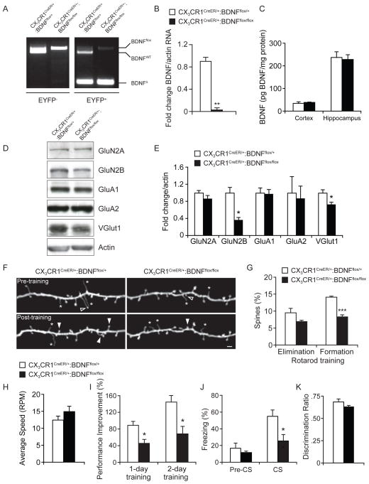
The above results show that microglia deletion causes a reduction in several synaptic proteins involved in synaptic plasticity and function. However, the molecular mechanisms underlying the interaction between microglia and neurons remain unknown. To address this question, we investigated the role of brain-derived neurotrophic factor (BDNF) in mediating microglial-neuron interactions. We choose to study microglial BDNF for the following two reasons: First, BDNF exists in many cell types including microglia, and BDNF from microglia has been shown to modulate neuronal plasticity in a mouse model of neuropathic pain (Coull et al., 2005). Second, BDNF is a potent regulator of synaptic development and plasticity (Chao, 2003) and increases dendritic spine plasticity in the adult cortex (Chakravarthy et al., 2006), GluN2B activation (Levine et al., 1998), and the level of VGlut1 (Melo et al., 2013). Given our findings that microglia depletion causes a reduction of GluN2B and VGlut1 as well as dendritic spine plasticity, it is thus possible that lack of microglial BDNF may underlie some of the phenotypes associated with microglia depletion. To test this possibility, we crossed CX3CR1CreER mice with mice containing a floxed allele of BDNF (BDNFflox) (Rios et al., 2001) to remove BDNF from microglia. We first validated this approach by sorting CX3CR1-EYFP− cells or CX3CR1-EYFP+ microglia from the brains of CX3CR1CreER/+:BDNFfl/+ (BDNFfl/+), or CX3CR1CreER/+:BDNFfl/fl (BDNFfl/fl) mice that had been given tamoxifen (Fig. S5A), and assayed for recombination of the conditional BDNF allele using a PCR-based strategy. As expected, no recombination was observed in CX3CR1-EYFP− cells. In contrast, robust recombination of the floxed allele was observed in CX3CR1-EYFP+ microglia from BDNFfl/+ or BDNFfl/fl mice (Fig. 5A). After administration of tamoxifen, the level of BDNF mRNA in microglia from CX3CR1CreER/+:BDNFfl/fl mice was markedly reduced as compared to CX3CR1CreER/+:BDNFfl/+ controls (Fig. 5B). Interestingly, the overall levels of BDNF protein in the cortex and the hippocampus of CX3CR1CreER/+:BDNFfl/fl mice were unchanged compared to CX3CR1CreER/+:BDNFfl/+ controls (Fig. 5C). Taken together, these results indicate that BDNF can be specifically removed from microglia without causing a significant alteration in the total level of BDNF protein in the cortex or hippocampus.
Figure 5. Loss of microglial BDNF results in altered synaptic protein levels, synaptic structural plasticity, and performance improvement after learning.
(A) PCR-based analysis of wild-type (BDNFWT), conditional undeleted (BDNFflox), or conditional deleted (BDNFΔ) BDNF alleles from CX3CR1-EYFP− and CX3CR1-EYFP+ cells sorted from the CNS of CX3CR1CreER/+:BDNFflox/+ or CX3CR1CreER/+:BDNFflox/flox after tamoxifen treatment. (B) Quantitative real-time PCR analysis of BDNF mRNA isolated from CX3CR1-EYFP+ microglia purified from BDNFflox/+ or BDNFflox/flox mice (**p<0.01, n=3). (C) Average protein levels of total BDNF in the cortex or hippocampus of CX3CR1CreER/+:BDNFflox/+ or CX3CR1CreER/+:BDNFflox/flox mice as measured by ELISA (n=4). (D) Synaptosome fractions from the brains of CX3CR1CreER/+:BDNFflox/+ or CX3CR1CreER/+:BDNFflox/flox mice probed with indicated antibodies. (E) Densitometric quantification of western blots in (D) (*p<0.05, n=6). (F) Transcranial two-photon imaging of dendritic spines in Thy1 YFP mice crossed with CX3CR1CreER/+:BDNFflox/+ or CX3CR1CreER/+:BDNFflox/flox mice before or after rotarod training. Filled and empty arrowheads indicate spines formed or eliminated between two views. Asterisk indicates filopodia. Scale bar= 2 μm. (G) Percentage of existing spines eliminated, or new spines formed over 2 days of training in the motor cortex of BDNFflox/+ or BDNFflox/flox mice (***p<0.001, n=4). (H) Average speed reached during the first rotarod training session (n=5–7). (I) Performance increase in motor learning task over 1 or 2 days of rotarod training (*p<0.05, error bars= SEM, n=5–7). (J) Percentage of freezing in control CX3CR1CreER/+:BDNFflox/+ or CX3CR1CreER/+:BDNFflox/flox mice before (pre-CS) and during (CS) presentation of the conditioned stimulus in the recall test (*p<0.05, n=6–7) (K) Discrimination ratio of time spent interacting with a novel object vs. a familiar object in a novel object recognition assay was significantly altered in mice depleted of microglial BDNF (*p<0.05, n=8). Data are represented as mean +/− SEM. See also Figure S5.
To investigate the potential impact of microglial BDNF removal, we first stained the motor cortex and hippocampal CA1 region for NeuN, cleaved caspase 3, or SV2 but found no significant difference in these parameters between microglial BDNF-deleted and control mice (Fig. S5B–D). Thus, microglial BDNF removal does not alter the overall densities of neurons or synapses in the cortex or hippocampus. We next asked whether removal of BDNF from microglia had any effects on the level of synaptic protein expression, motor learning-induced synaptic plasticity and performance improvement after learning. We performed Western blot analysis of synaptosomes and found a significant decrease in the levels of GluN2B and VGlut1, but not GluA2, in mice lacking microglial BDNF (BDNFfl/fl) as compared to control mice (BDNFfl/+) (Fig. 5D–E). Importantly, BDNF removal from microglia resulted in a significant decrease in motor learning-induced spine formation, but not spine elimination, over 2 days (Fig. 5F–G). Furthermore, similar to microglia-depleted animals, while BDNF removal from microglia had no effect on baseline rotarod performance, mice lacking microglial BDNF demonstrated a reduction in performance improvement after motor training as compared to controls (Fig. 5H–I). Mice lacking microglial BDNF also showed significantly reduced freezing fear response to the conditioned auditory cue stimulus during the recall test (Fig. 5J). In the novel object recognition task, however, mice lacking microglial BDNF did not show a significant difference from the control mice (Fig. 5K). Taken together, these results demonstrate that loss of microglial BDNF recapitulates several defects observed in microglia-depleted mice and suggest that microglial BDNF is an important regulator of learning-induced synaptic formation and behavioral performance.
Microglial BDNF affects synaptic plasticity via the TrkB signaling pathway
As microglial processes are located in close proximity to synapses, microglial BDNF could be released to directly affect the structure and function of nearby synapses. Alternatively, microglial BDNF could have a role in microglial differentiation or homeostasis and the removal of BDNF from microglia might act in an autocrine fashion and indirectly affect synaptic plasticity. To distinguish between these possibilities, we first compared the overall number, rate of proliferation, and tissue distribution of microglia between CX3CR1CreER/+:BDNFfl/fl and CX3CR1CreER/+:BDNFfl/+ controls, but found no significant differences in any of these parameters (Fig. S6A–E). To examine whether loss of microglial BDNF might alter the association of microglial processes with dendritic spines, we measured the percentage of spines of layer V pyramidal neurons in the motor cortex that had Iba1+ microglial processes within 1 μm and found no significant difference between CX3CR1CreER/+:BDNFfl/fl and CX3CR1CreER/+:BDNFfl/+ control mice (Fig. S6F–G). These results demonstrate that loss of BDNF from microglia does not alter the number, proliferation and distribution of microglia, nor the spatial association of microglial processes with dendritic spines.
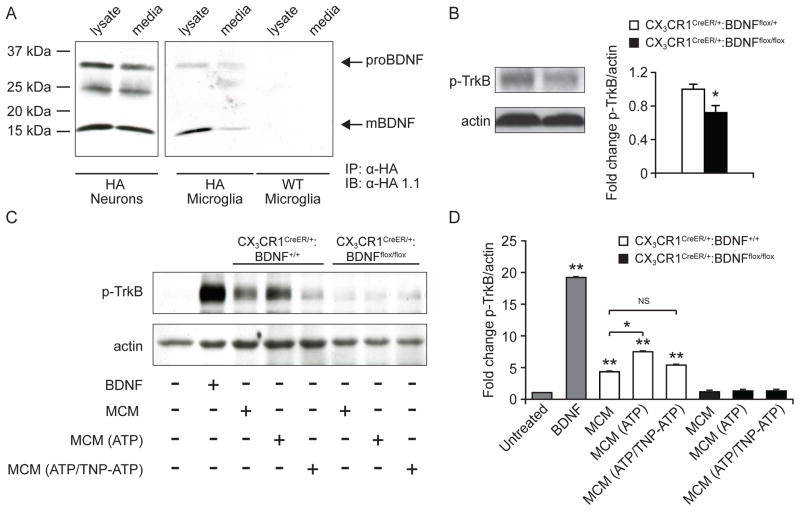
The above findings suggest that microglial BDNF may be released to directly affect the plasticity of nearby synapses. BDNF is produced within neurons as a pro-peptide (proBDNF) that is subsequently cleaved to generate mature BDNF (mBDNF). Because the pro and mature forms of BDNF bind different receptors on the surface of neurons (p75/sortilin and tropomyosin-related kinase receptor B (TrkB) respectively) to affect different cellular processes (Chao, 2003), we asked whether and what forms of BDNF might be produced and secreted by microglia. Because endogenous BDNF protein is difficult to detect due to low abundance (Herzog et al., 1994), we utilized BDNF-HA knock-in mice (BDNF-HA) in which the BDNF gene is replaced by haemagglutinin-tagged BDNF (Yang et al., 2009b). We generated purified cultures of hippocampal neurons or microglia from P1 BDNF-HA/HA mice (microglial purity 98.6 ± 0.1%, Fig S6H) and immunoprecipitated HA-tagged BDNF from either the cell lysates or culture media of both cell types. In agreement with previous results, significant quantities of both proBDNF and mBDNF could be detected in both cell lysates and culture media from BDNF-HA neurons. Notably, both forms of BDNF were also detected in cell lysates and culture media from pure microglia cultures although the distribution of pro- to mBDNF in microglial lysates appeared to be weighted towards the mature form (Fig. 6A). These results demonstrate that microglia are capable of producing and secreting both pro- and mature-BDNF.
Figure 6. Microglia produce both pro and mature BDNF to phosphorylate neuronal TrkB.
(A) Neurons or microglia were cultured from P1 mice from BDNF-HA or WT animals. Cell lysates or culture media were immmunoprecipitated (IP) with a rabbit antibody to HA. Pro-BDNF and mature BDNF were detected by immunoblotting with a second antibody to HA (mouse HA1.1). (B) Synaptosome westerns for p-TrkB from CX3CR1CreER/+:BDNFflox/+ or CX3CR1CreER/+:BDNFflox/flox mice (*p<0.05, n=6). (C) Representative immunoblots of E18 rat neurons at DIV 8 treated as indicated. (D) Densitometric quantification of p-TrkB western blots in (C) (*p<0.05, **p<0.005, n=9). Data are represented as mean +/− SEM. See also Figure S6.
It is well established that mature BDNF binds to TrkB receptors located on neurons, and binding of mBDNF to TrkB triggers a signaling cascade that promotes auto-phosphorylation of TrkB and synaptic plasticity (Chao, 2003). Consistent with an interaction between microglia BDNF and TrkB receptors, Western blot analysis of synaptosomes showed a significant decrease in the levels of phosphorylated-TrkB (p-TrkB) in CX3CR1 CreER/+:BDNFfl/fl mice lacking microglial BDNF as compared to control CX3CR1 CreER/+:BDNFfl/+ mice (Fig. 6B). To investigate microglia BDNF and TrkB signaling further, we tested the ability of the media from cultures of purified microglia (microglia conditioned media, MCM) from CX3CR1CreER/+:BDNF+/+ or CX3CR1CreER/+:BDNFflox/flox P1 mice to phosphorylate TrkB in cultures of purified cortical neurons. Within 30 minutes, MCM from CX3CR1CreER/+:BDNF+/+ mice was able to significantly increase the phosphorylation of TrkB compared to baseline levels in untreated neuronal cultures. Importantly, MCM generated from microglial cultures devoid of BDNF (Fig. S6I) failed to increase the phosphorylation of TrkB (Figs. 6C–D). Previous studies have demonstrated that ATP acting through the purinergic receptor P2X4 is able to increase both the synthesis and release of BDNF from microglia in vitro (Trang et al., 2009). To test whether ATP and/or P2X4R are also involved in the ability of microglia conditioned media to phosphorylate TrkB, we treated purified cultures of microglia with ATP or with ATP and the P2X4R antagonist 2′,3′-O-(2,4,6-Trinitrophenyl)adenosine-5′-triphosphate (TNP-ATP) before harvesting MCM. Pretreatment of microglia cultures with ATP significantly increased the ability of MCM to phosphorylate TrkB, and this increase was blocked by the addition of TNP-ATP to the cultures (Figs. 6C–D). Taken together, these findings suggest that microglial BDNF promotes synaptic structural plasticity involving activation of TrkB signaling.
DISCUSSION
The lack of tools to specifically perturb microglia at defined time points in the living animal has made it difficult to delineate the precise functions of microglia in the physiological brain. In this work, we have generated a tool that allows, for the first time, the specific and inducible manipulation of microglial numbers and functions within the CNS. Using this tool, we have revealed a novel physiological role of microglia in learning and learning-associated synaptic structural remodeling. Moreover, use of CX3CR1CreER mice has allowed us to uncover an important role of microglial BDNF in these processes.
Interactions Between Microglia and Neurons: Promoting Learning-induced Synapse Formation
Microglial processes are constantly motile in the physiological brain, and are found in close proximity to synapses in both the early postnatal and adult cortex (Davalos et al., 2005; Tremblay et al., 2010; Wake et al., 2009). The functional outcome of microglial interaction with neurons appears to depend on the developmental stage of the CNS. In the embryonic brain with high levels of programmed neuronal cell death, microglia have been observed to phagocytose apoptotic neurons (Peri and Nusslein-Volhard, 2008), or actively participate in the killing of neurons (Marín-Teva et al., 2004). In the embryonic zebrafish optic tectum, microglia appear to play a role in the homeostasis of neuronal activity by silencing neurons through a contact-dependent mechanism (Li et al., 2012). Recent work has shown that mice lacking CX3CR1 have delayed invasion of the cortical barrel fields by microglia, and a transient delay in the normally observed AMPA receptor subunit switching in thalamocortical barrel cortex synapses (Hoshiko et al., 2012). Furthermore, it has been recently proposed that microglia are involved in the active engulfment of synapses in the hippocampus and the dorsal lateral geniculate nucleus (dLGN) during early postnatal development (Paolicelli et al., 2011; Schafer et al., 2012). Pruning of synapses within dLGN is thought to depend upon the presence of CR3, which may interact with synaptically localized C3 protein that acts as a positive signal for phagocytosis of pre-synaptic axonal terminals.
By taking advantage of CX3CR1CreER mice and transcranial two-photon imaging, we observed a significant decrease in learning-induced formation of postsynaptic dendritic spines in the absence of microglia or microglial BDNF in young and mature adult mice. Two lines of evidence suggest that the main synaptic function of microglia in the mature brain is the promotion of dendritic spine formation rather than spine elimination. First, removal of BDNF from microglia causes a significant decrease in learning-induced spine formation, but not spine elimination over two days. Second, previous studies have shown that motor learning-induced spine formation occurs two days before spine elimination and that the degrees of spine formation and elimination are strongly correlated (Liston et al., 2013; Yang et al., 2009a). Therefore, the decreased spine elimination observed in the absence of microglia may simply be a result of fewer spines being formed after motor learning. It is worth noting that we also observed a decrease in spine formation and elimination at P19. This raises the possibility that microglia have an important function in promoting synapse formation not only during the late postnatal stage and adulthood but also during the early postnatal period (e.g., the first postnatal week) when both synaptogenesis and pruning are extensive.
Previous studies have shown that learning-dependent synapse formation strongly correlates with the performance improvement after learning (Liston et al., 2013; Yang et al., 2009a). Concomitant with the reduction of learning-induced spine formation, we observed a significant decrease in performance improvement after rotarod learning in the absence of microglia or microglial BDNF. Additionally we found that microglia/microglia BDNF have important roles in fear learning and novel object recognition tasks. These results underscore the important function of microglia in forming structural correlates of learning experiences that are critical for behavioral performance. These findings also suggest that microglial dysfunctions may cause a reduction of learning-dependent synaptic formation and contribute to learning deficits in neurological diseases. Furthermore, because activation of microglia is common in many neurodegenerative diseases and accompanies alterations of gene expression, microglia activation may compromise the physiological function of microglia and contribute to the disease pathophysiology. Future use of CX3CR1CreER mice to activate and inactivate important signaling pathways in microglia will aid in addressing these questions.
The Role of Microglial BDNF in Synaptic Plasticity and Function
The neurotrophin BDNF is a critical mediator of neuronal survival, differentiation, and plasticity (Chao, 2003). Although the major source of BDNF in the adult brain appears to be neurons (Rauskolb et al., 2010), BDNF can also be detected in oligodendrocytes, astrocytes, and microglia (Dougherty et al., 2000). Through conditional gene inactivation, we have determined that microglial BDNF also has an important role in the healthy brain by regulating learning-induced synapse formation. Our results showing that the overall levels of BDNF protein do not change in the absence of microglial BDNF are in agreement with the findings that small changes in BDNF levels or the presence of a single nucleotide polymorphism in the BDNF gene could have significant neurological effects (Greenberg et al., 2009). As microglial processes are highly motile and located in close proximity to synaptic terminals (Davalos et al., 2005; Tremblay et al., 2010; Wake et al., 2009), our data raises the possibility that the release of BDNF from microglia may be regulated in a localized and/or activity dependent fashion. Indeed, the majority of BDNF release by neurons in the adult brain appears to be triggered by neuronal activity (Balkowiec and Katz, 2000). It is attractive to speculate that BDNF may be secreted from microglial processes to specifically modulate a subset of synaptic connections involved in a particular learning task.
Through what mechanisms does microglial BDNF exert its effects on the CNS? Our results suggest that similar to neuronal BDNF, microglial BDNF could act on neuronal TrkB and modulate glutamatergic synaptic transmission and plasticity (Rex et al., 2007). Microglial BDNF could also affect inhibitory synaptic transmission via TrkB signaling as it has been shown in the spinal cord (Coull et al., 2005) and in hippocampus (Zheng et al., 2011). In addition, microglia themselves express activated TrkB, and BDNF released from microglia has been shown to increase their proliferation (Spencer-Segal et al., 2011). Although we are unable to detect changes in the number or spatial distribution of microglia in mice lacking microglial BDNF, it would be interesting to investigate whether microglial BDNF may act in an autocrine manner to influence microglial function. An equally interesting area for further investigation is the signals that trigger BDNF release from microglia within the cortex. Several lines of evidence suggest that a major stimulus for BDNF release from microglia is the binding of adenosine triphosphate (ATP) to the purinergic receptor P2X4R. As ATP is released at sites of active synaptic transmission (Khakh and North, 2012) and acts as a robust chemoattractant for microglial processes (Davalos et al., 2005), one intriguing possibility is that ATP recruits microglial processes to active synapses where they subsequently release BDNF. Our in vitro experiments suggest that P2X4 receptors could play an important role in promoting BDNF release and TrkB phosphorylation. However, it remains to be determined whether the basal level of P2X4 receptors are sufficient to promote BDNF release in the normal CNS, or whether other factors also contribute. Lastly, microglia may modulate synaptic functions in the healthy CNS that are independent of BDNF. Our proteomics screen identified numerous proteins with known synaptic functions that were altered in the absence of microglia (Table S1). Future studies using CX3CR1CreER mice are required to better understand how microglia impact synaptic plasticity and function via BDNF and other signaling pathways in both the developing and adult brain.
EXPERIMENTAL PROCEDURES
Mice
All mice were maintained in the Skirball Institute specific pathogen-free animal facility and handled in accordance with the institutional guidelines for animal care and use. Detailed information on various mouse strains is provided in the Extended Experimental Procedures.
Isolation of tissues and flow cytometry
Single-cell suspensions were generated from harvested organs, stained, and analyzed by polychromatic flow cytometry.
Histology, qRT-PCR, and ELISA
These standard experiments were performed with commercially available kits and reagents. See supplemental experimental procedures for more information.
Mass spectrometry
Mass spectrometry sample processing and analysis was performed as previously reported (Butko et al., 2013), except that the samples were analyzed on a LTQ-Orbitrap XL with 9 MS/MS per MS. The false discovery rate at the protein identification level was less than or equal to 1 percent for each analysis, based on target-decoy strategy.
Isolation of synaptosome fractions and western blot
Mice were deeply anesthetized and perfused with 40 ml of Ca2+/Mg2+-free DPBS. Brain tissue was removed and synaptosomes were isolated as previously described (GRAY and WHITTAKER, 1962).
Electrophysiology
Layer V pyramidal neurons were recorded in acute slices as previously described (Pattwell et al., 2012). All electrophysiological recording experiments were done in double-blind fashion.
In Vivo Transcranial Two-Photon Imaging
Dendritic spines in the mouse motor cortex were imaged with a two-photon microscope through a thinned-skull window. This procedure has been described in detail previously (Yang et al., 2009a). All the imaging experiments and data analysis were performed in double-blind fashion.
Behavioral testing protocols
Rotarod training, cue-based fear conditioning, and novel object recognition behavioral testing was performed as previously described (Bevins and Besheer, 2006; Lai et al., 2012; Yang et al., 2009a). All behavioral experiments were performed in double-blind fashion.
Detailed information on all experimental procedures can be found in Extended Experimental Procedures within the Supplemental Information.
Supplementary Material
HIGHLIGHTS.
CX3CR1CreER mice allow for the specific manipulation of microglial function
Microglia are important for learning and learning-dependent synaptic remodeling
Microglial BDNF is an important regulator of synaptic plasticity and function
Acknowledgments
The authors would like to thank Takeshi Egawa for assistance in construction of the CX3CR1CreER mouse line, members of the Fishell laboratory for reagents and advice, Hernandez Moura Silva for assistance with the microglial proliferation assay, members of the Chao lab for assistance with generation of neuronal cultures, and members of the Gan lab for critical readings of the manuscript. All raw MS data in this paper are available at http://fields.scripps.edu/published/MG_deletion upon publication. This work was supported by NIH grants R01 NS047325, P01 NS074972, and the McKnight memory disorder award (to W.B.G.), NIH fellowship F30 MH096370 (to C.N.P.), research grants from the Whitehall Foundation (to G.Y.), and Alzheimer’s Association (NIRG-11-205362) (to G.Y.), RO1 MH96899 (to I.N.), National Institute of Aging Fellowship F32 AG039127 (to J.N.S.); and NIH Grants P41 GM103533, P01 AG031097, and R01 MH067880 (to J.R.Y.), NIH Grant NS 030687 (to BLH), and D.R.L. is a Howard Hughes Medical Institute Investigator.
References
- Ajami B, Bennett JL, Krieger C, Tetzlaff W, Rossi FMV. Local self-renewal can sustain CNS microglia maintenance and function throughout adult life. Nat Neurosci. 2007;10:1538–1543. doi: 10.1038/nn2014. [DOI] [PubMed] [Google Scholar]
- Bailey CH, Kandel ER. Structural changes accompanying memory storage. Annu Rev Physiol. 1993;55:397–426. doi: 10.1146/annurev.ph.55.030193.002145. [DOI] [PubMed] [Google Scholar]
- Balkowiec A, Katz DM. Activity-dependent release of endogenous brain-derived neurotrophic factor from primary sensory neurons detected by ELISA in situ. Journal of Neuroscience. 2000;20:7417–7423. doi: 10.1523/JNEUROSCI.20-19-07417.2000. [DOI] [PMC free article] [PubMed] [Google Scholar]
- Bevins RA, Besheer J. Object recognition in rats and mice: a one-trial non-matching-to-sample learning task to study ‘recognition memory’. Nature Protocols. 2006;1:1306–1311. doi: 10.1038/nprot.2006.205. [DOI] [PubMed] [Google Scholar]
- Buch T, Heppner FL, Tertilt C, Heinen TJAJ, Kremer M, Wunderlich FT, Jung S, Waisman A. A Cre-inducible diphtheria toxin receptor mediates cell lineage ablation after toxin administration. Nat Meth. 2005;2:419–426. doi: 10.1038/nmeth762. [DOI] [PubMed] [Google Scholar]
- Butko MT, Savas JN, Friedman B, Delahunty C, Ebner F, Yates JR, Tsien RY. In vivo quantitative proteomics of somatosensory cortical synapses shows which protein levels are modulated by sensory deprivation. Proc Natl Acad Sci USA. 2013;110:E726–E735. doi: 10.1073/pnas.1300424110. [DOI] [PMC free article] [PubMed] [Google Scholar]
- Chakravarthy S, Saiepour MH, Bence M, Perry S, Hartman R, Couey JJ, Mansvelder HD, Levelt CN. Postsynaptic TrkB signaling has distinct roles in spine maintenance in adult visual cortex and hippocampus. Proc Natl Acad Sci USa. 2006;103:1071–1076. doi: 10.1073/pnas.0506305103. [DOI] [PMC free article] [PubMed] [Google Scholar]
- Chao MV. Neurotrophins and their receptors: A convergence point for many signalling pathways. Nat Rev Neurosci. 2003;4:299–309. doi: 10.1038/nrn1078. [DOI] [PubMed] [Google Scholar]
- Chen N, Luo T, Raymond LA. Subtype-dependence of NMDA receptor channel open probability. Journal of Neuroscience. 1999;19:6844–6854. doi: 10.1523/JNEUROSCI.19-16-06844.1999. [DOI] [PMC free article] [PubMed] [Google Scholar]
- Chen SK, Tvrdik P, Peden E, Cho S, Wu Sen, Spangrude G, Capecchi MR. Hematopoietic Origin of Pathological Grooming in Hoxb8 Mutant Mice. Cell. 2010;141:775–785. doi: 10.1016/j.cell.2010.03.055. [DOI] [PMC free article] [PubMed] [Google Scholar]
- Coull JAM, Beggs S, Boudreau D, Boivin D, Tsuda M, Inoue K, Gravel C, Salter MW, De Koninck Y. BDNF from microglia causes the shift in neuronal anion gradient underlying neuropathic pain. Nature. 2005;438:1017–1021. doi: 10.1038/nature04223. [DOI] [PubMed] [Google Scholar]
- Dantzer R, O’Connor JC, Freund GG, Johnson RW, Kelley KW. From inflammation to sickness and depression: when the immune system subjugates the brain. Nat Rev Neurosci. 2008;9:46–56. doi: 10.1038/nrn2297. [DOI] [PMC free article] [PubMed] [Google Scholar]
- Davalos D, Grutzendler J, Yang G, Kim JV, Zuo Y, Jung S, Littman DR, Dustin ML, Gan WB. ATP mediates rapid microglial response to local brain injury in vivo. Nat Neurosci. 2005;8:752–758. doi: 10.1038/nn1472. [DOI] [PubMed] [Google Scholar]
- Derecki NC, Cronk JC, Lu Z, Xu E, Abbott SBG, Guyenet PG, Kipnis J. Wild-type microglia arrest pathology in a mouse model of Rett syndrome. Nature. 2012;484:105–109. doi: 10.1038/nature10907. [DOI] [PMC free article] [PubMed] [Google Scholar]
- Dougherty KD, Dreyfus CF, Black IB. Brain-derived neurotrophic factor in astrocytes, oligodendrocytes, and microglia/macrophages after spinal cord injury. Neurobiol Dis. 2000;7:574–585. doi: 10.1006/nbdi.2000.0318. [DOI] [PubMed] [Google Scholar]
- English CN, Vigers AJ, Jones KR. Genetic evidence that brain-derived neurotrophic factor mediates competitive interactions between individual cortical neurons. Proc Natl Acad Sci USA. 2012;109:19456–19461. doi: 10.1073/pnas.1206492109. [DOI] [PMC free article] [PubMed] [Google Scholar]
- Fogg DK, Sibon C, Miled C, Jung S, Aucouturier P, Littman DR, Cumano A, Geissmann F. A clonogenic bone marrow progenitor specific for macrophages and dendritic cells. Science. 2006;311:83–87. doi: 10.1126/science.1117729. [DOI] [PubMed] [Google Scholar]
- Ginhoux F, Greter M, Leboeuf M, Nandi S, See P, Gokhan S, Mehler MF, Conway SJ, Ng LG, Stanley ER, et al. Fate mapping analysis reveals that adult microglia derive from primitive macrophages. Science. 2010;330:841–845. doi: 10.1126/science.1194637. [DOI] [PMC free article] [PubMed] [Google Scholar]
- GRAY EG, WHITTAKER VP. The isolation of nerve endings from brain: an electron-microscopic study of cell fragments derived by homogenization and centrifugation. J Anat. 1962;96:79–88. [PMC free article] [PubMed] [Google Scholar]
- Greenberg ME, Xu B, Lu B, Hempstead BL. New insights in the biology of BDNF synthesis and release: implications in CNS function. Journal of Neuroscience. 2009;29:12764–12767. doi: 10.1523/JNEUROSCI.3566-09.2009. [DOI] [PMC free article] [PubMed] [Google Scholar]
- Grutzendler J, Kasthuri N, Gan WB. Long-term dendritic spine stability in the adult cortex. Nature. 2002;420:812–816. doi: 10.1038/nature01276. [DOI] [PubMed] [Google Scholar]
- Herzog KH, Bailey K, Barde YA. Expression of the BDNF gene in the developing visual system of the chick. Development. 1994;120:1643–1649. doi: 10.1242/dev.120.6.1643. [DOI] [PubMed] [Google Scholar]
- Hoshiko M, Arnoux I, Avignone E, Yamamoto N, Audinat E. Deficiency of the microglial receptor CX3CR1 impairs postnatal functional development of thalamocortical synapses in the barrel cortex. Journal of Neuroscience. 2012;32:15106–15111. doi: 10.1523/JNEUROSCI.1167-12.2012. [DOI] [PMC free article] [PubMed] [Google Scholar]
- Jung S, Aliberti J, Graemmel P, Sunshine MJ, Kreutzberg GW, Sher A, Littman DR. Analysis of fractalkine receptor CX(3)CR1 function by targeted deletion and green fluorescent protein reporter gene insertion. Mol Cell Biol. 2000;20:4106–4114. doi: 10.1128/mcb.20.11.4106-4114.2000. [DOI] [PMC free article] [PubMed] [Google Scholar]
- Khakh BS, North RA. Neuromodulation by Extracellular ATP and P2X Receptors in the CNS. Neuron. 2012;76:51–69. doi: 10.1016/j.neuron.2012.09.024. [DOI] [PMC free article] [PubMed] [Google Scholar]
- Lai CSW, Franke TF, Gan WB. Opposite effects of fear conditioning and extinction on dendritic spine remodelling. Nature. 2012;483:87–91. doi: 10.1038/nature10792. [DOI] [PubMed] [Google Scholar]
- Lawson LJ, Perry VH, Gordon S. Turnover of resident microglia in the normal adult mouse brain. Neuroscience. 1992;48:405–415. doi: 10.1016/0306-4522(92)90500-2. [DOI] [PubMed] [Google Scholar]
- Levine ES, Crozier RA, Black IB, Plummer MR. Brain-derived neurotrophic factor modulates hippocampal synaptic transmission by increasing N-methyl-D-aspartic acid receptor activity. Proc Natl Acad Sci USa. 1998;95:10235–10239. doi: 10.1073/pnas.95.17.10235. [DOI] [PMC free article] [PubMed] [Google Scholar]
- Li Y, Du XF, Liu CS, Wen ZL, Du JL. Reciprocal Regulation between Resting Microglial Dynamics and Neuronal Activity In Vivo. Developmental Cell. 2012;23:1189–1202. doi: 10.1016/j.devcel.2012.10.027. [DOI] [PubMed] [Google Scholar]
- Liston C, Cichon JM, Jeanneteau F, Jia Z, Chao MV, Gan WB. Circadian glucocorticoid oscillations promote learning-dependent synapse formation and maintenance. Nat Neurosci. 2013;16:698–705. doi: 10.1038/nn.3387. [DOI] [PMC free article] [PubMed] [Google Scholar]
- Luche H, Weber O, Nageswara Rao T, Blum C, Fehling HJ. Faithful activation of an extra-bright red fluorescent protein in “knock-in” Cre-reporter mice ideally suited for lineage tracing studies. Eur J Immunol. 2007;37:43–53. doi: 10.1002/eji.200636745. [DOI] [PubMed] [Google Scholar]
- Marín-Teva JL, Dusart I, Colin C, Gervais A, Van Rooijen N, Mallat M. Microglia promote the death of developing Purkinje cells. Neuron. 2004;41:535–547. doi: 10.1016/s0896-6273(04)00069-8. [DOI] [PubMed] [Google Scholar]
- Melo CV, Mele M, Curcio M, Comprido D, Silva CG, Duarte CB. BDNF regulates the expression and distribution of vesicular glutamate transporters in cultured hippocampal neurons. PLoS ONE. 2013;8:e53793. doi: 10.1371/journal.pone.0053793. [DOI] [PMC free article] [PubMed] [Google Scholar]
- Michaelson MD, Bieri PL, Mehler MF, Xu H, Arezzo JC, Pollard JW, Kessler JA. CSF-1 deficiency in mice results in abnormal brain development. Development. 1996;122:2661–2672. doi: 10.1242/dev.122.9.2661. [DOI] [PubMed] [Google Scholar]
- Paloneva J, Kestilä M, Wu J, Salminen A, Böhling T, Ruotsalainen V, Hakola P, Bakker AB, Phillips JH, Pekkarinen P, et al. Loss-of-function mutations in TYROBP (DAP12) result in a presenile dementia with bone cysts. Nat Genet. 2000;25:357–361. doi: 10.1038/77153. [DOI] [PubMed] [Google Scholar]
- Paolicelli RC, Bolasco G, Pagani F, Maggi L, Scianni M, Panzanelli P, Giustetto M, Ferreira TA, Guiducci E, Dumas L, et al. Synaptic Pruning by Microglia Is Necessary for Normal Brain Development. Science. 2011 doi: 10.1126/science.1202529. [DOI] [PubMed] [Google Scholar]
- Pattwell SS, Bath KG, Perez-Castro R, Lee FS, Chao MV, Ninan I. The BDNF Val66Met polymorphism impairs synaptic transmission and plasticity in the infralimbic medial prefrontal cortex. Journal of Neuroscience. 2012;32:2410–2421. doi: 10.1523/JNEUROSCI.5205-11.2012. [DOI] [PMC free article] [PubMed] [Google Scholar]
- Peri F, Nusslein-Volhard C. Live Imaging of Neuronal Degradation by Microglia Reveals a Role for v0-ATPase a1 in Phagosomal Fusion In Vivo. Cell. 2008;133:916–927. doi: 10.1016/j.cell.2008.04.037. [DOI] [PubMed] [Google Scholar]
- Rauskolb S, Zagrebelsky M, Dreznjak A, Deogracias R, Matsumoto T, Wiese S, Erne B, Sendtner M, Schaeren-Wiemers N, Korte M, et al. Global deprivation of brain-derived neurotrophic factor in the CNS reveals an area-specific requirement for dendritic growth. Journal of Neuroscience. 2010;30:1739–1749. doi: 10.1523/JNEUROSCI.5100-09.2010. [DOI] [PMC free article] [PubMed] [Google Scholar]
- Rex CS, Lin CY, Kramár EA, Chen LY, Gall CM, Lynch G. Brain-derived neurotrophic factor promotes long-term potentiation-related cytoskeletal changes in adult hippocampus. Journal of Neuroscience. 2007;27:3017–3029. doi: 10.1523/JNEUROSCI.4037-06.2007. [DOI] [PMC free article] [PubMed] [Google Scholar]
- Rios M, Fan G, Fekete C, Kelly J, Bates B, Kuehn R, Lechan RM, Jaenisch R. Conditional deletion of brain-derived neurotrophic factor in the postnatal brain leads to obesity and hyperactivity. Mol Endocrinol. 2001;15:1748–1757. doi: 10.1210/mend.15.10.0706. [DOI] [PubMed] [Google Scholar]
- Roumier A, Bechade C, Poncer JC, Smalla KH, Tomasello E, Vivier E, Gundelfinger ED, Triller A, Bessis A. Impaired Synaptic Function in the Microglial KARAP/DAP12-Deficient Mouse. Journal of Neuroscience. 2004;24:11421–11428. doi: 10.1523/JNEUROSCI.2251-04.2004. [DOI] [PMC free article] [PubMed] [Google Scholar]
- Savas JN, Toyama BH, Xu T, Yates JR, Hetzer MW. Extremely long-lived nuclear pore proteins in the rat brain. Science. 2012;335:942. doi: 10.1126/science.1217421. [DOI] [PMC free article] [PubMed] [Google Scholar]
- Schafer DP, Lehrman EK, Kautzman AG, Koyama R, Mardinly AR, Yamasaki R, Ransohoff RM, Greenberg ME, Barres BA, Stevens B. Microglia sculpt postnatal neural circuits in an activity and complement-dependent manner. Neuron. 2012;74:691–705. doi: 10.1016/j.neuron.2012.03.026. [DOI] [PMC free article] [PubMed] [Google Scholar]
- Spencer-Segal JL, Waters EM, Bath KG, Chao MV, Mcewen BS, Milner TA. Distribution of phosphorylated TrkB receptor in the mouse hippocampal formation depends on sex and estrous cycle stage. Journal of Neuroscience. 2011;31:6780–6790. doi: 10.1523/JNEUROSCI.0910-11.2011. [DOI] [PMC free article] [PubMed] [Google Scholar]
- Swinnen N, Smolders S, Avila A, Notelaers K, Paesen R, Ameloot M, Brône B, Legendre P, Rigo JM. Complex invasion pattern of the cerebral cortex by microglial cells during development of the mouse embryo. Glia. 2013;61:150–163. doi: 10.1002/glia.22421. [DOI] [PubMed] [Google Scholar]
- Trang T, Beggs S, Wan X, Salter MW. P2X4-receptor-mediated synthesis and release of brain-derived neurotrophic factor in microglia is dependent on calcium and p38-mitogen-activated protein kinase activation. Journal of Neuroscience. 2009;29:3518–3528. doi: 10.1523/JNEUROSCI.5714-08.2009. [DOI] [PMC free article] [PubMed] [Google Scholar]
- Tremblay M-È, Lowery RL, Majewska AK. Microglial interactions with synapses are modulated by visual experience. Plos Biol. 2010;8:e1000527. doi: 10.1371/journal.pbio.1000527. [DOI] [PMC free article] [PubMed] [Google Scholar]
- van Furth R, Cohn ZA. The origin and kinetics of mononuclear phagocytes. J Exp Med. 1968;128:415–435. doi: 10.1084/jem.128.3.415. [DOI] [PMC free article] [PubMed] [Google Scholar]
- Wake H, Moorhouse AJ, Jinno S, Kohsaka S, Nabekura J. Resting Microglia Directly Monitor the Functional State of Synapses In Vivo and Determine the Fate of Ischemic Terminals. Journal of Neuroscience. 2009;29:3974–3980. doi: 10.1523/JNEUROSCI.4363-08.2009. [DOI] [PMC free article] [PubMed] [Google Scholar]
- Washburn MP, Wolters D, Yates JR. Large-scale analysis of the yeast proteome by multidimensional protein identification technology. Nat Biotechnol. 2001;19:242–247. doi: 10.1038/85686. [DOI] [PubMed] [Google Scholar]
- Yang G, Pan F, Gan WB. Stably maintained dendritic spines are associated with lifelong memories. Nature. 2009a;462:920–924. doi: 10.1038/nature08577. [DOI] [PMC free article] [PubMed] [Google Scholar]
- Yang J, Siao CJ, Nagappan G, Marinic T, Jing D, McGrath K, Chen ZY, Mark W, Tessarollo L, Lee FS, et al. Neuronal release of proBDNF. Nat Neurosci. 2009b;12:113–115. doi: 10.1038/nn.2244. [DOI] [PMC free article] [PubMed] [Google Scholar]
- Zheng K, An JJ, Yang F, Xu W, Xu ZQ, Wu J, Hökfelt TG, Fisahn A, Xu B, Lu B. TrkB signaling in parvalbumin-positive interneurons is critical for gamma-band network synchronization in hippocampus. Proc Natl Acad Sci U S A. 2011;108:17201–6. doi: 10.1073/pnas.1114241108. [DOI] [PMC free article] [PubMed] [Google Scholar]
Associated Data
This section collects any data citations, data availability statements, or supplementary materials included in this article.


