Abstract
Purpose
Dynamic stability of the knee joint is a research topic of increasing focus after ACL injury, stroke, and incomplete spinal cord injury. Since rehabilitation programs use functional weight-bearing tasks to improve neuromuscular control of the knee, it is important to understand the adaptability of muscle control strategies during weight-bearing exercise. The purpose of this study was to compare muscle activation patterns during a single leg squat (SLS) exercise performed before and after feedback-controlled training.
Methods
This was a cross-sectional comparative study. Fifteen young, healthy individuals performed the SLS exercise while tracking a sinusoidal target with flexion and extension of the knee. The SLS instrument provided bidirectional resistance that was normalized to body weight. Six trials of 10 repetitions of the SLSs were performed to quantify improved performance (learning). Electromyographic activity from five muscles that cross the knee was analyzed. Accuracy of performance was measured by calculating the error between the target and actual knee displacement.
Results
Reduction in error measurements verified that individuals increased the accuracy of performance in each trial and retained this improvement across trials (p < 0.05). Modulation in muscle activity as a result of learning was reflected mainly in the biceps femoris, rectus femoris, and vastus lateralis muscles.
Conclusion
Increased accuracy with the SLS exercise was accompanied by a decrease in coactivation of selected musculature around the knee. This study presents a novel approach to quantify the effect of performance on muscle synergistic activation patterns during weight-bearing exercise. Controlled strengthening, as defined in this study, emphasizes accuracy of performance in conjunction with principles of strength training and has implications to knee control.
Keywords: single leg squat, neuromuscular control, knee joint, muscle coactivation, motor learning
INTRODUCTION
Neuromuscular control of the knee is a topic of increasing interest to those specializing in lower extremity motor control. Neuromuscular strategies to control the knee joint are highly varied and likely play a role in protecting the knee during unexpected perturbations. Rehabilitation treatments focus on improving “dynamic stability” of the knee joint during functional tasks.1–3 Weight-bearing exercises are often included in a knee rehabilitation program4 because they (1) implicate a broad patient base ranging from those with neurological deficits (stroke) to those with sports-related injuries (anterior cruciate ligament [ACL injury]), (2) are purported to minimize strain on the ACL,5,6 (3) produce lower patellofemoral compressive forces compared to non–weight-bearing exercises,7 and (4) involve synergistic muscle activation consistent with functional activities like standing and stair climbing.3,8 Weight-bearing exercises, like single leg squats, necessitate a coactivation of hamstring and quadriceps activity leading to dynamic stability of the knee. As weight-bearing is a necessary prerequisite to ambulation, improving this ability is also one of the foremost goals in the physical rehabilitation of adults with neurological impairment from stroke.9
The single leg squat (SLS) exercise is often a precursor to more aggressive activities such as walking, jogging, and running in those with knee ligament injury. During the performance of this exercise in the clinic, elastic tubing is commonly used to provide resistance during the knee extension phase of the SLS,10,11 whereas clinical observation is relied on to judge the quality of the performed task. Previous studies have examined the synergistic activation patterns of the quadriceps and hamstrings during various types of SLS exercise1,12,13; however, the quality of the SLS exercise (accuracy) and the magnitude of resistance with the task was not controlled. Specifically, the interactions between accuracy of performance and resistance during the SLS exercise have not, to our knowledge, been examined. The implication is that certain synergistic strategies will emerge when accuracy is achieved, whereas others remain in the absence of accurate movement. Behavioral studies of limb movement control have shown that cocontraction decreases gradually over the learning of a novel task,14 whereas some studies have shown that cocontraction increases with movement accuracy.15 Accordingly, it is important to know whether measured accuracy during a weight-bearing task increases or decreases the coactivation of muscles surrounding the knee.
The present study examined the neuromuscular control of the knee joint during an SLS task that involved tracking a sinusoidal target (flexion and extension) at a normalized bidirectional resistance level. The purposes of this study were to (1) examine the extent to which error can be reduced with training during the controlled SLS at a given level of resistance and (2) determine whether muscle activation strategies change as a result of improved performance during the weight-bearing exercise (SLS). We hypothesize that individuals can quickly improve performance with the SLS and that coactivation of the quadriceps and hamstrings will be reduced with more accurate performance.
METHODS
Subjects
Fifteen young, healthy subjects (seven females, eight males, age = 24.5 ± 3.1 years) with no previous hip or knee injury or surgery were recruited to participate in the study. Exclusion criteria also included any history of grade 2 or greater ligament injury, meniscal tears, degenerative joint diseases, patellar dislocations, or any fractures of the lower extremity. These subjects were students who participated in sports only for recreational purposes and did not compete intercollegiately. After a brief description of the protocol, subjects signed an informed consent document, which was approved by University of Iowa’s Human Subjects Review Board.
Experimental Protocol
The main task was to perform a controlled SLS on the dominant leg (preferred leg to kick a ball). The SLS was performed in a custom mechanical device that enabled the subject’s linear knee joint excursion to follow a sinusoidal target projected on the computer screen. Resistance to knee motion was normalized to each subject and set at 8% of body weight. One complete squat took 2.5 seconds to complete, with 1.25 seconds for knee flexion and 1.25 seconds for knee extension. During the task, knee joint excursion was on average from zero degrees to 40 degrees of knee flexion. Subjects performed six trials of 10 repetitions in each set for 60 total squats. A two-minute rest period was given between each set of 10 repetitions to minimize fatigue. After every set, subjects were asked to report their rating of perceived exertion (RPE) of the task on the Borg scale.16 This scale is found to be sensitive to perceived levels of exertion in isolated muscle.17,18 Subjects reported their RPE to be very light throughout the entire session, suggesting that the task was not fatiguing to any subject. Subjects completed a supervised warm-up session on a stationary bike for five minutes before the experiment began.
Instrumentation
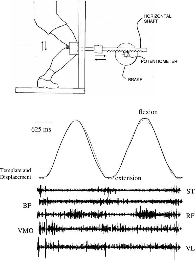
The SLS exercise testing and training system used in this study has been previously described.12 Briefly, the system consists of a rack and pinion gear system that attaches to the anterior knee (Figure 1). The linear displacement of the rack during the SLS is measured by a potentiometer calibrated to convert angular displacement into linear displacement (cm). Pilot studies showed that this horizontal forward and backward translation of the knee has a strong correlation to knee angular position as measured by an electrogoniometer attached to the knee during the SLS task (R2 = 0.97). Subjects went through a linear displacement of 20 cm for each flexion and extension cycle which lead to, on average, 40 (±2.6) degrees of knee flexion. An electromagnetic braking system with an associated shaft controls the resistance to the gear. Thus, the brake is directly controlled by a microcomputer through digital to analog control. The resistance of the brake was input according to the subject’s body weight (8%). Linearity, repeatability, and hysteresis of the brake and potentiometer system were within 0.5% of full scale. Subjects were instructed to follow a sinusoidal tracking pattern that appeared on a computer at a frequency of 0.4 Hz. Thus, one complete flexion and extension cycle took about 2500 milliseconds for the subjects to perform.
FIGURE 1.
Representative example of the target template, knee linear displacement (top of sinusoid is knee flexion; bottom of sinusoid is knee extension), and electromyographic (EMG) signal from a single subject during the SLS task. Note that the displacement trace is overlapping the target template. The bottom traces represent raw EMG from the semitendinosis (ST), biceps femoris (BF), rectus femoris (RF), vastus medialis oblique (VM), and vastus lateralis (VL).
Surface electromyographic (EMG) recordings were collected from five muscles, the rectus femoris (RF), vastus medialis oblique (VMO), vastus lateralis (VL), biceps femoris (BF), and semitendinosis (ST), of the exercised limb. Before fixing the electrodes, the skin was cleaned with alcohol to ensure adequate contact. Silver-silver chloride electrodes (eight millimeters in diameter) with on-site pre-amplification (gain * 35), further amplified at the main frame by 10 K, were placed by a single investigator according to the landmarks described by Cram et al.19 The amplifier uses a high-impedance circuit with a common mode rejection ratio of 87 dB at 60 Hz and a bandwidth of 15 to 4000 Hz (Model 544, Therapeutics Unlimited, Iowa City, IA).
Data Collection
Before the start of the protocol, three maximum voluntary isometric contractions (MVICs) of each muscle were obtained. Subjects were positioned in sitting as described by Kendall et al.20 and were asked to hold each MVIC for three seconds. Subjects were then placed in the experimental apparatus and their dominant knee strapped to the movable segment of the device. We provided the subjects with a detailed verbal description of the protocol and instructed them to follow the template as closely as possible. The noninvolved leg was kept off the ground by flexing slightly at the knee and the nondominant finger was allowed make contact with a support surface to assist with balance. Subjects were instructed to avoid leaning or rotating during the task and were given verbal cues if there were any deviations in the technique or form of exercise during the learning sessions. The foot placement was marked so that any change in the position could be easily detected and corrected.
Data Analysis
For purposes of analysis, knee displacement was divided into 10% bins in each flexion and extension cycle (125 milliseconds; approximately every four degrees of knee angle), creating 10 bins for flexion and 10 bins for extension. Pilot analysis of data showed that maximum learning occurred in six trials of 10 repetitions. For clarity in presentation, the data were reduced to trials 1, 3, and 6, and repetitions 1, 5, and 10. Summary data are presented during flexion and extension cycles.
The degree of improved task performance was examined by calculating absolute error measurements in these trials. Errors were calculated by taking the difference between the target template and knee displacement. Absolute error was calculated by taking the absolute value of this difference. Errors were then averaged within each 10% bin. Error across an entire repetition or trial was computed by taking the mean of these averaged bins.
The EMG signals were sampled at a rate of 2000 Hz and analyzed with Datapac II software (Version 3.0 RUN Technologies Inc.). We obtained the average RMS EMG within 10% bins of each knee flexion and extension cycle to correspond to the error bins. We analyzed the MVICs by finding the average rectified EMG during one second of peak contraction and chose the maximum average value among the three contractions. EMG data were normalized as a percentage of MVIC. Coactivation ratio during the task was calculated by dividing the average muscle activity of the quadriceps by the average muscle activity of the hamstrings in each repetition of the SLS exercise (RF + VMO + VL/ST + BF).
The data from the flexion and extension phases during the study were analyzed independently. We performed a repeated-measures analysis of variance with two factors, trial and repetition, on the errors and EMG activity using an α = 0.05 level to test for significant differences. Tukey’s follow-up test was used to test for simple effects in the event of a significant interaction.
RESULTS
A representative example from a single subject is seen in Figure 1. The uppermost trace shows the knee displacement curve overlapping the sine wave target that was projected on the screen. The ascending part of the sine wave target represents knee flexion and the descending part is when the knee was being returned to the extended position. The subjects went through a knee displacement of 20 cm, which is approximately 40 degrees of knee flexion. All the subjects actively recruited their hamstrings and quadriceps during the performance of the task.
Improved Performance of the Task
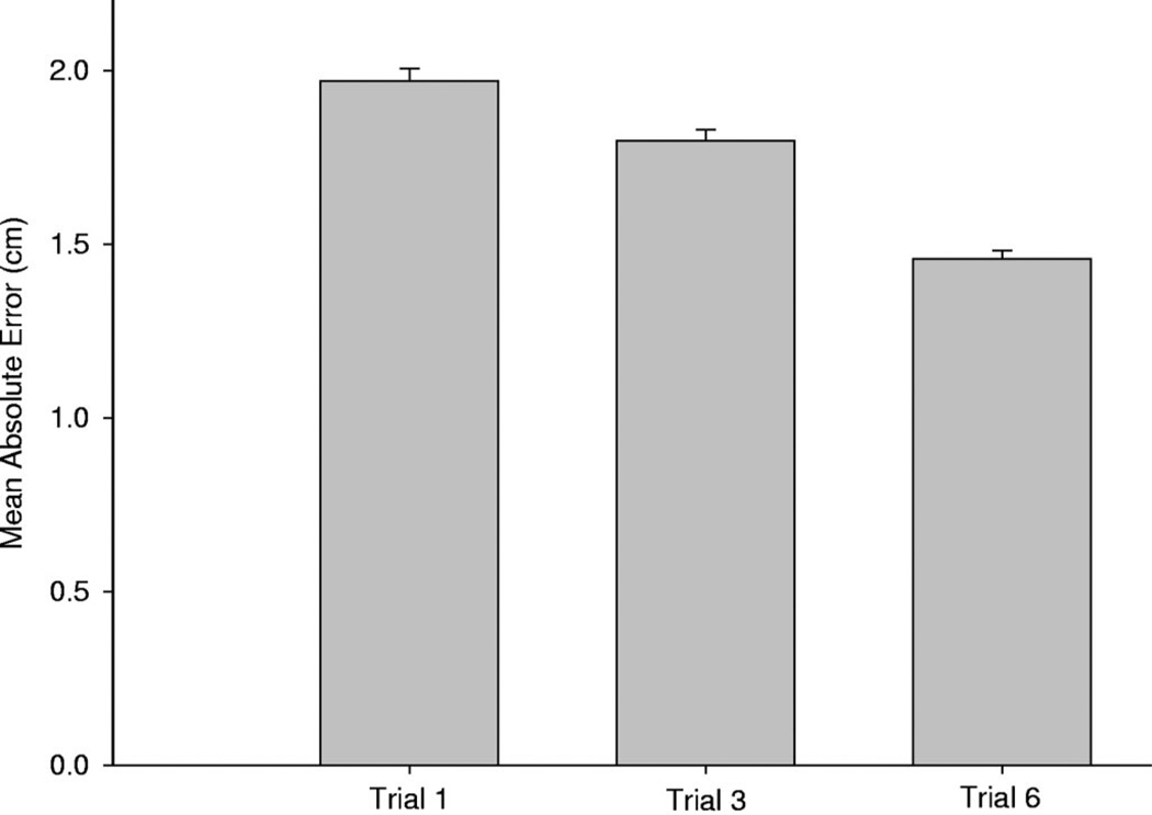
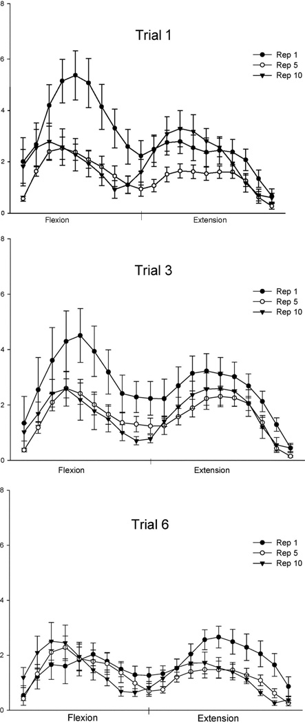
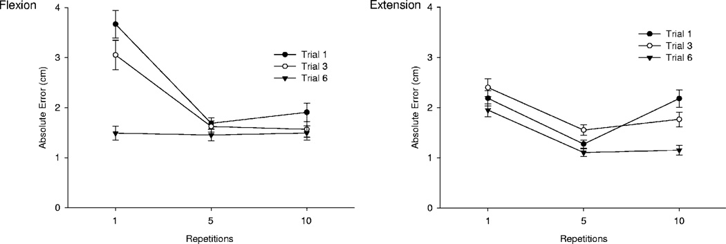
Total mean absolute error was decreased overall by trial 6 of the training protocol (F2,28 = 77.31, p < 0.0001) (Fig. 2). Subjects made the most errors when the target was changing most rapidly midway between knee extension and knee flexion during trial 1 (Fig. 3). The peak absolute error during knee flexion was reduced by more than 50% (approximately six centimeters to approximately three centimeters) by trial 6 (Fig. 3; F4,56 = 5.22, p = 0.0229). A summary of the absolute errors grouped according to the flexion and extension phases are presented in Figs. 3 and 4, respectively. Subjects displayed a significant decrease in absolute error during the extension phase in each trial (repetition effect) (F2,28 = 6.29, P < 0.0056) but not a significant improvement between trials (Figure 4).
FIGURE 2.
The vertical bars represent total mean absolute errors during trials 1, 3, and 6 of all 15 subjects. The error bars represent standard error. The absolute error has been aggregated over both flexion and extension phases and across all repetitions in each trial.
FIGURE 3.
The mean absolute error of all 15 subjects during repetitions 1, 5, and 10 of trials 1, 3, and 6 are presented. Each data point represents the average error over a 100-millisecond period during knee flexion and extension. The error bars are standard errors. Note the significant decrease in error during mid-flexion from trial 1 to trial 3.
FIGURE 4.
The mean absolute error during the flexion (top) and extension (bottom) phases of the SLS during repetitions 1, 5, and 10 of trials 1, 3, and 6 are presented. Data points represent the means of all 15 subjects and error bars are standard errors. Note the significant decrease in error for trial and repetition during the flexion phase and a significant improvement in each trial in the extension phase of the SLS.
Muscle Activity and Accuracy
During flexion, the greatest modulation of EMG activity between the accurate and less accurate conditions occurred in the biceps femoris and the rectus femoris muscles (Fig. 5). A significant trial × repetition interaction was seen for the biceps femoris (F4,56 = 3.96, p = 0.0067). Analysis of simple effects showed that reduced BF activity occurred in trials 3 and 6 when the task was performed more accurately. The RF showed a progressive increase in activity for all repetitions. An increase in RF activity was seen by trials 3 and 6 for repetition 10, causing a significant trial × repetition interaction (F4,56 = 3.24, p = 0.0183). Thus, the elevated RF activity may have contributed to reducing the error observed by repetition 10 (trials 3 and 6). The ST and VL showed no change in activity with each trial, but did show a decrease in activity with repetition (Table 1) leading to a significant repetition effect (F2,28 = 19.71, p < 0.001). The VL also showed no change in activity with each trial, but did show an increase in activity with repetition (Table 1) leading to a significant repetition effect (F2,28 = 6.97, p < 0.0035). Activity of the ST was very low for all conditions. The VMO did not show any significant effect of trial or repetition or interaction and thus appears to be minimally modulated during the performance of this task.
FIGURE 5.
Muscle activity (normalized to MVIC) of the BF and RF, the two muscles that showed significant trial × repetition effect, during the flexion phase of knee displacement are shown. Data points represent means of all 15 subjects and error bars are standard errors.
TABLE 1.
Summary of Muscle Activity (Normalized to % MVIC) Averaged in the Flexion and Extension Phases of the SLS Exercise Is Represented. Values are Mean (SD).
| Flexion (% MVIC) |
Extension (% MVIC) |
Combined (% MVIC) |
|
|---|---|---|---|
| Trial 1 | |||
| Repetition 1 | |||
| ST | 10.6 (14.8) | 6.0 (5.6) | |
| BF | 13.4 (9.4) | 10.1 (7.4) | |
| Hamstrings | 12.0 (12.5) | 8.0 (6.9) | 10.8 (10.2) |
| RF | 11.6 (12.67) | 18.0 (14.3) | |
| VMO | 19.4 (9.47) | 24.7 (10.2) | |
| VL | 20.0 (25.2) | 27.6 (19.8) | |
| Quadriceps | 17.0 (17.55) | 23.4 (15.6) | 18.5 (16.9) |
| Repetition 5 | |||
| ST | 6.5 (5.96) | 5.0 (3.18) | |
| BF | 13.9 (8.5) | 8.8 (8.3) | |
| Hamstrings | 10.2 (8.2) | 7.0 (6.6) | 8.8 (7.6) |
| RF | 15.8 (16.5) | 20.0 (14.7) | |
| VMO | 22.3 (18.8) | 27.7 (19.7) | |
| VL | 25.2 (28.4) | 26.7 (18.7) | |
| Quadriceps | 21.1 (20.2) | 24.9 (18.2) | 21.9 (19.3) |
| Repetition 10 | |||
| ST | 5.5 (4.52) | 4.8 (12.8) | |
| BF | 17.3 (11.9) | 9.6 (14.4) | |
| Hamstrings | 11.4 (10.8) | 7.4 (11.4) | 9.6 (9.2) |
| RF | 17.3 (15.0) | 21.0 (20.1) | |
| VMO | 24.6 (13.2) | 29.9 (20.2) | |
| VL | 26.8 (28.5) | 28.4 (20.9) | |
| Quadriceps | 22.9 (20.5) | 26.3 (20.6) | 24.3 (21.0) |
| Trial 3 | |||
| Repetition 1 | |||
| ST | 9.3 (8.7) | 13.4 (12.2) | |
| BF | 12.2 (8.1) | 15.6 (9.3) | |
| Hamstrings | 10.7 (8.5) | 14.5 (10.9) | 11.9 (9.9) |
| RF | 18.8 (18.9) | 7.6 (3.9) | |
| VMO | 23.9 (6.1) | 18.6 (14.4) | |
| VL | 12.7 (7.0) | 16.7 (11.8) | |
| Quadriceps | 18.5 (13.0) | 14.3 (12.0) | 16.4 (12.7) |
| Repetition 5 | |||
| ST | 5.5 (5.2) | 7.3 (6.6) | |
| BF | 11.8 (11.7) | 13.0 (10.0) | |
| Hamstrings | 8.7 (9.6) | 10.1 (9.0) | 9.4 (9.3) |
| RF | 18.4 (15.6) | 14.3 (5.4) | |
| VMO | 24.0 (13.4) | 26.7 (12.8) | |
| VL | 25.9 (19.8) | 39.3 (48.5) | |
| Quadriceps | 22.8 (15.2) | 26.8 (30.7) | 22.4 (24.3) |
| Repetition 10 | |||
| ST | 6.2 (4.0) | 8.2 (6.1) | |
| BF | 10.1 (7.3) | 15.8 (15.7) | |
| Hamstrings | 8.2 (6.2) | 12.0 (12.0) | 10.2 (10.4) |
| RF | 38.1 (30.4) | 21.1 (16.1) | |
| VMO | 25.1 (3.2) | 39.5 (34.1) | |
| VL | 26.0 (11.7) | 34.8 (27.7) | |
| Quadriceps | 29.7 (19.7) | 31.6 (28.2) | 33.19 (25.6) |
| Trial 6 | |||
| Repetition 1 | |||
| ST | 9.3 (8.1) | 11.7 (10.4) | |
| BF | 11.0 (10.2) | 13.1 (8.7) | |
| Hamstrings | 10.2 (9.3) | 12.4 (9.6) | 10.9 (9.5) |
| RF | 19.7 (18.3) | 7.4 (4.0) | |
| VMO | 23.6 (6.8) | 19.7 (14.7) | |
| VL | 12.7 (8.1) | 15.0 (10.3) | |
| Quadriceps | 18.7 (12.9) | 14.0 (11.8) | 16.6 (12.6) |
| Repetition 5 | |||
| ST | 5.3 (5.0) | 6.5 (3.2) | |
| BF | 12.2 (11.7) | 10.1 (8.1) | |
| Hamstrings | 8.8 (9.6) | 8.3 (6.5) | 9.09 (8.1) |
| RF | 18.3 (11.5) | 13.8 (6.0) | |
| VMO | 24.1 (13.5) | 24.9 (11.9) | |
| VL | 26.2 (18.2) | 34.5 (39.4) | |
| Quadriceps | 22.9 (13.2) | 24.5 (25.3) | 21.0 (20.2) |
| Repetition 10 | |||
| ST | 5.8 (3.7) | 6.9 (4.3) | |
| BF | 14.3 (19.0) | 13.1 (11.3) | |
| Hamstrings | 10.0 (14.2) | 10.1 (9.4) | 10.5 (12.5) |
| RF | 34.5 (33.3) | 17.7 (11.4) | |
| VMO | 24.9 (3.7) | 41.7 (39.1) | |
| VL | 23.2 (13.8) | 28.5 (19.2) | |
| Quadriceps | 27.4 (21.2) | 29.2 (27.7) | 32.1 (26.1) |
During extension, the RF and VL showed increased activity as the accuracy with the task increased (Fig. 6). This effect was supported by a significant trial × repetition interaction during the extension phase (F4,56 = 5.55, p < 0.008 for RF and F4,56 = 2.80, p < 0.0344 for VL). Both of these muscles were recruited more as the number of repetitions increased in each trial, with a greater increase during trials 3 and 6. The ST showed a significant effect of trial regardless of repetition (F2,28 = 5.14, p < 0.0125). Although the BF and VMO were recruited actively during the extension cycle of the exercise, their activity did not show significant pattern change as a result of learning.
FIGURE 6.
Muscle activity (normalized to MVIC) of the RF and VL, the two muscles that showed significant trial × repetition effect, during the extension phase of knee displacement are shown. Data points represent means of all 15 subjects and error bars are standard errors.
Quadriceps hamstrings coactivation was found to decrease in each trial from repetition 1 to repetition 10 (Table 1). The coactivation ratio (quadriceps/hamstrings) increased from ~1.35 during repetition 1 to ~3.15 in repetition 10 supporting less coactivation. In addition, at the end of trial 6, all subjects were found to have significantly higher quadriceps to hamstrings ratios than trial 1 (p < 0.005).
DISCUSSION
The goals of this study were to examine the neuromuscular activity of synergists controlling the knee joint during a weight-bearing exercise that (1) provided a quantifiable bidirectional level of resistance (body weight) during the exercise and (2) assessed the accuracy of the performed task. This study verified that individuals improved the accuracy of this weight-bearing exercise with feedback both within trials as well as between trials in a single session. The primary muscles that modulate their activity with this improved control were the BF and RF during the flexion phase and the RF and VMO during the extension phase. The VL and ST showed consistent changes in activation in a trial, but did not appear to contribute to any between trial accuracy. The VMO was minimally modulated as a function of increased accuracy with the task. Quadriceps hamstrings ratio was found to be increased with improvements in performance. Collectively, these results support our hypothesis that coactivating both the quadriceps and hamstrings is decreased after learning during a resisted SLS task. Consequently, these findings support the notion that synergistic coactivation is reduced as the goal for accuracy is increased. This finding may form the basis for why increased knee joint stiffness (through cocontraction of the quadriceps and hamstrings) is not an effective strategy for high-level tasks requiring accuracy during human performance.
The importance of this study is that two conflicting principles, often difficult to reconcile, are examined using a novel method to perform weight-bearing exercise. The first is the ability to measure the prescribed dose of load, and the second is the ability to examine the accuracy of the performed exercise. Given the many degrees of freedom that are possible during synergistic control of the knee, rehabilitation scientists are constantly striving to understand muscle activation patterns that promote joint stability while maximizing performance. The combination of promoting neuromuscular strengthening exercise while maintaining joint control under visual feedback is not a routine part of preseason strengthening regimens or inpatient rehabilitation after stroke. Consequently, we present the term controlled strengthening to delineate strengthening programs that also quantify the accuracy of performance during weight-bearing exercise. Although this is the goal of many therapeutic exercises, few of these clinical exercises actually quantify performance and resistance. In this study, the subject was required to control the rate and excursion of the knee by tracking a sinusoidal target, while at the same time exerting a force equivalent to eight percent of body weight acting in a horizontal direction.
Weight-bearing exercise, like the SLS and the lateral step up, are increasingly preferred in lower extremity rehabilitation programs because they replicate functional activities.3 The results of this study showed more than a 50% reduction in mean absolute error of performance (from 0.60 cm in repetition 1, trial 1 to 0.24 cm in repetition 1, trial 6) during the flexion phase of this weight-bearing exercise. The improvement in the overall accuracy of performance and decrease in variability across the session suggests that the subjects were able to perform the task with fewer errors in a trial and extend that performance to consecutive trials. The notion that a “less than optimal” pattern of training could be adopted during rehabilitative or preventive exercise programs appears to be fertile ground for future investigations. In this context, it is unknown whether the SLS exercise can be performed accurately while maintaining high levels of coactivation across the joint, which theoretically would provide the greatest stiffness across the knee.
Consistent quadriceps and hamstrings coactivation was noted during the entire task for all subjects. In particular, the average activity of the BF during the session was found to be approximately 13% of MVIC. The hamstrings are thought to act as a proponent for ligamentous and soft-tissue structures (ACL and posterior capsule), decreasing anterior shear forces acting on the ligament and thus reducing the risk of injury, making the SLS task safe to be used for knee rehabilitation.7 The level of quadriceps activation achieved by subjects in this study was 30% MVIC. This exercise could be very effective in improving neuromuscular control after ACL injury or reconstruction where one of the main consequences is quadriceps muscle weakness.21–23 Although there were clearly certain muscles that modulated their activity as a result of improved performance, it also appeared that each subject likely had an individualized synergistic pattern as well. The muscles that showed the greatest modulation of activity between trials 1 and 6 were the BF, RF, and VL. To be more accurate in performance during flexion, the subjects had to reduce their activity of the BF and increase activation of the RF. Increased activity of the RF and VL was also noticed during the extension phase as the subjects became more accurate. These findings suggest that reducing the coactivation of the hamstrings and quadriceps during this resisted SLS exercise helps to gain accuracy in performance. Thus, the goal to increase stiffness around the knee joint to prevent injury may not be consistent with the goal of being accurate. However, more extensive research is required before this generalization can be confirmed.
Several studies have nicely performed EMG and joint force analysis during single-leg weight-bearing exercises.1,13,24 Very important differences, however, exist between this study and these previous studies. Previous reports did not include accuracy of knee control and performance during the task. Proper training of neuromuscular control around the knee would appear to require that some measure of error be included so that the quality of the movement being trained is monitored. Many noncontact ACL injuries have been attributed to the lack of neuromuscular control.2,25
The trend in rehabilitation after knee injury is toward accelerated training programs with early return to functional activities. Neuromuscular training is aimed at improving the nervous system’s ability to recruit muscles to improve coordination and dynamic stability. Muscle strength, coordination, and overall proprioceptive ability are necessary for complete functional knee stability. Liu-Ambrose et al26 determined the effects of proprioceptive training programs on neuromuscular function after ACL reconstruction and found that proprioceptive training can improve muscle strength and is beneficial for restoring functional ability. They suggest that proprioceptive training should involve adequate practice of desired motor patterns. Hewett et al.27 support that recruiting and activating muscles in functional patterns may improve proprioception and coordination leading to decreased rates of injury. Hence, it appears that functional rehabilitation activities should strive to quantify the quality of the performance as well as the load being lifted. When both are quantified during training, then the extent of controlled strengthening can be documented.
CONCLUSIONS
This study presents a novel method that examines the neuromuscular control strategies during a weight-bearing exercise. Unique components to this study were that the performance of the task was quantified, the resistance offered was bidirectional and normalized to body weight, and the synergistic strategies were compared before and after learning. A significant reduction in error and significant change in the BF and RF muscle activity levels as a result of improved performance supports that both control and strengthening are important components of neuromuscular training programs. The findings in this study support that tasks that promote neuromuscular strengthening while simultaneously quantifying accuracy will continue to be important considerations when developing safe and accurate strategies to control the knee during weight-bearing tasks.
REFERENCES
- 1.Beutler A, Cooper L, Kirkendall D, et al. Electromyographic analysis of single-leg, closed chain exercises. J Athletic Train. 2002;37:13. [PMC free article] [PubMed] [Google Scholar]
- 2.Parkkari J, Kujala UM, Kannus P. Is it possible to prevent sports injuries? Review of controlled clinical trials and recommendations for future work. Sports Med. 2001;31:985–995. doi: 10.2165/00007256-200131140-00003. [DOI] [PubMed] [Google Scholar]
- 3.Wilk KE, Reinold MM, Hooks TR. Recent advances in the rehabilitation of isolated and combined anterior cruciate ligament injuries. Orthop Clin North Am. 2003;34:107–137. doi: 10.1016/s0030-5898(02)00064-0. [DOI] [PubMed] [Google Scholar]
- 4.Fitzgerald GK. Open versus closed kinetic chain exercise: issues in rehabilitation after anterior cruciate ligament reconstructive surgery. Phys Ther. 1997;77:1747–1754. doi: 10.1093/ptj/77.12.1747. [DOI] [PubMed] [Google Scholar]
- 5.Ebben WP, Jensen RL. Electromyographic and kinetic analysis of traditional, chain, and elastic band squats. J Strength Cond Res. 2002;16:547–550. [PubMed] [Google Scholar]
- 6.Toutoungi DE, Lu TW, Leardini A, et al. Cruciate ligament forces in the human knee during rehabilitation exercises. Clin Biomech. 2000;15:176–187. doi: 10.1016/s0268-0033(99)00063-7. [DOI] [PubMed] [Google Scholar]
- 7.More R, Karras B, Neiman RF, et al. Hamstrings—an anterior cruciate ligament protagonist. An in vitro study. Am J Sports Med. 1993;21:231. doi: 10.1177/036354659302100212. [DOI] [PubMed] [Google Scholar]
- 8.Palmitier R, An K, Scott S, et al. Kinetic chain exercise in knee rehabilitation. Sports Med. 1991:402. doi: 10.2165/00007256-199111060-00005. [DOI] [PubMed] [Google Scholar]
- 9.Brunt D, Vander Linden DW, Behrman AL. The relation between limb loading and control parameters of gait initiation in persons with stroke. Arch Phys Med Rehabil. 1995;76:627–634. doi: 10.1016/s0003-9993(95)80631-8. [DOI] [PubMed] [Google Scholar]
- 10.Mikesky AE, Topp R, Wigglesworth JK, et al. Efficacy of a home-based training program for older adults using elastic tubing. Eur J App Physiol Occup Physiol. 1994;69:316–320. doi: 10.1007/BF00392037. [DOI] [PubMed] [Google Scholar]
- 11.Patterson RM, Stegink Jansen CW, Hogan HA, et al. Material properties of Thera-Band tubing. Phys Ther. 2001;81:1437–1445. doi: 10.1093/ptj/81.8.1437. [DOI] [PubMed] [Google Scholar]
- 12.Shields RK, Madhavan S, Gregg E, et al. Neuromuscular control of the knee during a resisted single-limb squat exercise. Am J Sports Med. 2005;33:1520–1526. doi: 10.1177/0363546504274150. [DOI] [PMC free article] [PubMed] [Google Scholar]
- 13.Zeller BL, McCrory JL, Kibler WB, et al. Differences in kinematics and electromyographic activity between men and women during the single-legged squat. Am J Sports Med. 2003;31:449–456. doi: 10.1177/03635465030310032101. [DOI] [PubMed] [Google Scholar]
- 14.Thoroughman KA, Shadmehr R. Electromyographic correlates of learning an internal model of reaching movements. J Neurosci. 1999;19:8573–8588. doi: 10.1523/JNEUROSCI.19-19-08573.1999. [DOI] [PMC free article] [PubMed] [Google Scholar]
- 15.Gribble PL, Mullin LI, Cothros N, et al. Role of cocontraction in arm movement accuracy. J Neurophysiol. 2003;89:2396–2405. doi: 10.1152/jn.01020.2002. [DOI] [PubMed] [Google Scholar]
- 16.Borg GA. Psychophysical bases of perceived exertion. Med Sci Sports Exerc. 1982;14:377–381. [PubMed] [Google Scholar]
- 17.Hunter SK, Critchlow A, Enoka RM. Influence of aging on sex differences in muscle fatigability. J Appl Physiol. 2004;97:1723–1732. doi: 10.1152/japplphysiol.00460.2004. [DOI] [PubMed] [Google Scholar]
- 18.Hunter SK, Lepers R, MacGillis CJ, et al. Activation among the elbow flexor muscles differs when maintaining arm position during a fatiguing contraction. J Appl Physiol. 2003;94:2439–2447. doi: 10.1152/japplphysiol.01038.2002. [DOI] [PubMed] [Google Scholar]
- 19.Cram JR, Kasman GS, Holtz J. Introduction to Surface Electromyography. Gaithersburg, Md: Aspen Publishers Inc.; 1998. pp. 360–375. [Google Scholar]
- 20.Kendall F, McCreary E, Provance P. Muscles, Testing and Function. 4th ed. Baltimore, Md: Williams & Wilkins; 1993. pp. 161–185. [Google Scholar]
- 21.Ikeda H, Kurosawa H, Kim SG. Quadriceps torque curve pattern in patients with anterior cruciate ligament injury. Int Orthop. 2002;26:374–376. doi: 10.1007/s00264-002-0402-0. [DOI] [PMC free article] [PubMed] [Google Scholar]
- 22.Konishi Y, Fukubayashi T, Takeshita D. Mechanism of quadriceps femoris muscle weakness in patients with anterior cruciate ligament reconstruction. Scand J Med Sci Sports. 2002;12:371–375. doi: 10.1034/j.1600-0838.2002.01293.x. [DOI] [PubMed] [Google Scholar]
- 23.Williams GN, Barrance PJ, Snyder-Mackler L, et al. Specificity of muscle action after anterior cruciate ligament injury. J Orthop Res. 2003;21:1131–1137. doi: 10.1016/S0736-0266(03)00106-2. [DOI] [PubMed] [Google Scholar]
- 24.Wilk KE, Escamilla RF, Fleisig GS, et al. A comparison of tibiofemoral joint forces and electromyographic activity during open and closed kinetic chain exercises. Am J Sports Med. 1996;24:518–527. doi: 10.1177/036354659602400418. [DOI] [PubMed] [Google Scholar]
- 25.Bjordal JM, Arnly F, Hannestad B, et al. Epidemiology of anterior cruciate ligament injuries in soccer. Am J Sports Med. 1997;25:341–345. doi: 10.1177/036354659702500312. [DOI] [PubMed] [Google Scholar]
- 26.Liu-Ambrose T, Taunton J, MacIntyre D, et al. The effects of proprioceptive or strength training on the neuromuscular function of the ACL reconstructed knee: a randomized clinical trial. Scand J Med. 2003;13:115–123. doi: 10.1034/j.1600-0838.2003.02113.x. [DOI] [PubMed] [Google Scholar]
- 27.Hewett TE, Lindenfeld TN, Riccobene JV, et al. The effect of neuromuscular training on the incidence of knee injury in female athletes. A prospective study. Am J Sports Med. 1999;27:699–706. doi: 10.1177/03635465990270060301. [DOI] [PubMed] [Google Scholar]


