The V-SVZ Neurogenic Niche
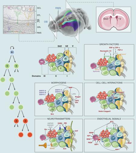
The wall of the lateral ventricles (the ventricular-subventricular zone; V-SVZ) is the site of the birth of neurons and glial cells in the brain of juvenile and adult mammals. Neural stem cells (NSCs) correspond to a subpopulation of specialized astrocytes (B1 cells) derived from radial glia. These primary progenitors give rise to intermediate progenitor cells (IPCs or C cells), which in turn generate large numbers of neuroblasts (A cells) that migrate tangentially through the rostral migratory stream (RMS) to the olfactory bulb (OB). Cell cycle and population analysis in mice suggests that after the initial division of B1 cells, C cells divide three times and A cells once, possibly twice. V-SVZ NSCs are heterogeneous, generating at least six different subtypes of OB interneurons depending on their position along the dorsoventral and anterioposterior axes. Live imaging of NSCs isolated from the adult mouse V-SVZ revealed that NSCs exclusively generate oligodendroglia or neurons, but never both within a single lineage.
Properties of V-SVZ NSCs
B1 cells retain the apical-basal polarity of their predecessors, radial glia. Most B1 cells contact the ventricle through small, specialized apical processes that contain a single primary cilium. They also have long basal processes with specialized endings contacting blood vessels (BVs). When viewed en face, from the ventricular side, the V-SVZ is organized as pinwheels; the small apical endings of B1 cells are surrounded by a rosette of ependymal cells (E1 cells) with larger apical surfaces. B1 cells have homotypic cell-cell contacts (gap junctions) with each other, as well as heterotypic adherent junctions with ependymal cells.
Signaling Pathways that Regulate V-SVZ Neurogenesis
Growth Factors
Fibroblast growth factor-2 (FGF-2 or bFGF) may maintain the self-renewing V-SVZ population. The epidermal growth factor receptor (EGFR) is primarily expressed on C cells and a subpopulation of B1 cells. Elevated EGF signaling biases V-SVZ cells toward the oligodendrocytic lineage. The most likely endogenous ligand for the EGFR pathway is transforming growth factor-alpha (TGF-a). ErbB4 and its ligands, neuregulin 1 and 2, are also expressed in the V-SVZ and are implicated in progenitor proliferation and the initiation of neuroblast migration. PDGF increases proliferation in V-SVZ cells, and many of these may be oligodendrocyte progenitors. Also expressed within the V-SVZ are the receptors EphA and EphB. Ephrin signaling appears to impact B1 cell proliferation, A cell migration, and V-SVZ cell apoptosis.
Morphogens
Bone morphogenetic proteins (BMPs) in the cerebrospinal fluid (CSF) inhibit V-SVZ neurogenesis. E1 cells secrete the BMP antagonist noggin, which promotes V-SVZ progenitor proliferation and neuroblast generation. E1 cells also express LRP2, a receptor that sequesters BMP4. Wnt signaling may promote the proliferation of C cells. The Insulin-like growth factor 2 (IGF2) in the CSF also regulates progenitor proliferation. Sonic hedgehog (Shh) signaling occurs specifically in the ventral subregion of the V-SVZ to promote production of ventrally derived neuron types. The apical primary cilium of B1 cells may directly integrate signaling of CSF factors, although this remains to be demonstrated.
Cell-Cell Interactions
Canonical Notch signaling is highly active in V-SVZ NSCs and regulates their maintenance by inhibiting the production of IPCs. IPCs express high levels of achaete-scute complex homolog 1 (ASCL1 or Mash1), which is repressed by Hes1, a downstream effector of Notch signaling. ASCL1 promotes the expression of Notch ligands, suggesting a feedback mechanism of lateral inhibition between IPCs and NSCs. It is unclear how Notch signaling is regulated in the V-SVZ, but expression of the Notch ligands Delta1 and Jagged1 is observed in IPCs and neuroblasts. Delta-like1 (Dll1) is induced in activated NSCs and segregates to one daughter cell during mitosis.
Neurotransmitters
Neuroblasts spontaneously release gamma-aminobutyric acid (GABA), depolarizing progenitors by activation of GABAA receptors. This inhibits progenitor cell-cycle progression and neuronal production. The diazepam binding inhibitor protein (DBI) is secreted by B1 cells and IPCs, but not neuroblasts, and competes with GABA for binding to its receptor, resulting in increased progenitor proliferation. Glutamate may positively regulate neurogenesis, possibly by increasing C cell numbers. Dopamine (DA) is released into the SVZ via axonal projections from the ventral tegmental area and the substantia nigra. Activation of D2-like receptors on IPCs increases their proliferation. Serotonin (5HT) derived from the raphe nuclei and cholinergic inputs also modulates V-SVZ neurogenesis.
Endothelial Signals
The chemokine stromal cell-derived factor 1 (SDF1) secreted by endothelial cells induces the recruitment of activated B1 cells and IPCs to the vascular plexus. This effect is mediated by a6b1-integrin. The transition of IPCs into neuroblasts is accompanied by a decrease in b1-integrin expression, allowing the differentiating progeny to migrate away from the niche. Pigment epithelium-derived factor (PEDF) secreted by endothelial and E1 cells increases the number of dividing glial fibrillary acidic protein (GFAP)+ cells. Endothelial-derived betacellulin (BTC) stimulates V-SVZ progenitor proliferation and neurogenesis. Vascular endothelial growth factor (VEGF) signaling is implicated in progenitor survival and proliferation and in neuroblast migration and maturation. The vasculature also secretes IGF1 and brain-derived neurotrophic factor (BDNF), which promote proliferation and differentiation of progenitor cells, respectively.
ACKNOWLEDGMENTS
This SnapShot reflects many excellent contributions from many laboratories. Due to space constraints we could not cite all of them but references to this work can be found within the articles listed below. We thank Kenneth X. Probst for some of the illustrations. Our laboratory is funded by the U.S. National Institutes of Health (NS 28478 and HD 32116). A.A.-B. is the Heather and Melanie Muss Endowed Chair of Neurological Surgery at UCSF. C.K.T. is supported by the Singapore Agency for Science, Technology, and Research.
REFERENCES
- Aguirre A, Rubio ME, Gallo V. Nature. 2010;467:323–327. doi: 10.1038/nature09347. [DOI] [PMC free article] [PubMed] [Google Scholar]
- Alfonso J, Le Magueresse C, Zuccotti A, Khodosevich K, Monyer H. Cell Stem Cell. 2012;10:76–87. doi: 10.1016/j.stem.2011.11.011. [DOI] [PubMed] [Google Scholar]
- Bath KG, Lee FS. Dev. Neurobiol. 2010;70:339–349. doi: 10.1002/dneu.20781. [DOI] [PMC free article] [PubMed] [Google Scholar]
- Fuentealba LC, Obernier K, Alvarez-Buylla A. Cell Stem Cell. 2012;10:698–708. doi: 10.1016/j.stem.2012.05.012. [DOI] [PMC free article] [PubMed] [Google Scholar]
- Lehtinen MK, Zappaterra MW, Chen X, Yang YJ, Hill AD, Lun M, Maynard T, Gonzalez D, Kim S, Ye P, et al. Neuron. 2011;69:893–905. doi: 10.1016/j.neuron.2011.01.023. [DOI] [PMC free article] [PubMed] [Google Scholar]
- Mirzadeh Z, Merkle FT, Soriano-Navarro M, Garcia-Verdugo JM, Alvarez-Buylla A. Cell Stem Cell. 2008;3:265–278. doi: 10.1016/j.stem.2008.07.004. [DOI] [PMC free article] [PubMed] [Google Scholar]
- Ortega F, Gascón S, Masserdotti G, Deshpande A, Simon C, Fischer J, Dimou L, Chichung Lie D, Schroeder T, Berninger B. Nat. Cell Biol. 2013;15:602–613. doi: 10.1038/ncb2736. [DOI] [PubMed] [Google Scholar]
- Shen Q, Wang Y, Kokovay E, Lin G, Chuang SM, Goderie SK, Roysam B, Temple S. Cell Stem Cell. 2008;3:289–300. doi: 10.1016/j.stem.2008.07.026. [DOI] [PMC free article] [PubMed] [Google Scholar]
- Tavazoie M, Van der Veken L, Silva-Vargas V, Louissaint M, Colonna L, Zaidi B, Garcia-Verdugo JM, Doetsch F. Cell Stem Cell. 2008;3:279–288. doi: 10.1016/j.stem.2008.07.025. [DOI] [PMC free article] [PubMed] [Google Scholar]


