Abstract
Tuberculosis is a systemic disease that still affects many people. While pleural involvement is frequently observed in extrapulmonary tuberculosis, multiple skeletal system and articular involvements are quite rare. FDG PET imaging could be a promising diagnostic and treatment monitoring method, especially in complicated cases and if the other methods are inadequate. In this case study, we report a patient who was admitted with suspected malignancy and then diagnosed with tuberculosis pleuritis, lymphadenitis, spondylodiscitis, and sacroiliitis with specific symptoms; the response to anti-tuberculosis therapy was shown using FDG PET/CT.
Keywords: Tuberculosis, Spondylodiscitis, Sacroiliitis, PET/CT
Introduction
Tuberculosis is a rare disease in individuals with healthy immune systems living in developed countries, but it is still a serious infection that affects many people in the world. Extrapulmonary involvement occurs in 15–20 % of tuberculosis infections. The pleura is a frequent site of extrapulmonary tuberculosis, whereas bone or joint involvement is uncommon. While osteoarticular tuberculosis most commonly occurs in the vertebral column, less frequently affected sites are the hip, knee, and sacroiliac joints [1]. The multifocal form of skeletal tuberculosis is exceptional, even in countries where the disease is endemic [2]. The diagnosis of disease is frequently delayed because of vague symptoms, and also it is difficult to differentiate multifocal skeletal tuberculosis from other multiple bone lesions, including bone metastases.
18F-fluoro-2-deoxy-D-glucose positron emission tomography (FDG-PET)/computed tomography (CT) is a metabolic imaging technique that has been used for diagnosing cancers of various organs. In addition, FDG-PET has become a promising imaging modality in the field of infection and inflammation, especially to determine the extension and severity of disease. The purpose of the presented case is to evaluate FDG PET/CT imaging in the diagnosis and follow-up of patients with multifocal skeletal tuberculosis.
Case Report
A chest X-ray of a 20-year old male with complaints of back pain and shortness of breath showed pleural effusion. Further examination with thoracic CT revealed massive pleural fluid and nodular pleural thickenings on the right side. In addition, on thoracic vertebrae 6 and 7, lytic and sclerotic lesions associated with soft tissue densities were found. Laboratory findings showed a high erythrocyte sedimentation rate (62/h, normal range: 0–20) and C-reactive protein level (12.1, normal range: 0–0.8). For sputum, fiberoptic bronchoscopy/aspiration, and pleural fluid samples, acid-resistant bacilli were negative, and there was no culture positivity. PET/CT was ordered for this patient with suspected malignancy.
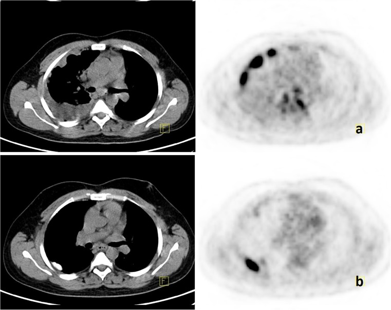
PET/CT examination revealed areas of pleural thickening, which were more prominent at the right maximum thickness of 1.7 cm; maximum standardized uptake values (SUVmax) were 15.9 (Fig. 1a). There were also increased FDG activities at the right upper paratracheal (SUVmax: 3.4), subcarinal (SUVmax: 4.6), peripancreatic (SUVmax: 9.3), and perisplenic (SUVmax: 4.8) lymph nodes.
Fig. 1.
a Initial FDG PET/CT images showing increased FDG activity in the areas of nodular pleural thickening. b Follow-up PET/CT images showing hyperdense pleural thickening representing a talc deposition area on which intense FDG uptake is seen
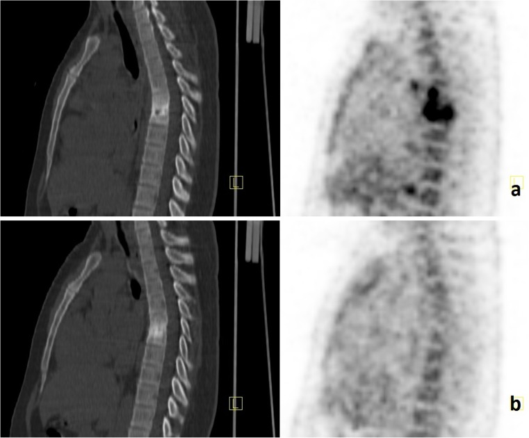
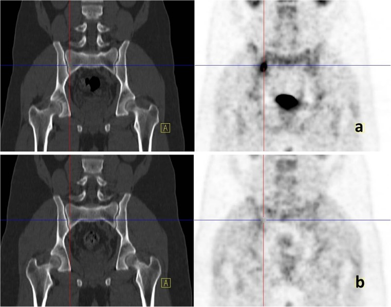
Lytic sclerotic views and intense uptake (SUVmax: 18.6) were found at the bodies of the T6 and T7 vertebrae, associated with paravertebral soft tissue densities (Fig. 2a). Increased FDG uptake was also present at the right sacroiliac joint (SUVmax: 12.4) without any corresponding CT finding (Fig. 3a).
Fig. 2.
a Increased FDG uptake representing tuberculosis spondylodiscitis at the level of T6 and T7 is seen on initial PET/CT images. b A complete metabolic response is observed on follow-up examination after 4 months of therapy
Fig. 3.
a Increased focal FDG uptake at the right sacroiliac joint without any abnormal findings on CT images. b No FDG accumulation at the same localization after 4 months of therapy
To determine the etiology, a pleural biopsy was performed. Then talc pleurodesis was applied in the same procedure using the video-assisted thoracoscopic surgery method. Granulomatous pleuritis with caseification necrosis was the final pathological finding, indicating tuberculosis. Antituberculosis therapy was started combining isoniazid, rifampicin, ethambutol, and pyrazinamide.
A follow-up PET/CT examination 4 months after the initiation of treatment showed decreased SUVmax (18.6 versus 4.0) levels, consistent with complete metabolic response at the T6 and T7 vertebrae and right sacroiliac joint (Figs. 2b and 3b). There were areas of increased FDG activity (SUVmax: 14.5) on the pleural membranes on the right side, but this time they were attributed to the inflammatory reaction due to talc pleurodesis (Fig. 1b). The follow-up erythrocyte sedimentation rate was 23/h.
Discussion
Early diagnosis and adequate treatment of tuberculosis are important for public health and infection control. Isolation of tubercle bacilli in the fluid of infected patients or reproduction in cultures is not always possible. Because of the non-specific character of the disease, diagnosing is generally time-consuming and difficult. Moreover, bone tuberculosis usually has an insidious start and slow course.
In 50 % of skeletal tuberculosis cases, it appears as spinal tuberculosis. Frequently thoracic vertebrae are affected (33–67 %), and usually two adjacent vertebral bodies are involved together with the intervertebral disc (tuberculosis spondylodiscitis). A so-called “cold abscess formation” may be seen as paravertebral soft tissue thickening (Pott’s disease). In particular, sacroiliac joint involvement has been reported in 9.7 % of patients with skeletal tuberculosis [3, 4].
As seen in tuberculosis, FDG uptake was correlated with the severity of the inflammatory process. Also, in acute infections more intense uptake is observed than in chronic active infection or the healing process [5, 6]. In the literature, after 4 months of antituberculosis treatment, in the evaluation of treatment response in patients with tuberculosis lymphadenopathy, the SUVmax cutoff value taken is 4.5; sensitivity and specificity have been reported as 95 % and 85 %, respectively [7]. It is emphasized that FDG PET can be a method for providing additional information for the detection of disease foci and the evaluation of the effectiveness of tuberculostatic treatment [8]. Especially in multiple drug resistance cases, FDG PET appears to be a promising method facilitating the decision on the treatment strategy by determining the active disease [9].
For prognosis and detection of residual disease in patients with spinal infection, FDG PET/CT also shows encouraging results, particularly when magnetic resonance images (MRI) are unconvincing in the distinction between degenerative changes and infection [10].
Multiple skeletal and joint involvements due to tuberculosis are extremely rare. In our case, the thoracic spine and sacroiliac joints were involved. Detection of FDG uptake, although there were no pathological findings on CT images of the right sacroiliac joint, supports the idea that FDG PET may be more sensitive, especially at early stages of inflammation before the occurrence of anatomical changes. In this case, a complete metabolic response was obtained at all involved parts of the body after 4 months of tuberculostatic treatment. This indicates that the use of this method is promising for treatment monitoring.
On post-treatment study, FDG uptake was only observed in the pleural talc deposits, as reported in the literature, thought to be due to inflammation secondary to talc [11].
Use of FDG for the localization of inflammatory foci in solid organs, soft-tissue, or bone structures and the additional information provided by CT will likely be very useful in the evaluation of patients with tuberculosis. Therefore, our experience with the presented case supports the idea that integration of FDG PET/CT into routine diagnostic and follow-up methods may be promising, especially in resistant and complicated cases.
Acknowledgments
Conflict of Interest
On behalf of all authors, the corresponding author states that there is no conflict of interest.
References
- 1.Moore SL, Rafii M. Imaging of musculoskeletal and spinal tuberculosis. Radiol Clin N Am. 2001;39(2):329–342. doi: 10.1016/S0033-8389(05)70280-3. [DOI] [PubMed] [Google Scholar]
- 2.Moujtahid M, Essadki B, Lamine A, Bennouna D, Zryouil B. Multifocal bone tuberculosis: apropos of a case. Rev Chir Orthop Reparatrice Appar Mot. 1995;81(6):553–556. [PubMed] [Google Scholar]
- 3.Rowe LJ, Yochum TR. Infection. In: Rowe LJ, Yochum TR, editors. Essentials of skeletal radiology. Philadelphia: Lippincott Williams and Wilkins; 2005. pp. 1373–1426. [Google Scholar]
- 4.Pouchot J, Vinceneux P, Barge J, Boussougant Y, Grossin M, Pierre J, et al. Tuberculosis of the sacroiliac joint: clinical features, outcome, and evaluation of closed needle biopsy in 11 consecutive cases. Am J Med. 1988;84(3 Pt 2):622–628. doi: 10.1016/0002-9343(88)90146-5. [DOI] [PubMed] [Google Scholar]
- 5.Mamede M, Higashi T, Kitaichi M, Ishizu K, Ishimori T, Nakamoto Y, et al. [18F]FDG uptake and PCNA, Glut-1, and Hexokinase-II expressions in cancers and inflammatory lesions of the lung. Neoplasia. 2005;7(4):369–379. doi: 10.1593/neo.04577. [DOI] [PMC free article] [PubMed] [Google Scholar]
- 6.Ichiya Y, Kuwabara Y, Sasaki M, Yoshida T, Akashi Y, Murayama S, et al. FDG-PET in infectious lesions: the detection and assessment of lesion activity. Ann Nucl Med. 1996;10(2):185–191. doi: 10.1007/BF03165391. [DOI] [PubMed] [Google Scholar]
- 7.Sathekge M, Maes A, D’Asseler Y, Vorster M, Gongxeka H, Van de Wiele C. Tuberculous lymphadenitis: FDG PET and CT findings in responsive and nonresponsive disease. Eur J Nucl Med Mol Imaging. 2012;39(7):1184–1190. doi: 10.1007/s00259-012-2115-y. [DOI] [PubMed] [Google Scholar]
- 8.Demura Y, Tsuchida T, Uesaka D, Umeda Y, Morikawa M, Ameshima S, et al. Usefulness of 18F-fluorodeoxyglucose positron emission tomography for diagnosing disease activity and monitoring therapeutic response in patients with pulmonary mycobacteriosis. Eur J Nucl Med Mol Imaging. 2009;36(4):632–639. doi: 10.1007/s00259-008-1009-5. [DOI] [PubMed] [Google Scholar]
- 9.Kosterink JG. Positron emission tomography in the diagnosis and treatment management of tuberculosis. Curr Pharm Des. 2011;17(27):2875–2880. doi: 10.2174/138161211797470183. [DOI] [PubMed] [Google Scholar]
- 10.Kim SJ, Kim IJ, Suh KT, Kim YK, Lee JS. Prediction of residual disease of spine infection using F-18 FDG PET/CT. Spine (Phila Pa 1976) 2009;34(22):2424–2430. doi: 10.1097/BRS.0b013e3181b1fd33. [DOI] [PubMed] [Google Scholar]
- 11.Nguyen NC, Tran I, Hueser CN, Oliver D, Farghaly HR, Osman MM. F-18 FDG PET/CT characterization of talc pleurodesis-induced pleural changes over time: a retrospective study. Clin Nucl Med. 2009;34(12):886–890. doi: 10.1097/RLU.0b013e3181bece11. [DOI] [PubMed] [Google Scholar]


