Abstract
18F-NaF was used as a bone-seeking PET tracer for skeletal imaging until the introduction of the widely available 99mTc-labeled bone agents. However, there is renewed clinical interest in 18F-NaF since prior technical and logistic limitations to its routine use are no longer present, and, as a consequence, it is likely that uptake unrelated to bone and non-osseous findings will be encountered more frequently. As a result of tumoral necrosis, soft tissue metastases may demonstrate 18F-NaF avidity due to dystrophic calcification. On the other hand, all non-osseous findings, whether 18F-NaF avid or not, may provide important diagnostic information that may alter the course of the disease, including treatment options. Herein we present a patient with ductal carcinoma of the breast in whom findings unrelated to the skeletal system in 18F-NaF PET/CT altered the treatment strategy.
Keywords: 18F-NaF PET/CT, 18F-FDG PET/CT, Bone metastasis, Breast carcinoma
Introduction
Recently, 18F-NaF has been introduced for imaging of skeletal metastases in various oncologic patients. Several studies have demonstrated the diagnostic properties of 18F-NaF PET/CT for bone metastasis. Fluoride ions exchange with hydroxy groups in hydroxyapatite crystal bone to form fluoroapatite, which makes 18F-NaF a highly sensitive bone-seeking PET tracer to detect active bone formation and mineralization. Soft tissue calcifications that may be caused by tumoral necrosis, vascular, infectious or traumatic processes can demonstrate 18F-NaF uptake utilizing the same mechanism. Clinical interest in 18F-NaF PET/CT in view of its higher accuracy demonstrated by recent comparative studies has increased. In this regard, it is likely that nonosseous findings, particularly soft tissue metastases, will be encountered more frequently.
Case Report
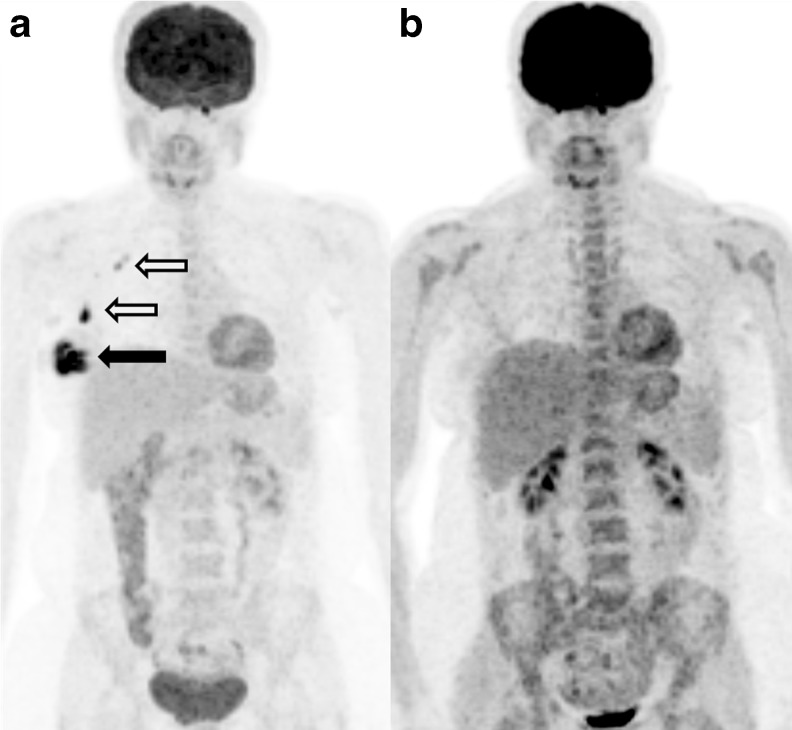
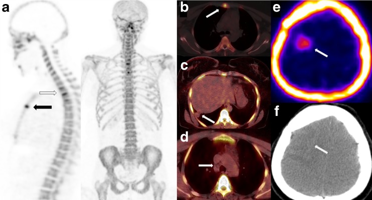
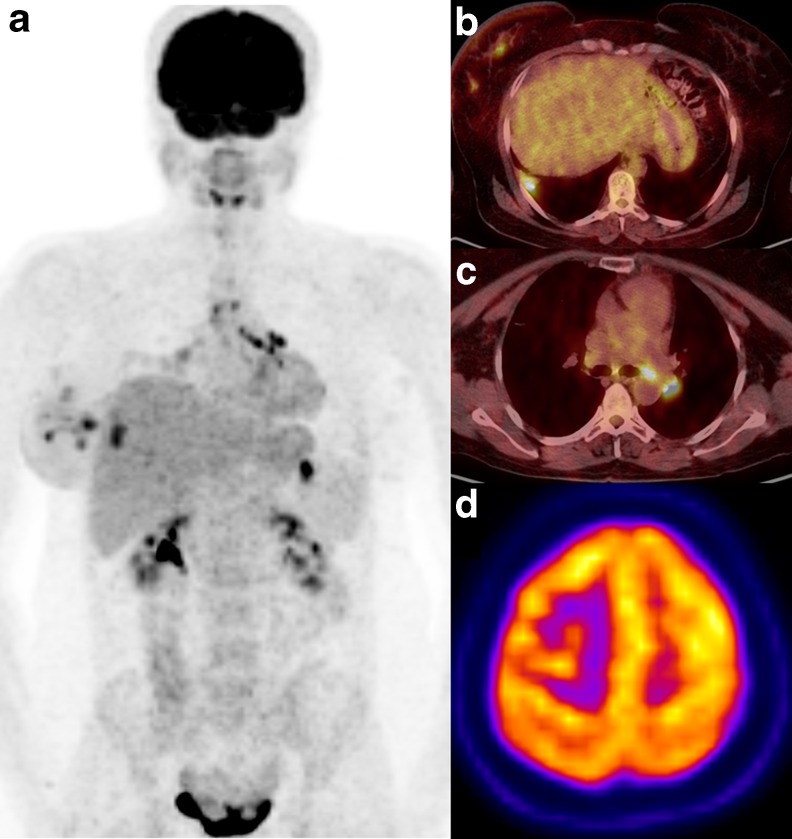
A 56-year-old female patient with newly diagnosed breast cancer underwent 18F-FDG PET/CT (FDG-PET), which demonstrated uptake in the primary tumoral lesion at the right breast and ipsilateral metastatic axillary lymph nodes. FDG-PET was performed following the second cycle of neoadjuvant chemotherapy and demonstrated near complete response to therapy (Fig. 1). However, following the fourth cycle of neoadjuvant chemotherapy, she underwent 18F-NaF PET/CT because of suspected bone metastasis presenting as back pain. 18F-NaF-avid metastases in the vertebrae and the sternum and nonosseous 18F-NaF uptake in the left frontal lobe of the cerebrum were observed (Fig. 2). The CT counterpart of the image demonstrated a hypodense 12 × 13-mm area with no visible calcification. The interpretation suggested tumoral necrosis. Nonosseous findings were also observed in CT images including pulmonary metastases and local recurrence of the breast cancer with no 18F-NaF uptake (Fig. 2). Further evaluation with FDG-PET (Fig. 3) revealed hypermetabolism in the lesions except for the brain metastasis.
Fig. 1.
Primary tumor (black arrow) in the right breast and metastatic lymph nodes (white arrows) are visualized on the maximum-intensity projection image of FDG PET/CT performed for staging (a), and near complete response to chemotherapy is demonstrated in the FDG PET/CT following the second cycle of chemotherapy (b). Bone marrow uptake due to reactive hyperplasia following chemotherapy is also demonstrated (b)
Fig. 2.
Sagittal and coronal MIP images (a) of 18F-NaF PET/CT performed because of severe back pain following the fourth cycle of chemotherapy demonstrate metastases in the thoracic vertebrae (black arrow) and sternum (white arrow). Sternum metastasis is also evident on axial PET/CT images (b). Metastatic pulmonary lesion in the posterolateral segment of the right lung (c) and metastatic right lower paratracheal lymph nodes (d) did not demonstrate any uptake on 18F-NaF PET/CT images. However, the metastatic brain lesion showed 18F-NaF uptake (e) with no visible calcification on the CT counterpart (f)
Fig. 3.
MIP images (a) of FDG PET/CT performed for restaging 1 week after 18F-NaF PET/CT revealed a metastatic pulmonary lesion (b) and hypermetabolic metastatic lymph nodes (c). Note the diminished FDG uptake (d) on the right frontal lobe lesion, which showed focal uptake on 18F-NaF PET/CT images
Discussion
18F-NaF was the first widely used agent for skeletal scintigraphy; however, technical limitations of the decade and the widespread availability of 99Mo/99mTc generators together with the introduction of 99mTc-labeled bone agents led 18F-NaF to fall into disuse [1]. There is renewed clinical interest in the use of 18F-NaF since prior technical and logistic limitations to its routine use are no longer present as the demand for 18F-FDG promotes commercial production and delivery of 18F-NaF. Studies have shown that 18F-NaF PET produces images with higher sensitivity and specificity than 99mTc-based bone scans; however, with the increasing use of 18FNaF PET/CT in routine clinical practice for skeletal imaging in oncology [2, 3], nonosseous uptake is likely to be more frequently encountered. In our case, 18F-NaF PET/CT was performed because of back pain and successfully revealed bone metastases. In addition, 18F-NaF uptake was observed at the metastatic cerebral lesion. However, the CT counterpart of the image did not demonstrate any calcification, which suggests cellular-level microscopic calcifications. Soft tissue calcific metastases concentrating bisphosphonates have been reported previously [4], and similar reports are also evident with 18F-NaF [5–7]. Nonosseous uptake of 18F-NaF due to heterotrophic ossification, brain metastases, metastatic lymph nodes [6] and liver metastasis [7], as well as atherosclerosis [8], has been reported. Evaluation of CT images of our patient also revealed metastatic axillary lymph nodes and pulmonary nodules that were not present in the patient's previous FDG-PET images. In this regard, in order to evaluate disease recurrence, the patient underwent FDG-PET, which revealed pulmonary metastases, local recurrence in the breast and metastatic lymph nodes. CT images help with accurate lesion localization and also provide a large amount of diagnostic information. Therefore, previously undetected, clinically significant nonosseous findings in the 18F-NaF PET/CT may have an impact on the treatment planning.
In conclusion, routine and careful reviewing of nonosseous uptake in 18F-NaF PET/CT as well as the CT portion is necessary since unexpected extra diagnostic information can alter the disease course.
Acknowledgments
All authors declare no conflict of interest.
References
- 1.Grant FD, Fahey FH, Packard AB, Davis RT, Alavi A, Treves ST. Skeletal PET with 18F-fluoride: applying new technology to an old tracer. J Nucl Med. 2008;49(1):68–78. doi: 10.2967/jnumed.106.037200. [DOI] [PubMed] [Google Scholar]
- 2.Tateishi U, Morita S, Taguri M, Shizukuishi K, Minamimoto R, Kawaguchi M, et al. A meta-analysis of (18)F-fluoride positron emission tomography for assessment of metastatic bone tumor. Ann Nucl Med. 2010;24(7):523–531. doi: 10.1007/s12149-010-0393-7. [DOI] [PubMed] [Google Scholar]
- 3.Nishiyama Y, Tateishi U, Shizukuishi K, Shishikura A, Yamazaki E, Shibata H, et al. Role of 18F-fluoride PET/CT in the assessment of multiple myeloma: initial experience. Ann Nucl Med. 2013;27(1):78–83. doi: 10.1007/s12149-012-0647-7. [DOI] [PubMed] [Google Scholar]
- 4.Pirayesh E, Rakhshan A, Amoui M, Rakhsha A, Poor AS, Assadi M. Metastasis of femoral osteosarcoma to the abdominal wall detected on 99mTc-MDP skeletal scintigraphy. Ann Nucl Med. 2013 doi: 10.1007/s12149-013-0708-6. [DOI] [PubMed] [Google Scholar]
- 5.Basu S, Kembhavi S. Serendipitous observation of hepatic metastases on (18)F-fluoride PET in a patient with infiltrating ductal carcinoma of breast: correlation with contrast enhanced CT. Clin Nucl Med. 2012;37(12):1176–1178. doi: 10.1097/RLU.0b013e318270820a. [DOI] [PubMed] [Google Scholar]
- 6.Sheth S, Colletti PM. Atlas of sodium fluoride PET bone scans: atlas of NaF PET bone scans. Clin Nucl Med. 2012;37(5):e110–e116. doi: 10.1097/RLU.0b013e31824c81ee. [DOI] [PubMed] [Google Scholar]
- 7.Swiętaszczyk C, Prasad V, Baum RP. Intense 18F-fluoride accumulation in liver metastases from a neuroendocrine tumor after peptide receptor radionuclide therapy. Clin Nucl Med. 2012;37(4):e82–e83. doi: 10.1097/RLU.0b013e3182478a50. [DOI] [PubMed] [Google Scholar]
- 8.Stewart VL, Herling P, Dalinka MK. Calcification in soft tissues. JAMA. 1983;250(1):78–81. doi: 10.1001/jama.1983.03340010060032. [DOI] [PubMed] [Google Scholar]


