

Abstract
The immense potential of nanobiotechnology makes it an intensely researched field in modern medicine. Green nanomaterial synthesis techniques for medicinal applications are desired because of their biocompatibility and lack of toxic byproducts. We report the toxic byproducts free phytosynthesis of stable silver nanoparticles (AgNPs) using the bark extract of the traditional medicinal plant Acacia leucophloea (Fabaceae). Visual observation, ultraviolet–visible spectroscopy, and transmission electron microscopy (TEM) were used to characterize the synthesized AgNPs. The visible yellow-brown color formation and surface plasmon resonance at 440 nm indicates the biosynthesis of AgNP. The TEM images show polydisperse, mostly spherical AgNP particles of 17–29 nm. Fourier transform infrared spectroscopy revealed that primary amines, aldehyde/ketone, aromatic, azo, and nitro compounds of the A. leucophloea extract may participate in the bioreduction and capping of the formed AgNPs. X-ray diffraction confirmed the crystallinity of the AgNPs. The in vitro agar well diffusion method confirmed the potential antibacterial activity of the plant extract and synthesized AgNPs against the common bacterial pathogens Staphylococcus aureus (MTCC 737), Bacillus cereus (MTCC 1272), Listeria monocytogenes (MTCC 657), and Shigella flexneri (MTCC 1475). This research combines the inherent antimicrobial activity of silver metals with the A. leucophloea extract, yielding antibacterial activity-enhanced AgNPs. This new biomimetic approach using traditional medicinal plant (A. leucophloea) barks to synthesize biocompatible antibacterial AgNPs could easily be scaled up for additional biomedical applications. These polydisperse AgNPs green-synthesized via A. leucophloea bark extract can readily be used in many applications not requiring high uniformity in particle size or shape.
Keywords: AgNPs, antibacterial activity, Acacia leucophloea, biogenic, FTIR, XRD, TEM
Introduction
Nanobiotechnology is a fast-growing interdisciplinary area where nanotechnology extends the tools and technology platforms for the investigation and transformation of biological systems and, in turn, biology contributes inspiring models and bioassembled components to nanotechnology.1 It is a field with great potential for countries rich in biological diversity, such as India, whose biodiversity may be used as a key resource for biotechnological products and processes that are suitable for large-scale synthesis.2 There is immense interest in the biomedical applications of nanoparticles (NPs) owing to their size and structural similarity to biological molecules.3 Currently, the chemical methods widely used for NP synthesis have associated risks, such as chemical precursor contamination, solvent toxicity, and hazardous byproduct formation, which make alternative synthetic methods imperative. The promising alternative of “green chemistry” exploits the intricate biological pathways and biological resources of living systems, including bacteria, fungi, algae, viruses, plants, and plant extracts, for the bioproduction of NPs.4 It has been suggested that to achieve NP synthesis, techniques employing natural reagents such as biodegradable polymers, microorganisms, plant extracts, and sugars and vitamins as reductants and capping agents would be attractive.5 Though both plants and microbes are used for NP synthesis, plant extracts are preferable because of production advantages such as the lack of elaborate culture maintenance and the possibility of large-scale production.6 Although the role of plant phytochemicals in nanostructure formation and causal chemical interactions is debatable, phytochemicals have been successfully used for the reduction of metals such as gold, silver, platinum, and zinc, and the subsequent synthesis of metallic NPs,7 which have the ability to fulfill the application-oriented demands of biomedicine and industry. These biological approaches employing plant extract-mediated NP synthesis are also considered creditable alternative tools since they ensure biocompatible, nontoxic, and in vivo-applicable nanomaterials.4 Moreover, the demand is increasing for bio-NP production via highly stable, eco-friendly processes with no toxic chemicals, even during large-scale production.8 Hence, opportunities are still available for developing large-scale green processes and employing natural products in the emerging areas of nanotechnology where NPs have important applications.5
Among the NPs exploited in nanomedicine, silver NPs (AgNPs) are very promising because of their unique properties. They exhibit high antimicrobial activity against Gram positive and negative bacteria, fungi, protozoa, and certain viruses. Recently, the antitumor activity of AgNPs against various cancerous cell lines was also reported.9 Various new biomedical applications of AgNPs have been found, including diagnostic applicative biological tags and biosensors, as well as antibacterial agents in apparel, cosmetics, footwear, wound dressings, paints, and plastics, in addition to thermal, electrical, and optical applications.10 Hence, a number of bio-inspired approaches employing plant extracts as bioreductants for the synthesis of AgNPs have been explored. The leaf extracts of plants such as neem (Azadirachta indica), rose geranium (Pelargonium graveolens), alfalfa (Medicago sativa), Aloe vera, Amla (Emblica officinalis), Acalypha indica, European Rowan (Sorbus aucuparia), camphor laurel (Cinnamomum camphora),8 Polyalthia longifolia, Morinda citrifolia L.,9 and the stem and bark extracts of Cinnamon zeylanicum,11 Indian Rosewood (Dalbergia sissoo Roxb.),12 and Ocimum sanctum2 have also been used for the synthesis of AgNPs.
As the antibacterial activity of medicinal plants becomes more recognized, an attempt is being made to establish the antibacterial activity of plant extracts used to synthesize NPs, and the combined effect of the metal and the plant extract.8 Acacia leucophloea (Fabaceae) is a large, thorny, tropical and subtropical tree found in geographical areas including India, Sri Lanka, Pakistan, Nepal, Bangladesh, Indonesia, Myanmar, Thailand, and Vietnam. Normally, the tree attains a height of 35 m and a diameter of 100 cm at breast height. It has a white to yellowish-gray smooth bark, which exfoliates in long strips, causing blackening and roughening of the trees upon aging. The bark of the plant has great value in the Indian traditional medicine system. It is used as an antimicrobial, anthelmintic, expectorant, blood purifier, and as a treatment for diabetes, fever, dry cough, dysentery, gum bleeding, mouth ulcers, skin diseases (leprosy), and ulcers, as well as snakebite. The gum and bark decoction is used as an anticontraceptive and menstrual complaint regulator.13 The aqueous extract of the tree contains phytochemicals such as amino acids, alkaloids, carbohydrates, flavonoids, glycosides, steroids, and tannins, whereas the ethanolic extract is found to contain amino acids, alkaloids, glycosides, flavonoids, tannins, and steroids.14 The tree extracts have also been found to contain phytoconstituents such as n-hexacosanol, β-amyrin, β-sitosterol, and tannin as major compounds. The presence of these biogenic multifunctional molecules assists in the self-assembly and capping of the metal NPs formed, thereby allowing a measure of shape control during metal ion reduction.2 For this reason, we synthesize AgNPs through a biomimetic route employing the bark extract of the readily available medicinal plant A. leucophloea, and investigate their antibacterial activity against common bacterial pathogens with the goal of biomedical applicative AgNP synthesis.
Materials and methods
Plant collection and extraction of their bioactive compounds
The stem barks of Acacia leucophloea were collected from the foot of the Kolli Hills (latitude 10°12′–11°7′ north; longitude 76°–77°17′ east), Namakkal, Tamil Nadu, India. The prepared herbarium of the same was submitted for authentication to Dr Saravanababu (Department of Botany, CN College, Erode, Tamil Nadu), who confirmed the identity of the same. The collected A. leucophloea stem barks were shade-dried for 10 days after the removal of adhered soil and after surface sterilization with sterilized distilled water, and were finally dried at 50°C in a hot-air oven for 6 hours. The dried stem barks were chopped into small pieces and powdered using a pulverizer, and the active compounds were extracted by a reflux method in a Soxhlet apparatus with water. The extraction was repeated until a colorless solvent appeared in the siphon tube. At this point, the extract was vacuum-dried and concentrated using a hot-air drier.
Biogenic synthesis of AgNPs
The 1% A. leucophloea aqueous extract silver organosol was prepared with milli-Q deionized water (EMD Millipore, Billerica, MA, USA), incubated at 60°C for 5 minutes,15 and used for the bioreduction of the silver nitrate (AgNO3) starting material (Sigma-Aldrich Co., St Louis, MO, USA). About 5 mL of the organosol was added to 95 mL of 1 Mm aqueous AgNO3 solution, and the mixture was heated gradually in a water bath at temperatures varying between 30°C and 95°C. The color shift from light yellow to dark brown was monitored as an indicator of metallic silver formation in the solution. It takes 1 hour and 10 minutes at 90°C for the development of this dark brown color and thereafter no color change was observed. Free entities were separated by repeated centrifugation of the brown organosol at 10,000 rpm for 20 minutes at 4°C.
Characterization of AgNPs
Ultraviolet–visible (UV–Vis) spectroscopy
The bioreductive synthesis of AgNP was monitored using a Shimadzu UV-1601 PC scanning double beam UV–Vis spectrophotometer (Shimadzu Corporation, Kyoto, Japan). The UV–Vis spectra were recorded between 200–800 nm as a function of temperature for the bioreductive property of aqueous A. leucophloea extracts. The increase in 433 nm-centered strong resonance bands was noted.16
Fourier transform infrared (FTIR) spectroscopy
The surface free biomass residue or unbound extracts of the synthesized AgNPs were removed by washing them three times with distilled water. The AgNPs were then separated from the bulk by centrifugation at 10,000 rpm for 15 minutes, dried, and ground with potassium bromide (KBr) pellets, after which their infrared spectra were recorded via FTIR. The characteristic infrared (IR) absorption spectra of the different functional groups were recorded in the frequency range of 4,000–400 cm−1 using a Thermo Nicolet Nexus 670 FT-IR (Thermo Fisher Scientific, Waltham, MA, USA). The spectra were obtained by operating the same equipment in the diffuse reflectance mode at 4 cm−1 resolution. Likewise, the FTIR spectra of the KBr encapsulated dried powder of the control plant extract was also obtained. Reference blank KBr pellets were used for background correction.17
X-ray diffraction (XRD) analysis
XRD patterns were recorded from a thin film of powdered AgNPs using a D8 X-ray diffractometer (Bruker Biosciences Corporation, Billerica, MA, USA) at a scan rate of 1 step/second. The powdered sample was irradiated with Cu-Kα radiation and the analysis was performed from 20°–90° (2θ) with a step size of 0.001°.
Transmission electron microscopy (TEM)
The bioreductive formation and existence of AgNPs was analyzed via TEM by placing a drop of the AgNP solution on carbon-coated TEM grids and air-drying. The images were captured using a high-resolution TEM (Philips TECHNAI 10; FEI Co., Hillsboro, OR, USA). The NP sizes were measured from the TEM micrographs.
Determination of antibacterial activity
Bacterial strains
The Staphylococcus aureus (MTCC 737), Bacillus cereus (MTCC 1272), Listeria monocytogenes (MTCC 657), and Shigella flexneri (MTCC 1475) obtained from Microbial Type Culture Collection (MTCC, Chandigarh, India) were used for determining the antibacterial activity of the plant extract and the synthesized NPs.
Determination of the antibacterial activity of the plant extracts and synthesized AgNPs
The agar well diffusion method was used to determine the antibacterial activity of the aqueous A. leucophloea extract and of the NPs synthesized via the bioreductive ability of the extract. Briefly, the Mueller-Hinton agar (HiMedia, Mumbai, India) plates were inoculated with 1 mL (1.0×10⁷ colony-forming units) of 18-hour-young bacterial cultures by spread plating. After drying for 5 minutes, 5 mm diameter wells were made in the plates using a sterile borer. Then, 50 μL of various concentrations of A. leucophloea extracts (25–100 μL/0.1 mg mL−1) and the synthesized AgNPs (25 μL, 50 μL, 75 μL, and 100 μL) were introduced into the well, and the plates were incubated at 37°C for 24 hours. The aminoglycoside antibiotic streptomycin was used as a positive control. After incubation, bacterial growth inhibition was determined by measuring the diameter of the inhibitory zones around the wells in millimeters. The growth inhibitory assays were carried out in triplicate.
Statistical analysis
The statistical significance of the antimicrobial activity results was determined using one-way analysis of variance (ANOVA; v 15; Minitab; Pennsylvania State University, University Park, PA, USA) via Student’s t-tests. Statistically significant differences were determined using the Duncan’s new multiple range test at P≤0.05. The graphs were analysed using Microcal Origin (v6.0; OriginLab, Corporation, Northampton, MA, USA).
Results
The A. leucophloea extract was used for the green-synthesis of AgNPs showing antibacterial action. Visual observation of the color change of the AgNO3 solution/plant extract indicated the bioreductive formation of AgNPs. Blending AgNO3 with A. leucophloea extract at a temperature of 90°C resulted in the formation of AgNO3 NPs (AgNP), as indicated by a prominent color change to dark yellow-brown due to excitation of the AgNP surface plasmon vibration. It is well known that AgNPs exhibit a dark brown color in aqueous solution. The colorless AgNO3/extract solution initially changed to light yellow, and finally to a yellow-brown, indicating the formation of the AgNPs. This color change took place more rapidly at 90°C than at lower temperatures, including at room temperature. The color shift to dark yellow-brown took 1 hour and 10 minutes at 90°C, after which no color change took place. With decreasing temperature, the time taken for the same color change increased from 2 hours to 7 hours. The control AgNO3 solution without plant extract did not show any color changes under a similar set of conditions. In addition, the bioreductive formation of AgNPs was ascertained by UV–Vis and FTIR, and by TEM analysis.
The formation and stability of AgNPs synthesized via A. leucophloea extracts were initially examined by UV–Vis analysis, which is an important technique used to monitor metal NPs in aqueous solution. The UV–Vis absorption spectra obtained were typical and revealed the bioreductive formation of AgNPs; the plots are shown in Figure 1. A high-intensity surface plasmon resonance band is seen at 433 nm, along with the synthesized AgNPs characteristic wavelength range. The large number of weak absorption peaks at shorter wavelengths reveals the presence of many participating organic compounds that can interact to reduce the silver ions.
Figure 1.
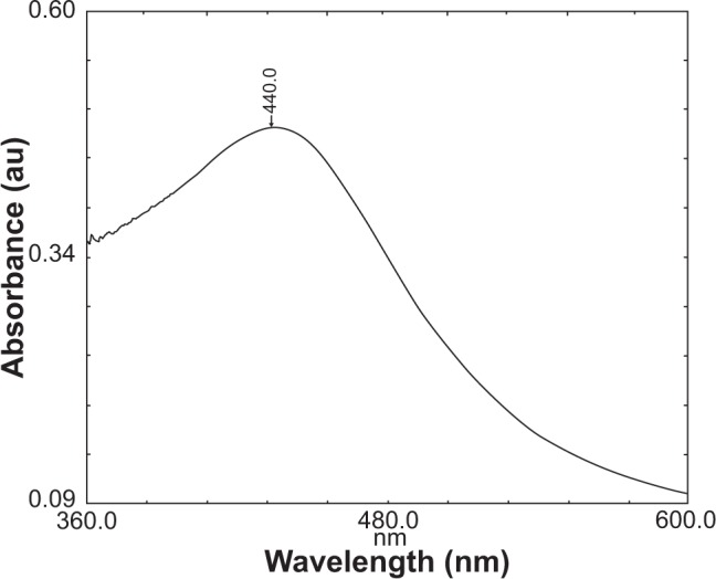
Ultraviolet–visible absorption spectra of Acacia leucophloea synthesized silver nanoparticles.
The FTIR analysis of the synthesized AgNPs divulged the two-fold function of the A. leucophloea extract as a bioreductant whose biomolecules participate in the reduction of the Ag+ ions, and as a capping agent that stabilizes the bioreduced AgNPs. The surface chemistry of the AgNPs synthesized via A. leucophloea is revealed by the appearance in the FTIR spectra of IR bands at 3,420, 2,922, 1,622, 1,427, 1,377, 1,260, 1,196, 1,088, 834, 725, and 691 cm−1 and by the peaks centered at 3,420, 2,922, 1,622, and 1,260 cm−1, which correspond to various groups (Figure 2). The A. leucophloea AgNP FTIR absorption spectra indicates the presence of the N–H stretching hydrogen-bonded primary amine (3,420 cm−1), C–H stretching hydrogen (2,922 cm−1), N–H bonding (1,622 cm−1), and C=O stretching (1,260 cm−1). The absorption peaks situated around 1,260 and 1,196 cm−1 are the C=O characteristic peaks, indicating the presence of aldehyde/ketone and aromatic compounds. The peak at 1,427 cm−1 indicates the presence of N=N stretching of the azo compound, and the peak at 1,377 cm−1 indicates N=O symmetry stretching typical of the nitro compound.
Figure 2.
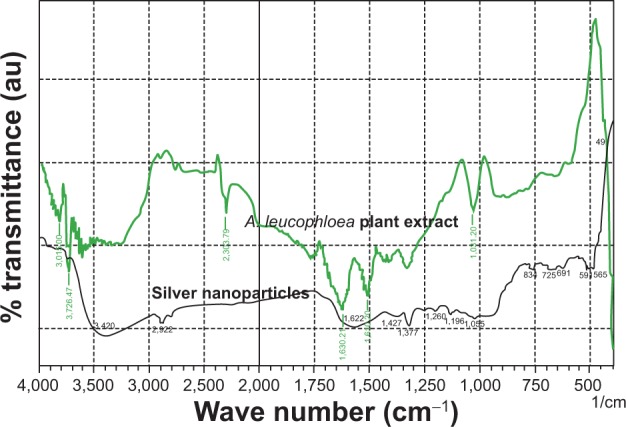
FTIR spectra of the Acacia leucophloea aqueous extract and the synthesized AgNPs.
Abbreviations: AgNPs, silver nanoparticles; FTIR, fourier transform infrared.
The XRD pattern of the AgNPs synthesized via A. leucophloea bark extract was compared and interpreted using standard data. The major peaks at 38°, 46°, 65°, and 78° (2θ values) correspond to the reflections from the (111), (200), (220), and (311) planes, respectively, and confirm the crystalline phase of the AgNPs. The polycrystalline nature of extract-synthesized AgNPs can be inferred from the fact that the XRD data suggest smaller crystallite sizes than the particle sizes observed in TEM images (Figure 3).
Figure 3.
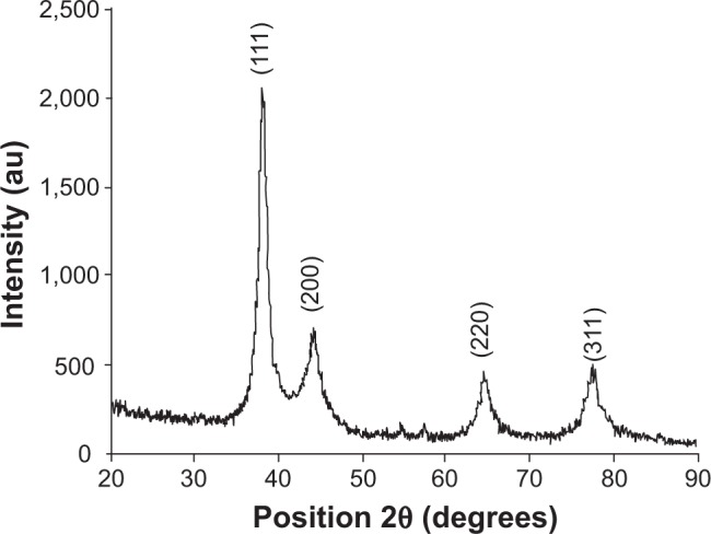
X-ray diffraction spectrum of Acacia leucophloea bark extract synthesized AgNPs.
Abbreviation: AgNPs, silver nanoparticles.
The TEM micrographs of AgNPs green-synthesized via A. leucophloea extract reveal their morphology and size, showing polydisperse AgNPs, most of which have a spherical morphology, with a size range of 17–29 nm (Figure 4).
Figure 4.
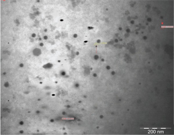
Transmission electron micrograph showing AgNPs synthesized using Acacia leucophloea bark extract.
Abbreviation: AgNPs, silver nanoparticles.
Varying the concentration (5–20 μg/mL) of the A. leucophloea aqueous extract reveals in vitro bactericidal activity that is linear with increasing concentration (Figure 5). The data indicate that this species exhibits broad-spectrum antibacterial activity against the common pathogens S. aureus, B. cereus, L. monocytogenes, and S. flexneri (Figures 5A–D). The extract exhibits the highest activity against S. aureus and B. cereus, whereas it shows only moderate activity against S. flexneri. The obtained results support, at least in part, the use of this plant as traditional medicine against Gram positive bacteria.
Figure 5.
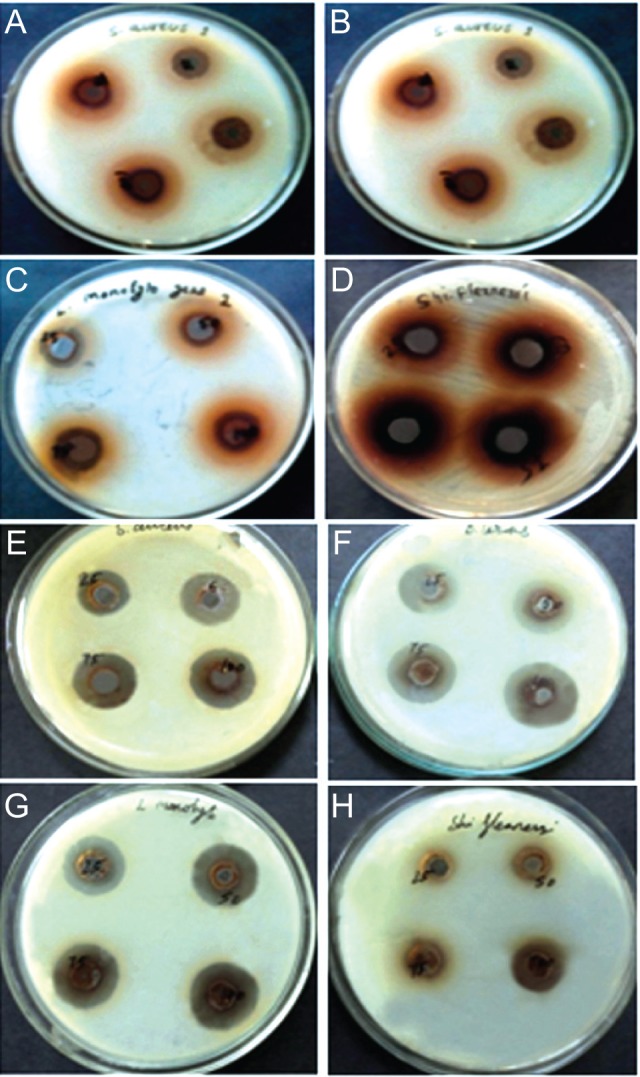
Antibacterial activity of Acacia leucophloea extract and its synthesized AgNPs. Plates (A–D) showing A. leucophloea extract diameters of zones of inhibition of Staphylococcus aureus (A), Bacillus cereus (B), Listeria monocytogenes (C), and Shigella flexneri (D). Plates (E–H) showing AgNP inhibitory activity on S. aureus (E), B. cereus (F), L. monocytogenes (G), and S. flexneri (H).
Abbreviation: AgNP, silver nanoparticle.
The antibacterial activity assay of A. leucophloea aqueous extract-biosynthesized AgNPs showed effective inhibitory action against common pathogens such as S. aureus, B. cereus, L. monocytogenes, and S. flexneri. Agar well diffusion studies showed effective growth inhibitory activity of AgNPs on common bacterial pathogens (Figures 5E–H). Greater bactericidal activity was observed against the Gram positive rods B. cereus and L. monocytogenes, and the Gram positive cocci S. aureus. The Gram negative S. flexneri was found to be less susceptible to AgNPs than the Gram positive bacteria. The comparative analysis on the antibacterial activity of A. leucophloea extract and its synthesized AgNPs is statistically significant at the threshold set at P<0.05. These results demonstrate that the synergistic combination of the antibacterial activity of the medicinal plant with AgNPs enhances the antimicrobial effects (Figures 6A and B).
Figure 6.

A comparative analysis on antibacterial activity of (A) Acacia leucophloea extract and (B) its synthesized AgNPs.
Note: Mean ± standard deviation, statistically significant at P≤0.05.
Abbreviation: AgNP, silver nanoparticle.
Discussion
Modern times have witnessed many potential biomedicinal applications of nanobiotechnology. NPs, for instance, have immense applications in the early diagnosis and management of diseases, including those caused by emerging multidrug-resistant pathogens. AgNPs have found wide usage in various industries and are known to inhibit a number of microorganisms. These AgNPs are employed widely in the manufacture of burn and wound infection-preventing ointments and creams.18 Worldwide attempts have been made to increase their scope further with the synthesis of an NP that is free of toxic byproducts and is biocompatible. Currently, reports are available on the use of a variety of plants for AgNP synthesis, such as Acalypha indica, Aloe vera, Arbutus unedo, Azadirachta indica, Capsicum annuum, Catharanthus roseus, Chenopodium album, Cinnamomum camphora, Coriandrum sativum, Dalbergia sissoo, Ficus benghalensis, Gliricidia sepium, Magnolia kobus, Mentha piperita, Murraya koenigii, Nicotiana tabacum, Ocimum sanctum, Pelargonium graveolens, Piper longum, Rosa rugosa, and Stevia rebaudiana leaf extracts.6,19 The bioreduction that reduces metal ions into zero-valance metal NPs exploits the reducing ability of the different biochemicals, such as reducing sugars, proteins, terpenoids, and other phenolic compounds of the plant extracts. In addition, these bioreduction solutions also contain natural capping agents that hinder aggregation of the synthesized metal NPs and control the particle size.20 The studies mentioned have been performed on leaf extracts, but available literature on the nanotechnological exploration of plant stem and barks are very few, though these phytochemicals are distributed throughout the plant. Among natural extracts, stem bark extracts have many advantages. For instance, stem bark collection does not destroy the parent plant. In addition, the multifunctional molecules present in stem barks reduce the metal ions and stabilize the newly formed zero-valence NPs, thereby requiring no additional reducing or stabilizing agents.12 The bark of the thorny tropical and subtropical tree A. leucophloea is an important component in the Indian traditional medicine system, where it is used for various treatments, including wound care.13 The extracts from this bark are found to contain various bioactive components14 that not only scientifically validate their use in traditional medicine, but also the study of their participation in self-assembling and capping metal NPs.2 Hence, employing the medicinal plant A. leucophloea extract to biosynthesize AgNPs in this work may pave the way to increase the therapeutic value of this traditional medicinal plant.3
The visually observed color change from dark brown to yellowish brown during the bioreductive AgNP formation upon adding A. leucophloea extract may be due to the reduction of silver ions.15 The UV–Vis spectroscopy measurements of the bioreductive formation of AgNPs via reduction during the addition of A. leucophloea reveals a weak surface plasmon resonance from the leaf extract centered at 359 nm, and a peak characteristic of AgNPs at 433 nm. Mondal et al18 made similar observations while synthesizing AgNPs using extracts of various medicinal plants. However, other studies showed a peak at 240 nm while conducting extract-mediated AgNP synthesis, namely from extracts of Nicotiana tabacum,21 Ocimum,17 Allium cepa,16 Boswellia ovalifoliolata, and Shorea tumbuggaia.22
The plant biomolecules comprising the differently shaped polyol and water-soluble heterocyclic components are known for their protective and reductive activity, and are primarily accountable for the reduction of silver and gold ions using chemical and radiation methods.23 The FTIR analysis of AgNPs bioreduced via A. leucophloea confirms the capping of synthesized NPs by A. leucophloea biomolecules. Various reports are available on the stabilization and capping of NPs synthesized via plant extract mediation. Many plant phytochemicals, such as quercetin, terpenoids, verbascoside,3 saponins, phenolic compounds, and quinines,23 are known as capping and stabilizing agents of AgNPs. Therefore, we conclude that the aldehyde/ketone, aromatic, azo, and nitro compounds of the A. leucophloea extract participate in the bioreduction and stabilization of AgNPs by coating them, thereby hindering agglomeration.
The TEM images show that the phytosynthesized AgNPs have sizes ranging from 11–29 nm and are largely spherical in shape. The images also show that the AgNPs do not aggregate and are mostly dispersed, thereby confirming the NP-stabilizing nature of the A. leucophloea extracts. The TEM images also reveal that the AgNPs are embedded in a dense matrix, confirming the capping and stabilizing components of the A. leucophloea extract. The antibacterial activity measured also indicates the potentiation of the antibacterial activity of A. leucophloea AgNPs. Currently, AgNPs showing inhibitory activities toward several microorganisms are widely employed in the pharmaceutical industry. These NPs find extensive use in balms and ointments to prevent burn and wound infections. Earlier reports on the use of AgNPs as potential antifungal, antibacterial, and antiviral agents are also available.24 Indeed, the antimicrobial activities of silver ions and compounds have been historically recognized. Silver have found extensive and varied applications, from medical devices and home appliance disinfection to water treatment because of their high microbicidal action against various species.25 Upon treatment with silver ions, microorganisms’ activities such as DNA replication and ribosomal subunit protein expression fail, along with the inactivation of other cellular proteins and enzymes necessary for adenosine triphosphate synthesis. Thus, these polydisperse AgNP particles green-synthesized via A. leucophloea extract can readily be used in many applications that do not require a high uniformity in particle size or shape.20
Conclusion
The unique physicochemical characteristics of AgNPs are believed to have increased medical applications when synthesized via environmentally benign methods free of toxic byproducts. The measured antibacterial activity of the green-synthesized AgNPs via A. leucophloea extracts clearly demonstrates the enhanced activity of these NPs against several pathogenic bacteria. This opens the possibility of various applications for these NPs of producing effective antibacterial agents for the management of emerging multidrug-resistant pathogenic bacteria. In addition, the synergistic combination of biocompatible medicinal plants with AgNPs may open new applications in medicine for therapeutic management of organisms that have developed resistance to current antibiotics. Further, the bark of trees found to possess comparatively more bioreductive phytochemicals can be considered good candidates for nanotechnological application investigation.
Acknowledgments
The authors extend their appreciation to the Deanship of Scientific Research at King Saud University for funding the work through the research group project No RGP-VPP-183.
Footnotes
Disclosure
The authors report no conflicts of interest in this work.
References
- 1.MubarakAli D, Thajuddin N, Jeganathan K, Gunasekaran M. Plant extract mediated synthesis of silver and gold nanoparticles and its antibacterial activity against clinically isolated pathogens. Colloids Surf B Biointerfaces. 2011;85(2):360–365. doi: 10.1016/j.colsurfb.2011.03.009. [DOI] [PubMed] [Google Scholar]
- 2.Ahmad N, Sharma S, Alam MK, et al. Rapid synthesis of silver nanoparticles using dried medicinal plant of basil. Colloids Surf B Biointerfaces. 2010;81(1):81–86. doi: 10.1016/j.colsurfb.2010.06.029. [DOI] [PubMed] [Google Scholar]
- 3.Murugan K, Selvanayaki K, Kalyanasundaram VB, Al-Sohaibani S. Nanotechnological approach for exploring the antibiofilm a potential of an ethanomedicinal herb Andrographis paniculata for controlling lung infection causing Pseudomonas aeruginosa. Dig J Nanomater Bios. 2013;8(1):117–126. [Google Scholar]
- 4.Faramarzi MA, Sadighi A. Insights into biogenic and chemical production of inorganic nanomaterials and nanostructures. Adv Colloid Interface Sci. 2013:189–190. 1–20. doi: 10.1016/j.cis.2012.12.001. [DOI] [PubMed] [Google Scholar]
- 5.Kharissova OV, Dias HV, Kharisov BI, Pérez BO, Pérez VM. The greener synthesis of nanoparticles. Trends Biotechnol. 2013;31(4):240–248. doi: 10.1016/j.tibtech.2013.01.003. [DOI] [PubMed] [Google Scholar]
- 6.Panda KK, Achary VM, Krishnaveni R, et al. In vitro biosynthesis and genotoxicity bioassay of silver nanoparticles using plants. Toxicol In Vitro. 2011;25(5):1097–1105. doi: 10.1016/j.tiv.2011.03.008. [DOI] [PubMed] [Google Scholar]
- 7.Annamalai A, Christina VL, Sudha D, Kalpana M, Lakshmi PT. Green synthesis, characterization and antimicrobial activity of Au NPs using Euphorbia hirta L. leaf extract. Colloids Surf B Biointerfaces. 2013;108:60–65. doi: 10.1016/j.colsurfb.2013.02.012. [DOI] [PubMed] [Google Scholar]
- 8.Prakash P, Gnanaprakasam P, Emmanuel R, Arokiyaraj S, Saravanan M. Green synthesis of silver nanoparticles from leaf extract of Mimusops elengi, Linn. for enhanced antibacterial activity against multi drug resistant clinical isolates. Colloids Surf B Biointerfaces. 2013;108:255–259. doi: 10.1016/j.colsurfb.2013.03.017. [DOI] [PubMed] [Google Scholar]
- 9.Jeyaraj M, Sathishkumar G, Sivanandhan G, et al. Biogenic silver nanoparticles for cancer treatment: an experimental report. Colloids Surf B Biointerfaces. 2013;106:86–92. doi: 10.1016/j.colsurfb.2013.01.027. [DOI] [PubMed] [Google Scholar]
- 10.Vijayaraghavan K, Nalini SP, Prakash NU, Madhankumar D. One step green synthesis of silver nano/microparticles using extracts of Trachyspermum ammi and Papaver somniferum. Colloids Surf B Biointerfaces. 2012;94:114–117. doi: 10.1016/j.colsurfb.2012.01.026. [DOI] [PubMed] [Google Scholar]
- 11.Sathishkumar M, Sneha K, Won SW, Cho CW, Kim S, Yun YS. Cinnamon zeylanicum bark extract and powder mediated green synthesis of nano-crystalline silver particles and its bactericidal activity. Colloids Surf B Biointerfaces. 2009;73(2):332–338. doi: 10.1016/j.colsurfb.2009.06.005. [DOI] [PubMed] [Google Scholar]
- 12.Roy N, Alam MN, Mondal S, et al. Exploring Indian rosewood as a promising biogenic tool for the synthesis of metal nanoparticles with tailor-made morphologies. Process Biochem. 2012;47(9):1371–1380. [Google Scholar]
- 13.Jain S, Sharma P, Jhade D, Sharma NK, Paliwal P, Ahirwar D. Pharmacognostic screening and phytochemical evaluation of Acacia leucophloea root. Int J Green Pharm. 2011;5(2):155–159. [Google Scholar]
- 14.Jhade D, Jain S, Jain A, Sharma P. Pharmacognostic screening, phytochemical evaluation and in-vitro free radical scavenging activity of Acacia leucophloea root. Asian Pac J Trop Biomed. 2012;2(2):S501–S505. [Google Scholar]
- 15.Antony JJ, Sivalingam P, Siva D, et al. Comparative evaluation of antibacterial activity of silver nanoparticles synthesized using Rhizophora apiculata and glucose. Colloids Surf B Biointerfaces. 2011;88(1):134–140. doi: 10.1016/j.colsurfb.2011.06.022. [DOI] [PubMed] [Google Scholar]
- 16.Saxena A, Tripathi RM, Singh RP. Biological synthesis of silver nanoparticles by using onion (Allium cepa) extract and their antibacterial activity. Dig J Nanomater Bios. 2010;5(2):427–432. [Google Scholar]
- 17.Mallikarjuna K, Narasimha G, Dillip GR, et al. Green synthesis of silver nanoparticles using Ocimum leaf extract and their characterization. Dig J Nanomater Bios. 2011;6(1):181–186. [Google Scholar]
- 18.Mondal AK, Mondal S, Samanta S, Mallick S. Synthesis of ecofriendly silver nanoparticle from plant latex used as an important taxonomic tool for phylogenetic inter-relationship. Adv Biores. 2011;2(1):122–133. [Google Scholar]
- 19.Awwad AM, Salem NM, Abdeen AO. Biosynthesis of silver nanoparticles using Loquat leaf extract and its antibacterial activity. Adv Materials Letters. 2013;4(5):338–342. [Google Scholar]
- 20.Vivekanandhan S, Schreiber M, Mason C, Mohanty AK, Misra M. Maple Leaf (Acer sp.) extract mediated green process for the functionalization of ZnO powders with silver nanoparticles. Colloids Surf B Biointerfaces. 2014;113:169–175. doi: 10.1016/j.colsurfb.2013.08.033. [DOI] [PubMed] [Google Scholar]
- 21.Prasad KS, Pathak D, Patel A, et al. Biogenic synthesis of silver nanoparticles using Nicotiana tobaccum leaf extract and study of their antibacterial effect. Afr J Biotechonol. 2011;10(41):8122–8130. [Google Scholar]
- 22.Savithramma N, Linga Rao M, Suvarnalatha Devi P. Evaluation of antibacterial efficacy of biologically synthesized silver nanoparticles using stem barks of Boswellia ovalifoliolata Bal. and Henry and Shorea tumbuggaia Roxb. J Biol Sci. 2011;11:39–45. [Google Scholar]
- 23.Arunachalam KD, Annamalai SK, Hari S. One-step green synthesis and characterization of leaf extract-mediated biocompatible silver and gold nanoparticles from Memecylon umbellatum. Int J Nanomedicine. 2013;3:1307–1315. doi: 10.2147/IJN.S36670. [DOI] [PMC free article] [PubMed] [Google Scholar]
- 24.Pasupuleti VR, Prasad TN, Shiekh RA, et al. Biogenic silver nanoparticles using Rhinacanthus nasutus leaf extract: synthesis, spectral analysis, and antimicrobial studies. Int J Nanomedicine. 2013;8:3355–3364. doi: 10.2147/IJN.S49000. [DOI] [PMC free article] [PubMed] [Google Scholar] [Retracted]
- 25.Jung WK, Koo HC, Kim KW, Shin S, Kim SH, Park YH. Antibacterial activity and mechanism of action of the silver ion in Staphylococcus aureus and Escherichia coli. Appl Environ Microbiol. 2008;74(7):2171–2178. doi: 10.1128/AEM.02001-07. [DOI] [PMC free article] [PubMed] [Google Scholar]