

Abstract
Background and Objectives:
Single-incision laparoscopic surgery is gaining popularity among minimally invasive surgeons and is now being applied to a broad number of surgical procedures. Although this technique uses only 1 port, the diameter of the incision is larger than in standard laparoscopic surgery. The long-term incidence of port-site hernias after single-incision laparoscopic surgery has yet to be determined.
Methods:
All patients who underwent a single-incision laparoscopic surgical procedure from May 2008 through May 2009 were included in the study. Single-incision laparoscopic surgical operations were performed either by a multiport technique or with a 3-trocar single-incision laparoscopic surgery port. The patients were seen at 30 to 36 months' follow-up, at which time they were examined for any evidence of port-site incisional hernia. Patients found to have hernias on clinical examination underwent repairs with mesh.
Results:
A total of 211 patients met the criteria for inclusion in the study. The types of operations included were cholecystectomy, appendectomy, sleeve gastrectomy, gastric banding, Nissen fundoplication, colectomy, and gastrojejunostomy. We found a port-site hernia rate of 2.9% at 30 to 36 months' follow-up.
Conclusion:
Port-site incisional hernia after single-incision laparoscopic surgical procedures remains a major setback for patients. The true incidence remains largely unknown because most patients are asymptomatic and therefore do not seek surgical aid.
Keywords: Single-incisional laparoscopic surgery, SILS, Port-site hernia
INTRODUCTION
In the past, most abdominal procedures were performed through large incisions. Most of these incisions were associated with multiple morbidities, such as surgical-site infections, incisional hernia, postoperative pain, and prolonged hospitalization. Unsatisfied with these morbidities, surgeons sought ways of minimizing their incision length without compromising the integrity of the surgical procedure performed within the intra-abdominal cavity.
With the use of smaller incisions facilitated by laparoscopy, most of the incision-related complications were significantly reduced. It was hoped that single-incision laparoscopy would add additional cosmesis to conventional laparoscopy. The concept of single-trocar laparoscopy is not new because it has been within the domain of gynecologists for several years. Reports of single-port laparoscopic sterilization were documented in the 1960s. In 1 report in 1969, single-trocar laparoscopy was performed with a 12-mm operative laparoscope with 1 operative channel.1,2 Despite these pioneering efforts, single-trocar laparoscopy was not widely embraced by the surgical community because of the lack of appropriately designed equipment. With improvements in optics and technology, single-trocar appendectomy and cholecystectomy were performed in the 1990s.3,4
Though known by different names, the term single-site laparoscopic surgery was coined by the Laparoendoscopic Single-Site Surgery Consortium for Assessment and Research for uniformity in academic publications in 2008.5
Single-incision laparoscopic surgery (SILS) represents a unique twist in minimally invasive surgery. This novel surgical approach was born out of the quest for scarless, highly cosmetic surgery. The primary benefit of SILS seems to be cosmetic related; the umbilicus is the preferred incision site because the scars can be easily hidden. Other reported benefits are improved postoperative pain and a quicker return to normal activities of daily living.6–8 The concept of a single-incision approach has been adopted for various surgical techniques ranging from sleeve gastrectomy, colectomy, and adrenalectomy to Nissen fundoplication. As SILS becomes more widely adopted, the incidence of port-site hernias (PSHs) becomes an important consideration in the overall risk-benefit discussion with the patient.
This study aims to determine the incidence of PSHs after SILS at a single institution performing a variety of SILS operations.
METHODS
We performed a retrospective study with prospective data collection of all SILS procedures performed in a high-volume tertiary center in Bronx, New York, between May 2008 and May 2009. Two fellowship-trained minimally invasive surgeons performed all procedures. The institutional review board approved the study, and informed consent was obtained from all patients.
We defined PSH as the development of a hernia at the single-incision skin site. PSH may be further characterized into early (dehiscence of fascial planes and peritoneum) or late (dehiscence of fascial plane with intact peritoneal hernia sac).
The inclusion criteria were all patients undergoing an SILS procedure during the study period. The exclusion criteria were as follows: history of mesh placement in the umbilical or upper abdomen; patients receiving steroid therapy, heparin, Coumadin: (Bristol-Myers Squibb, Princeton NJ, USA), Plavix (clopidogrel): (Bristol-Myers Squibb/Sanofi, Princeton NJ, USA), or aspirin; body mass index (BMI) >50 kg/m2; and appendicitis present for >36 hours. In addition, we excluded any patient who had a conversion from an SILS approach to a standard laparoscopic approach.
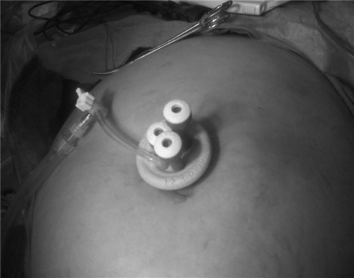
All patients operated on between May 2008 and February 2009 underwent a multiport technique (Figure 1). This consisted of standard ports placed through the umbilical skin incision into the fascia and used multiple points of fascial entry through the single skin incision.
Figure 1.
Multiport SILS technique.
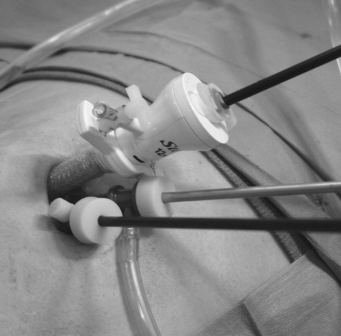
The 3-trocar SILS port (Covidien, North Haven, Connecticut) (Figure 2) was introduced in March 2009, and all procedures performed after that date used this port.
Figure 2.
Covidien SILS port.
Patient Positioning
When the SILS procedures were performed, the patients were placed in either a supine or modified lithotomy position, depending on the surgeon's preference.
Procedure
With the patient under general anesthesia, a 2-cm vertical transumbilical skin incision was made. In the early stage of the study, access to the abdomen was accomplished by introducing three 5-mm trocars through separate fascial punctures but contiguous skin incisions. At the end of the procedure, the separate fascial incisions were connected to form a single incision to facilitate the extraction of the resected specimen.
With the introduction of the SILS port, a 2-cm vertical skin incision was followed by a 2-cm fascial incision. The port was placed under direct visualization. Pneumoperitoneum was established to a pressure of 15 mm Hg. Once pneumoperitoneum was achieved, three 5-mm ports were inserted through the SILS port, and one of the 5-mm ports could be changeable to a 12-mm port.
To prevent port-site or incisional hernia, careful closure of the fascial incision was performed in all cases. The edges of the fascial incision were clearly identified and grasped with Kocher forceps. All fascial incisions were closed with 3 figure-of-8 No. 0 Vicryl sutures (polyglactin 910; Ethicon, Somerville, New Jersey).
The skin incision was infiltrated with a local anesthetic agent (bupivacaine) and was approximated with a running subcuticular No. 4–0 Monocryl suture (poliglecaprone 25; Ethicon).
Postoperatively, all patients were examined during their scheduled appointments 2 to 4 weeks after the procedure and then subsequently at 3 months depending on the procedure performed. All patients were contacted between 30 and 36 months after the procedure to return to the office for an additional evaluation. During this visit, each patient was questioned and examined by an attending surgeon looking specifically for evidence of port-site incisional hernia. Questions included any pain or bulging at the incision site. Patients who were found to have clinical evidence of incisional hernia underwent repair with mesh in all cases.
Statistical Analyses
Statistical analysis was performed with GraphPad Prism statistical software (GraphPad, San Diego, California). P values are considered significant at a level of .05.
RESULTS
A total of 211 patients underwent single-incision laparoscopic procedures during the study period. Table 1 show the patients' demographic data and indications for SILS. For the purpose of this study, patients were divided into 2 groups—standard multiport laparoscopy and SILS—based on the type of access port used. The 2 groups were identical in terms of age, BMI, and type of trocars used for the procedures.
Table 1.
Demographic Characteristics of Patients Who Underwent SILS and Indications for SILS Procedure
| Data | |
|---|---|
| Characteristics | |
| Total No. of patients | 211 |
| Male (n) | 64 |
| Female (n) | 147 |
| Median age (y) | 45 (range, 21–82) |
| BMI (kg/m2) | 32 (range, 28–47) |
| Indications (n) | |
| Cholecystitis | 130 |
| Biliary colic | 20 |
| Morbid obesity | 23 |
| Appendicitis | 29 |
| Sigmoid diverticulosis | 5 |
| GERDa | 3 |
| Other | 1 |
GERD = gastroesophageal reflux disease.
The mean operative time for all cases was 40 minutes (range, 21–120 minutes). The mean estimated blood loss was 30 mL (range, 10–150 mL). In 1 patient an additional port was placed because of bleeding. Multiports were used in 98 cases, and in the remaining patients, the SILS port was used (Table 2). No patient underwent conversion to laparotomy. In total, 291 bladed trocars were used in 98 cases. In the remainder of cases, 363 non-bladed trocars were used.
Table 2.
Type of SILS Procedures Performed
| SILS Port | Multiport | |
|---|---|---|
| Procedure (n) | ||
| Cholecystectomy | 70 | 80 |
| Appendectomy | 17 | 12 |
| Bariatric procedure | ||
| Gastric sleeve | 14 | 4 |
| Gastric banding | 3 | 2 |
| Hiatal hernia repair | 3 | 0 |
| Gastrojejunostomy | 1 | 0 |
| Colectomy | 5 | 0 |
| Operative time (min) | ||
| Mean | 40 | 43 |
| Range | 21–120 | 30–117 |
| Total (n) | 113 | 98 |
At 36 months, there was a 97% follow-up rate (n = 205). Six patients were lost to follow-up. The remaining 205 patients were deemed suitable for the final analysis. PSH developed in 6 patients, requiring surgical repair with mesh (PSH rate of 2.8% [n = 6]) (Table 4).
Table 4.
Subset of Reporting Variables Among Patients With PSH Based on Trocar Type and BMI
| Procedure | Trocar Type |
Duration (min) | BMI (kg/m2) | |
|---|---|---|---|---|
| Bladed | Non-Bladed | |||
| Sleeve gastrectomy | ||||
| Patient 1 | No | Yes | 90 | 45 |
| Patient 2 | No | Yes | 80 | 47 |
| Patient 3 | No | Yes | 75 | 42 |
| Cholecystectomy | ||||
| Patient 1 | Yes | No | 70 | 30 |
| Patient 2 | Yes | No | 87 | 32 |
| Patient 3 | Yes | No | 90 | 28 |
P = .11 (95% confidence interval, 0.0117–0.0621).
The interval between the SILS procedure and diagnosis of PSH varied from 24 months to 36 months (mean, 28.4 months). All patients were asymptomatic.
A subset analysis of the hernias showed that among patients who underwent SILS cholecystectomy, PSH was more common in patients who had multiport bladed trocars (2.5%, 2 of 98 patients). This finding was in sharp contrast to that in bariatric patients, in whom PSH occurred exclusively among SILS port patients with non-bladed trocars (3.5%, 4 of 113 patients) (Table 3).
Table 3.
Incidence of PSH by Procedure and Sex
| SILS Port | Multiport | Sex | |
|---|---|---|---|
| Procedure | |||
| Cholecystectomy | 1 | 2 | All female patients |
| Sleeve gastrectomy | 3 | 0 | All female patients |
| Total | 4 | 2 | 6 patients |
In addition, the pattern of PSH formation was different between bariatric and nonbariatric patients. All PSHs in bariatric patients occurred after the use of bladed trocars, whereas among nonbariatric patients, PSH followed the use of non-bladed trocars.
DISCUSSION
SILS appears to be finding greater acceptance and adoption among general surgeons. Over the past 5 years, the application of SILS has expanded to a growing number of surgical procedures. With this increasing adoption, we have learned a great deal about the benefits SILS has to offer, such as improved cosmesis, decreased postoperative pain, and a quicker return to general activity.6–10 However, because it is still a relatively new technique, long-term outcomes have yet to be determined. One of these long-term outcomes is port-site incisional hernia. In the laparoscopic literature, port-site incisional hernias typically occur as a late rather than an early postoperative complication, with a mean time to diagnosis of 9.2 months.8 Among the existing studies in the English-language literature, few have documented PSH beyond 1 year. Yet, there is mounting evidence to suggest that this complication occurs as late as 2 years and beyond. In our study we have >2 years' follow-up, which should encompass all postoperative PSHs.
Our study period, which began in May 2008, is relatively early in the lifespan of SILS. At that time, we were still developing our preferred technique within our own practice; moreover, no SILS port existed. Therefore multiports were used within the umbilicus to achieve the desired cosmetic effect of a single incision. After the development of the SILS port in 2009, it became our preferred SILS access technique, largely because of its ability to prevent air leak and its direct visual insertion into the abdominal cavity. Because many surgeons currently use the multiport SILS technique, we believed that it was important to include both patient populations in our study. We also recorded a similar rate of hernias among patients with multiports and those with SILS access ports. In this series we connected all fascial incisions in all patients with multiport access. We believe that by connecting these fascial incisions and turning them into 1 incision, the defect may be effectively repaired, thus reducing the risk of PSH.
In this study we achieved a 97% follow-up compliance rate at 36 months. Our PSH rate was 2.9%. This is comparable with other reported series.6,11–13 Of the patients in our series with PSHs, none had any obstructive symptoms. This is not unusual because other studies have shown that most patients have minimal symptoms, particularly those with the delayed-onset type.14,15
Six patients in our series were lost to follow-up. To the best of our knowledge, we are unaware of any late complications occurring in these patients.
Several factors, which can be categorized into operative or patient-related factors, are known to contribute to the development of PSH. Most laparoscopic surgeons agree that the diameter of the cannula or port is the single most common cause of port-site incisional hernia. Because, in SILS procedures, the fascial defect is larger than that in conventional laparoscopy and, in addition, multiple defects are converted to a single larger fascial defect, SILS procedures are inherently at risk of PSH development. Although it is unclear whether this technical maneuver translates into actual PSH, there are several reports in the literature that suggest that prolonged manipulation coupled with reinsertion of the port may be associated with an enhanced risk of PSH.11,16–19 In a multivariate analysis that examined the factors associated with PSH, Uslu et al12 determined that a prolonged duration of surgery is associated with increased risk of PSH. Other reports suggested that the act of extending an incision for the purpose of retracting a specimen was associated with an increased risk of PSH.11,19–21
The type of trocar used is also widely believed to be an important determinant of PSH. Bladed trocars generally require less force to insert but have a higher incidence of complications, such as bleeding, pain, and hernias. Conversely, bladeless trocars are radially dilating and associated with less pain and bleeding. In our experience PSH occurred among patients with bladed and non-bladed trocars alike. We believe that because PSH occurs in both groups of patients, other factors may be involved (P = .11; 95% confidence interval, 0.0117–0.0621). It is hoped that the new generation of hybrid trocars will address some of the limitations of traditional trocars.22
Other authors have reported on a variety of patient-related factors that are associated with an increased risk of PSH, such as the presence of pre-existing umbilical hernia, diabetes mellitus, chronic obstructive pulmonary disease, arterial insufficiency, immune deficiency, malnutrition, smoking, infection, obesity, and sex.19,23 Nassar et al19 observed that 12% of patients undergoing laparoscopic cholecystectomy had pre-existing umbilical or paraumbilical defects, of which 84% were asymptomatic. Although these defects were closed primarily, PSH occurred in 1.8% of patients. Interestingly, 25% of these hernias occurred in patients with pre-existing hernias with fascial closure at the time of the study. Azurin et al23 reported similar findings. In a retrospective review of 1300 patients who underwent laparoscopic cholecystectomy, Azurin et al observed that 9 of the 10 patients in whom PSHs developed were found at the time of surgery to have umbilical hernias, which were repaired at the time of surgery. In our series 2 patients had umbilical hernias, but PSH did not develop in either of these patients.
Studies have shown that obesity is a predisposing factor for PSH; however, definitive evidence in support of the effect of BMI on the incidence of PSH is lacking.12,20,23,24 One study showed evidence suggesting that a sudden weight gain rather than obesity per se may be the predisposing factor for hernia formation after surgery.11 However, only 1 study to date has reached significance in a multivariable analysis,12 and other studies have shown no statistical difference.19–21,23
In our study 50% of the patients with PSH were morbidly obese (BMI >40 kg/m2) and 83% were obese (BMI >30 kg/m2). Although we believe obesity may play a role in the development of PSH, because of the small size of our study, conclusions cannot be drawn from our observations. Other authors indicated that they believe that because of the substantially thickened preperitoneal space and increased intra-abdominal pressure, there is a tendency to improperly close the fascial defect in obese patients.7 This was not our experience.
Though not specifically addressed in our study, other studies that compared SILS with standard multiport laparoscopy have shown similar total adverse events but with an increased PSH rate among the SILS patients (8.4% for SILS vs 1.2% for multiport laparoscopy).25
There is conflicting evidence in the literature about the role of sex in the pathogenesis of PSH. In some reports, male sex appeared to be associated with a higher incidence of PSH, yet in another study, the incidence was higher among women on univariate analysis but not on multivariate analysis.19,26 All the patients in our series in whom PSH developed were women. Although the role of sex in the incidence of PSH in the literature is mixed, in our experience, it appears that the factors responsible for PSH go beyond sex and are multifactorial.12,19 Sex appears to be a source of bias in our study because most patients undergoing cholecystectomy and bariatric surgery are women, which may explain the female bias of PSH in our study.
Some reports have implicated wound infection in the pathogenesis of umbilical PSH.11,19,20,27,28 As shown by Callery et al,29 most umbilical incisions are infected during laparoscopic procedures and the subsequent development of late-onset–type PSHs may be related to this initial infection. In addition, a recently published randomized study investigating the effect of prophylactic topical rifamycin showed a reduction in the incidence of incisional hernia among patients in the study group,27 thus validating the observation of Callery et al. Other reports suggest that the placement of a drain through a port site may be a risk factor for PSH.24,30
There is evidence to support the notion that the site of trocar placement may have a bearing on the subsequent development of PSH. As Azurin et al23 and other authors have shown, most PSHs developed in the midline rather than at the lateral site. Plaus15 and Duron et al31 stated that because there is overlapping of muscles and 2 fascial layers, the lateral site is less susceptible to dehiscence.
There exists a unique factor related to PSH after SILS. The hernia and its subsequent mesh repair often negate the cosmetic benefit offered by this technique. The repairs in all patients necessitated enlargement of the skin incision to adequately expose all hernia edges and provide an adequate repair. Once the skin incision was enlarged, it extended beyond the umbilical borders and was no longer contained within the scar of the umbilicus, and it was therefore visible. It remains important to explain this potential complication to patients who are making the decision to undergo SILS based primarily on the cosmetic result.
CONCLUSION
As our study has shown, the development of PSH is a major setback for a procedure that is popularized based on its cosmetic superiority. SILS is still evolving, and it is unclear whether it will replace conventional laparoscopy in the future. Further studies are required to answer important questions about its safety profile and long-term outcome data.
Contributor Information
Emmanuel Atta Agaba, Department of Surgery, Montefiore Medical Center at Albert Einstein College of Medicine, New York, NY, USA..
Harvey Rainville, Department of Surgery, Montefiore Medical Center at Albert Einstein College of Medicine, New York, NY, USA..
Ojinika Ikedilo, Department of Surgery, Montefiore Medical Center at Albert Einstein College of Medicine, New York, NY, USA..
Pratibha Vemulapali, Department of Surgery, Montefiore Medical Center at Albert Einstein College of Medicine, New York, NY, USA..
References:
- 1. Wheeless CR. A rapid, inexpensive and effective method of surgical sterilization by laparoscopy. J Reprod Med. 1969;3(5):65–69 [Google Scholar]
- 2. Pelosi MA, Pelosi MA., III Laparoscopic hysterectomy with bilateral salpingo-oophorectomy using a single umbilical puncture. N J Med. 1991;88(10):721–726 [PubMed] [Google Scholar]
- 3. Cusati D, Swain JM, Kendrick M, Bingener J. Evaluation of commercially available port access devices for single incision laparoscopy. Surg Laparosc Endosc Percutan Tech. 2011;21(3):e134–e137 [DOI] [PubMed] [Google Scholar]
- 4. Hernandez J, Ross S, Morton C, et al. The learning curve of laparoendoscopic single-site surgery (LESS) cholecystectomy: definable, short, and safe. J Am Coll Surg. 2010;211(5):652–657 [DOI] [PubMed] [Google Scholar]
- 5. Gill IS, Advincula AP, Aron M, et al. Consensus statement of the consortium for laparoendoscopic single-site surgery. Surg Endosc. 2010;24(4):762–768 [DOI] [PubMed] [Google Scholar]
- 6. Wong JS, Cheung YS, Fong KW, et al. Comparison of postoperative pain between single-incision laparoscopic cholecystectomy and conventional laparoscopic cholecystectomy: prospective case-control study. Surg Laparosc Endosc Percutan Tech. 2012;22(1):25–28 [DOI] [PubMed] [Google Scholar]
- 7. Champagne BJ, Papaconstantinou HT, Parmar SS, et al. Single incision vs standard multiport laparoscopic colectomy: a multicenter case–controlled comparison. Ann Surg. 2012;255(1):66–69 [DOI] [PubMed] [Google Scholar]
- 8. Lai EC, Yang GP, Tang CN, et al. Prospective randomized comparative study of single incision laparoscopic cholecystectomy vs conventional four-port laparoscopic cholecystectomy. Am J Surg. 2011;202:254–258 [DOI] [PubMed] [Google Scholar]
- 9. Swank HA, Mulder IM, la Chapelle CF, et al. Systematic review of trocar-site hernia. Br J Surg. 2012;99:315–323 [DOI] [PubMed] [Google Scholar]
- 10. Bunting DM. Port-site hernia following laparoscopic cholecystectomy. JSLS. 2010;14:490–497 [DOI] [PMC free article] [PubMed] [Google Scholar]
- 11. Coda A, Bossotti M, Ferri F, et al. Incisional hernia and fascial defect following laparoscopic surgery. Surg Laparosc Endosc Percutan Tech. 2000;10:34–38 [PubMed] [Google Scholar]
- 12. Uslu HY, Erkek AB, Cakmak A, et al. Trocar site hernia after laparoscopic cholecystectomy. J Laparoendosc Adv Surg Tech A. 2007;17(5):600–603 [DOI] [PubMed] [Google Scholar]
- 13. Baird DR, Wilson JP, Mason EM, et al. An early review of 800 laparoscopic cholecystectomies at a university-affiliated community teaching hospital. Am Surg. 1992;58(3):206–210 [PubMed] [Google Scholar]
- 14. Sanz-Lopez R, Martinez-Ramos C, Nunez-Pena JR, et al. Incisional hernias after laparoscopic vs. open cholecystectomy. Surg Endosc. 1999;13:922–924 [DOI] [PubMed] [Google Scholar]
- 15. Plaus WJ. Laparoscopic trocar site hernias. J Laparoendosc Surg. 1993;3(6):567–570 [DOI] [PubMed] [Google Scholar]
- 16. Antoniou SA, Pointner R, Granderath FA. Single-incision laparoscopic cholecystectomy: a systematic review. Surg Endosc. 2011;25(2):367–377 [DOI] [PubMed] [Google Scholar]
- 17. Romanelli JR, Earle DB. Single-port laparoscopic surgery: an overview. Surg Endosc. 2009;23(7):1419–1427 [DOI] [PubMed] [Google Scholar]
- 18. Navarra G, La Malfa G, Bartolotta G, et al. The invisible cholecystectomy: a different way. Surg Endosc. 2008;22(9):2103. [DOI] [PubMed] [Google Scholar]
- 19. Nassar AH, Ashkar KA, Rashed AA, et al. Laparoscopic cholecystectomy and the umbilicus. Br J Surg. 1997;84:630–633 [PubMed] [Google Scholar]
- 20. Tonouchi H, Ohmori Y, Kobayashi M, et al. Trocar site hernia. Arch Surg. 2004;139:1248–1256 [DOI] [PubMed] [Google Scholar]
- 21. Mayol J, Garcia-Aguilar J, Ortiz-Oshiro E, et al. Risks of the minimal access approach for laparoscopic surgery: multivariate analysis of morbidity related to umbilical trocar insertion. World J Surg. 1997;21:529–533 [DOI] [PubMed] [Google Scholar]
- 22. Ahmad SA, Schuricht AL, Azurin DJ, et al. Complications of laparoscopic cholecystectomy: the experience of a university affiliated teaching hospital. J Laparoendosc Adv Surg Tech A. 1997;7:29–35 [DOI] [PubMed] [Google Scholar]
- 23. Azurin DJ, Go LS, Arroyo LR, et al. Trocar site herniation following laparoscopic cholecystectomy and the significance of an incidental preexisting umbilical hernia. Am Surg. 1995;61:718–720 [PubMed] [Google Scholar]
- 24. Hussain A, Mahmood H, Singhal T, et al. Long-term study of port-site incisional hernia after laparoscopic procedures. JSLS. 2009;13(3):346–349 [PMC free article] [PubMed] [Google Scholar]
- 25. Marks JM, Phillips MS, Tacchino R, et al. Single-incision laparoscopic cholecystectomy is associated with improved cosmesis scoring at the cost of significantly higher hernia rates: 1-year results of a prospective randomized, multicenter, single-blinded trial of traditional multiport laparoscopic cholecystectomy vs single-incision laparoscopic cholecystectomy. J Am Coll Surg. 2013;216:6:1037–1047 [DOI] [PubMed] [Google Scholar]
- 26. Lee JH, Kim W. Strangulated small bowel hernia through the port site: a case report. World J Gastroenterol. 2008;14(44):6881–6883 [DOI] [PMC free article] [PubMed] [Google Scholar]
- 27. Neri V, Fersini A, Ambrosi A, et al. Umbilical port-site complications in laparoscopic cholecystectomy: role of topical antibiotic therapy. JSLS. 2008;12(2):126–132 [PMC free article] [PubMed] [Google Scholar]
- 28. Leblanc F, Champagne BJ, Augestad KM, et al. Single incision laparoscopic colectomy: technical aspects, feasibility, and expected benefits. Diagn Ther Endosc. 2010;2010:913216. [DOI] [PMC free article] [PubMed] [Google Scholar]
- 29. Callery MP, Strasberg SM, Soper NJ. Complications of laparoscopic general surgery. Gastrointest Endosc Clin N Am. 1996;6:423–444 [PubMed] [Google Scholar]
- 30. Moreaux G, Estrade-Huchon S, Bader G, et al. Five-millimeter trocar site small bowel eviscerations after gynaecologic laparoscopic surgery. J Minim Invasive Gynecol. 2009;16(5):643–645 [DOI] [PubMed] [Google Scholar]
- 31. Duron JJ, Hay JM, Msika S, et al. Prevalence and mechanism of small intestinal obstruction following laparoscopic abdominal surgery: a retrospective multicenter study. French Association for Surgical Research. Arch Surg. 2000;135(2):208–212 [DOI] [PubMed] [Google Scholar]