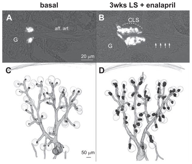
Figure 7.
Renin immunohistochemistry (white) in kidney sections of untreated Ren1d+/Cre–NO-sensitive guanylate cyclase (NO-GC)fl/ fl mice (A) and Ren1d+/Cre–NO-GCfl/fl mice treated with low-salt (LS) diet+enalapril for 3 weeks (B). aff. art. indicates afferent arteriole; CLS, renin cells forming cuff-like structures; and G, glomeruli. Arrows indicate afferent arteriole. 3D-reconstruction of α-smooth muscle actin (α-SMA; grey) and of renin-immunoreactive areas (black) in kidneys of mice lacking endothelial isoform of nitric oxide synthase (eNOS−/−) before (C) and after receiving a LS diet+enalapril for 3 weeks (D).

