Abstract
Purpose
To evaluate the repeatability of corneal epithelial thickness (ET) and corneal thickness (CT) measurements in normal and LASIK eyes using the optical coherence tomography (OCT, RTVue).
Methods
In 35 right eyes of 35 normal subjects and 45 right eyes of 45 subjects with prior myopic LASIK, corneal ET and CT were evaluated in 17 areas: 1) one central zone within 0-2.0 mm diameter, 2) eight paracentral zones from 2.0-5.0 mm diameter, and 3) eight peripheral zones from 5.0-6.0 mm diameter. The repeatability was assessed using within-subject standard deviation (SD), coefficient of variation (CoV), and intraclass correlation coefficient (ICC).
Results
At the central and paracentral zones, respectively, the SD values were 0.7 μm and 0.6-0.9 μm in normal eyes and 0.7 μm and 0.8-1.7 μm in LASIK eyes for ET, and 1.0 μm and 2.8-4.6 μm in normal eyes and 1.3 μm and 4.0-4.8 μm in LASIK eyes for CT. At the peripheral zones, in normal and LASIK eyes, respectively, SD values ranged from 0.8-1.2 μm and 1.4-2.2 μm for ET, and 4.1-6.4 μm and 6.0-9.1 μm for CT. The CoV values were low and ICC values were high in both groups for both ET and CT measurements.
Conclusion
The OCT produced excellent repeatability, especially at the central and paracentral zones up to 5-mm diameter for both corneal epithelial thickness and corneal thickness measurements.
Keywords: corneal epithelial thickness, optical coherence tomography, repeatability, normal and Post-LASIK/PRK eyes.
Introduction
The corneal epithelium is a critical element of the optical system of the eye. Corneal epithelial thickness can change in response to underlying corneal stroma changes to reduce corneal irregularity in response to corneal surgery or injury. Accurate and reproducible measurements of the corneal epithelial thickness (ET) provide important information for detecting early changes of keratoconus, assessing corneal remodeling changes after corneal refractive surgery, and monitoring development of corneal ectasia.1-2 Confocal microscopy and very high-frequency ultrasound have been used to map the corneal epithelium and stromal thickness.3-6 Optical coherence tomography (OCT) is a non-invasive powerful imaging technique that can produce cross-sectional images of the anterior and posterior segments of the eye.
Fourier-domain OCT has a higher speed, lower acquisition time and a higher signal-to-noise ratio compared to conventional time-domain OCT. It has been shown that the Fourier domain-based anterior segment OCT has a better reliability than time-domain OCT.7 In normal subjects, Vidal et al 8 reported that the repeatability and reproducibility of corneal ET measurements was poor using the Fourier-domain SOCT Copernicus HR system, and Prakash and colleagues 9 found reliable and reproducible measurements of corneal ET at vertex using the Fourier-domain based Cirrus HD-OCT. The purpose of our study was to evaluate in normal and post-LASIK corneas the repeatability of corneal ET measurements made by the Fourier-domain based RTVue system (Optovue, Inc., Fremont, CA).
Subjects and Methods
Subjects
Institutional Review Board approval was obtained for this study. Retrospectively, consecutive cases with three OCT scans at single visit between January 2011 and December 2012 were reviewed. Two groups of subjects were included: 1) normal subjects with virgin corneas and no history of corneal diseases (normal eyes), these were subjects who were scheduled to undergo corneal refractive surgery or cataract surgery; and 2) subjects who had undergone myopic LASIK (LASIK eyes) and presented for preoperative examination for cataract surgery. Inclusion criteria were no contact lens wear within 2 weeks of the OCT measurements and three OCT scans centered around the pupil.
In normal eyes, 35 right eyes of 35 subjects with mean age of 41 ± 15 years (range 21-75 years) were included. Of the 35 subjects, 16 were male and 19, female. In LASIK eyes, 45 right eyes of 45 subjects with prior myopic LASIK were enrolled. The mean age was 62 ± 8 years (range 42-78 years), and 27 were male and 189 were female. Subjects had LASIK 2-14 years ago and 18 eyes had historical data available with average amount of refractive correction of 4.36 ± 2.83 D (range 0.88 – 11.13D).
OCT measurements
We used the RTVue Fourier-domain OCT system with a cornea-anterior module long (CAM-L) adaptor lens. This device has a scan speed of 26000 axial scans per second using wavelength of 830-nm. 10 A “Pachymetry + Cpwr (corneal power)” scan pattern centered at the pupil center was used to map the cornea. Each eye was scanned 3 times. Subjects were asked to sit back after each scan, and the device was realigned before each scan.
Scans were reprocessed using the RTVue epithelial software (Version 6.8.0.27). Data were collected from corneal epithelial map and corneal pachymetry map. Average values in 17 areas were obtained: 1) one central zone within 0-2.0 mm diameter, 2) eight paracentral zones from 2.0-5.0 mm diameter, and 3) eight peripheral zones from 5.0-6.0 mm diameter.
Data analyses
The repeatability was assessed using three parameters: 1) within-subject standard deviation (SD), which is the pooled SD of repeated measurements (Sw).11 , 2) the coefficient of variation (CoV in %), which is defined as the ratio of the SD of the repeated measurements to the mean; a lower CoV value indicates higher repeatability, and 3)the intraclass correlation coefficient (ICC), which is a measure of correlation or consistency of repeated measurements.; the ICC values range from 0-1, with 1 being perfect consistency.
Microsoft Office Excel 2007 and SPSS software (version 15.0, SPSS, Inc.) were used for statistical analysis. Data were first checked if they were normally distributed. Then, a student t-test was used to compare normally distributed values between normal eyes and LASIK eyes. An F-test was performed to compare the standard deviation values. The Bonferroni correction was used to adjust the multiple comparisons. A P value smaller than 0.05 was considered statistically significant.
Results
Corneal epithelial thickness
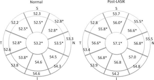
Compared to LASIK eyes, normal eyes had significantly smaller mean corneal ET at the center, 6 paracentral zones, and 1 peripheral zones (Figure 1) (all P<0.05).
Figure 1.

Average corneal epithelial thickness (μm) in normal and LASIK eyes (*significant difference between normal and LASIK eyes, all P<0.05 with Bonferroni correction)(S = superior, I = inferior, N = nasal, and T = temporal).
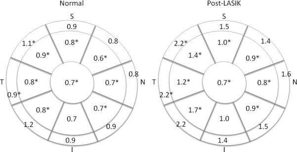
In normal and LASIK eyes, respectively, the SD values were 0.7 μm and 0.7 μm in the central zone, and 0.6 - 0.9 μm and 0.8 - 1.7 μm in paracentral zones (Figure 2); these values were significantly smaller in normal eyes than those in LASIK/PRK eyes, except the inferior zone (all P<0.05). In the peripheral zones, superior-temporal and temporal zones had smaller SD in normal eyes.
Figure 2.

Within-subject standard deviations (μm) of corneal epithelial thickness in normal and LASIK eyes (*significant difference between normal and LASIK eyes, all P<0.05 with Bonferroni correction)(S = superior, I = inferior, N = nasal, and T = temporal).
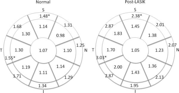
In the central and paracentral zones, the CoV values ranged from 0.98 - 1.30% in normal eyes, and 1.05 – 2.00% in LASIK eyes (Figure 3); there was no significant difference between the two groups. In the peripheral zones, superior and temporal zones had smaller CoV values in normal eyes.
Figure 3.

Coefficient of variance (%) of corneal epithelial thickness in normal and LASIK eyes (*significant difference between normal and LASIK eyes, all P<0.05 with Bonferroni correction)(S = superior, I = inferior, N = nasal, and T = temporal).
In the central and paracentral zones, the ICC values were ≥0.967 in normal eyes, and ≥0.957 in LASIK eyes (Supplemental Digital Content Figure 1). In the peripheral zones, the ICC values were ≥0.917 in normal eyes, and ≥0.891 in LASIK eyes. There were no significant differences between two groups.
Corneal thickness
Compared to LASIK eyes, normal eyes had significantly greater CT at the center and 4 zones at the paracentral zones (Figure 4) (all P<0.05).
Figure 4.

Average corneal thickness (μm) in normal and LASIK eyes (*significant difference between normal and LASIK eyes, all P<0.05 with Bonferroni correction)(S = superior, I = inferior, N = nasal, and T = temporal).
In normal and LASIK eyes, respectively, the SD values were 1.0 μm and 1.3 μm in the central zone, ≤4.6 μm and ≤4.8 μm in the paracentral zones, and ≤6.4 μm and ≤9.1 μm in the peripheral zones (Figure 5). There were no significant differences between the two groups.
Figure 5.

Within-subject standard deviations (μm) of corneal thickness in normal and LASIK eyes (*significant difference between normal and LASIK eyes, all P<0.05 with Bonferroni correction)(S = superior, I = inferior, N = nasal, and T = temporal).
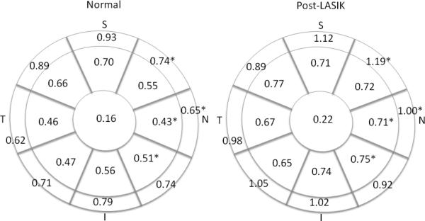
In the central and paracentral zones, the CoV values ranged from 0.16 - 0.70% in normal eyes, and 0.22 – 0.77% in LASIK eyes (Figure 6); there were significant differences in nasal and inferior-nasal zones. In the peripheral zones, nasal and superior-nasal zones had smaller CoV values in normal eyes.
Figure 6.

Coefficient of variance (%) of corneal thickness in normal and LASIK eyes (*significant difference between normal and LASIK eyes, all P<0.05 with Bonferroni correction)(S = superior, I = inferior, N = nasal, and T = temporal).
In all zones, the ICC values were ≥0.989 in normal eyes, and ≥0.983 in LASIK eyes (Supplemental Digital Content Figure 2). There were no significant differences between the two groups.
Discussion
OCT is a non-invasive technique for corneal epithelium measurements. There were a few studies in the literature that evaluated the reproducibility of central corneal ET measurements. 8-9, 12-13 In this study, we evaluated the repeatability of corneal ET and CT measurements at the center, paracentral and peripheral zones in normal and LASIK eyes using the RTVue.
For corneal ET measurements, the SD values were less than 1 μm at the central and paracentral zones in normal eyes. In LASIK eyes, the SD values were 0.7 μm at the central zone, and ≤1.7 μm in the paracentral zones. Although the SD values in normal eyes were significantly lower at the central and paracentral zones than those in LASIK eyes, the differences were not clinically significant. The CoV values were low and ICC values were high in both groups, indicating excellent repeatability.
Using a time-domain OCT (Zeiss OCT imager), for central corneal ET measurements in 18 normal subjects, Sin and Simpson13 reported ICC values of ≤ 0.73. Using the Fourier domain based device SOCT Copernicus HR, in 30 healthy eyes, Vidal et al8 found that repeatability for corneal epithelial measurements was poor with ICC value of 0.391. In contrast, the ICC values for central corneal ET found in current study using the RTVue were >0.98.
In the study investigating the role of the axial resolution of OCT on the measurements of corneal and epithelial thickness using 4 devices, Ge and colleagues 12 reported the repeatability of central CT and ET in 20 healthy subjects and 18 patients with prior LASIK. Using the RTVue, they found that the ICC values for the central ET (0-2 mm) were 0.74 -0.89 in healthy eyes and 0.84-0.88 in LASIK eyes, which were lower than the ICC values of 0.985 in normal eyes and 0.995 in LASIK eyes found in our study. One difference between these two studies is the software used to measure the corneal ET. In their study, they developed their own algorithm for segmenting the layers of the corneal images obtained from the RTVue, and we used the software developed for the RTVue by the manufacturer.
We also found excellent repeatability of corneal ET measurements at the paracentral and peripheral zones. Our results showed that, at 5-6 mm diameter zones, the SD of corneal ET was ≤1.2 μm in normal eyes and 1.3 μ in LASIK eyes, and ICC values of 0.99 in both groups. THese findings were in consistent with reports in the literature using various OCT devices. 7-8,10,14-21
Limitation of the study includes: 1) we did not evaluate the repeatability of corneal epithelial thickness measurements in PRK eyes, and the corneal epithelial responses may be different following LASIK and PRK; and 2)we included patients who underwent LASIK 2-14 years ago, and we assume that the corneal remodeling is stable after 2 years of the surgery. However, the purpose of this study was not to assess the long-term epithelial remodeling postoperatively; instead, using OCT, we wanted to evaluate the repeatability of corneal epithelial thickness measurements in post-LASIK eyes.
In summary, the Fourier-domain based RTVue device produced excellent corneal epithelial thickness and corneal thickness measurements at the central and paracentral zones up to 6-mm diameter.
Supplementary Material
Figure 1: Intraclass correlation coefficient of corneal epithelial thickness in normal and LASIK eyes (S = superior, I = inferior, N = nasal, and T = temporal).
Figure 2: Intraclass correlation coefficient of corneal thickness in normal and LASIK eyes (S = superior, I = inferior, N = nasal, and T = temporal).
Acknowledgments
Source of Funding: Supported in part by an unrestricted grant from Research to Prevent Blindness, New York, NY.
Footnotes
Conflict of Interest:
The authors received research support from Ziemer (Koch, Wang). In addition, Dr. Koch has a financial interest with Alcon Laboratories, Abbott Medical Optics, Optimedica, Revision Optics and Ziemer.
References
- 1.Li Y, Tan O, Brass R, et al. Corneal epithelial thickness mapping by fourier-domain optical coherence tomography in normal and keratoconic eyes. Ophthalmology. 2012;119:2425–33. doi: 10.1016/j.ophtha.2012.06.023. [DOI] [PMC free article] [PubMed] [Google Scholar]
- 2.Rocha KM, Perez-Straziota E, Stulting RD, et al. SD-OCT analysis of regional epithelial thickness profiles in keratoconus, postoperative corneal ectasia, and normal eyes. J Refract Surg. 2013;29:173–9. doi: 10.3928/1081597X-20130129-08. [DOI] [PMC free article] [PubMed] [Google Scholar]
- 3.Li HF, Petroll WM, Møller-Pedersen T, et al. Epithelial and corneal thickness measurements by in vivo confocal microscopy through focusing (CMTF). Curr Eye Res. 1997;16:214–21. doi: 10.1076/ceyr.16.3.214.15412. [DOI] [PubMed] [Google Scholar]
- 4.Erie JC, Patel SV, McLaren JW, et al. Effect of myopic laser in situ keratomileusis on epithelial and stromal thickness: a confocal microscopy study. Ophthalmology. 2002;109:1447–52. doi: 10.1016/s0161-6420(02)01106-5. [DOI] [PubMed] [Google Scholar]
- 5.Reinstein DZ, Archer TJ, Gobbe M, et al. Epithelial thickness in the normal cornea: three-dimensional display with Artemis very high-frequency digital ultrasound. J Refract Surg. 2008;24:571–81. doi: 10.3928/1081597X-20080601-05. [DOI] [PMC free article] [PubMed] [Google Scholar]
- 6.Reinstein DZ, Silverman RH, Raevsky T, et al. Arc-scanning very high-frequency digital ultrasound for 3D pachymetric mapping of the corneal epithelium and stroma in laser in situ keratomileusis. J Refract Surg. 2000;16:414–30. doi: 10.3928/1081-597X-20000701-04. [DOI] [PubMed] [Google Scholar]
- 7.Prakash G, Agarwal A, Jacob S, et al. Comparison of fourier-domain and time-domain optical coherence tomography for assessment of corneal thickness and intersession repeatability. Am J Ophthalmol. 2009;148:282–290. 11. doi: 10.1016/j.ajo.2009.03.012. [DOI] [PubMed] [Google Scholar]
- 8.Vidal S, Viqueira V, Mas D, et al. Repeatability and reproducibility of corneal thickness using SOCT Copernicus HR. Clin Exp Optom. 2013 Jan 14; doi: 10.1111/cxo.12002. doi: 10.1111/cxo.12002. [Epub ahead of print] [DOI] [PubMed] [Google Scholar]
- 9.Prakash G, Agarwal A, Mazhari AI, et al. Reliability and reproducibility of assessment of corneal epithelial thickness by fourier domain optical coherence tomography. Invest Ophthalmol Vis Sci. 2012;53:2580–5. doi: 10.1167/iovs.11-8981. [DOI] [PubMed] [Google Scholar]
- 10.Li Y, Tang ML, Zhang X, et al. Pachymetric mapping with Fourier-domain optical coherence tomography. J Cataract Refract Surg. 2010;36:826–31. doi: 10.1016/j.jcrs.2009.11.016. [DOI] [PMC free article] [PubMed] [Google Scholar]
- 11.Bland JM, Altman DG. Statistical notes: measurement error. BMJ. 1996;313:744. doi: 10.1136/bmj.313.7059.744. [DOI] [PMC free article] [PubMed] [Google Scholar]
- 12.Ge L, Yuan Y, Shen M, et al. The role of axial resolution of optical coherence tomography on the measurement of corneal and epithelial thicknesses. Invest Ophthalmol Vis Sci. 2013;54:746–55. doi: 10.1167/iovs.11-9308. [DOI] [PubMed] [Google Scholar]
- 13.Sin S, Simpson TL. The repeatability of corneal and corneal epithelial thickness measurements using optical coherence tomography. Optom Vis Sci. 2006;83:360–5. doi: 10.1097/01.opx.0000221388.26031.23. [DOI] [PubMed] [Google Scholar]
- 14.Kim E, Ehrmann K. Assessment of accuracy and repeatability of anterior segmentoptical coherence tomography and reproducibility of measurements using a customised software program. Clin Exp Optom. 2012;95:432–41. doi: 10.1111/j.1444-0938.2012.00724.x. [DOI] [PubMed] [Google Scholar]
- 15.Neri A, Malori M, Scaroni P, et al. Corneal thickness mapping by 3D swept-source anterior segment optical coherence tomography. Acta Ophthalmol. 2012;90:e452–7. doi: 10.1111/j.1755-3768.2012.02453.x. [DOI] [PubMed] [Google Scholar]
- 16.Muscat S, McKay N, Parks S, et al. Repeatability and reproducibility of corneal thickness measurements by optical coherence tomography. Invest Ophthalmol Vis Sci. 2002;43:1791–5. 12. [PubMed] [Google Scholar]
- 17.Hong JP, Nam SM, Kim TI, et al. Reliability of RTVue, Visante, and slit-lamp adapted ultrasonicpachymetry for central corneal thickness measurement. Yonsei Med J. 2012;53:634–41. doi: 10.3349/ymj.2012.53.3.634. [DOI] [PMC free article] [PubMed] [Google Scholar]
- 18.Correa-Pérez ME, López-Miguel A, Miranda-Anta S, et al. Precision of high definition spectral-domain optical coherence tomography for measuring central corneal thickness. Invest Ophthalmol Vis Sci. 53:1752–7. doi: 10.1167/iovs.11-9033. 20126. [DOI] [PubMed] [Google Scholar]
- 19.Huang JY, Pekmezci M, Yaplee S, et al. Intra-examiner repeatability and agreement of corneal pachymetry map measurement by time-domain and Fourier-domain optical coherence tomography. Graefes Arch Clin Exp Ophthalmol. 2010;248:1647–56. doi: 10.1007/s00417-010-1360-7. [DOI] [PubMed] [Google Scholar]
- 20.Mohamed S, Lee GK, Rao SK, et al. Repeatability and reproducibility of pachymetric mapping with Visante anterior segment-optical coherence tomography. Invest Ophthalmol Vis Sci. 2007;48:5499–504. doi: 10.1167/iovs.07-0591. [DOI] [PubMed] [Google Scholar]
- 21.Fukuda S, Kawana K, Yasuno Y, et al. Repeatability and reproducibility of anterior ocular biometric measurements with 2-dimensional and 3-dimensional optical coherence tomography. J Cataract Refract Surg. 2010;36:1867–73. doi: 10.1016/j.jcrs.2010.05.024. [DOI] [PubMed] [Google Scholar]
Associated Data
This section collects any data citations, data availability statements, or supplementary materials included in this article.
Supplementary Materials
Figure 1: Intraclass correlation coefficient of corneal epithelial thickness in normal and LASIK eyes (S = superior, I = inferior, N = nasal, and T = temporal).
Figure 2: Intraclass correlation coefficient of corneal thickness in normal and LASIK eyes (S = superior, I = inferior, N = nasal, and T = temporal).


