Abstract
Background
Effects of acupuncture stimulation on blood glucose concentration and body weight were investigated in the Otsuka Long-Evans Tokushima Fatty (OLETF) rat, a model for type-2 diabetes.
Material/Methods
Three groups of rats were used: OLETF, acupuncture-treated OLETF (AcOLETF), and Long-Evans Tokushima Otsuka (LETO) rats (as control for the OLETF rats). In AcOLETF rats, acupuncture stimulation was applied twice a week to 6 points (zhongwan, tianshu, qihai, ganshu, pishu, shenshu) and changes in blood glucose concentration and body weight were measured.
Results
Initially, at 6 weeks old, there was no significant difference in blood glucose levels between groups. Blood glucose levels increased with age in each group, reaching a maximum of about 430 mg/dl at 37 weeks in OLETF rats. In AcOLETF rats, blood glucose levels increased at a slower rate than in OLETF rats, reaching a maximum concentration of about 280 mg/dl at 37 weeks of age, significantly lower than that in OLETF rats. The concentration of blood glucose in LETO rats had stabilized at a maximum value of 120~140 mg/dl by 16 weeks, remaining at this level for up to 39 weeks. In each group, body weight increased with age and was not affected by acupuncture treatment.
Conclusions
In OLETF rats, acupuncture treatment significantly reduced blood glucose levels, but not their body weight, suggesting that acupuncture therapy was effective in preventing the development of type-2 diabetes mellitus.
MeSH Keywords: Acupuncture, Body Weight Changes, Diabetes Mellitus, Type 2, Hyperglycemia, Rats, Inbred OLETF
Background
There are 2 types of diabetes mellitus, type-1 and type-2, with the former being produced by the decreased production of insulin due to disorders of the pancreatic β-cells, while the latter is caused by the disorder of the regulation of blood glucose concentrations due to reduced release of insulin and also decreased sensitivity to insulin [1]. The majority of type-2 diabetic cases mainly result from a disorder of the metabolism of carbohydrate and fat [1]. The development of type-2 diabetes mellitus is causally related to daily life activities, and therefore an important factor in its treatment is a significant modification of daily life style [1]. However, strong regulation of daily food intake will produce a reduced quality of life and personal comfort.
Attempts have been made to apply acupuncture therapy for the treatment of diabetes mellitus, and the effectiveness of acupuncture treatment on the reduction of serum glucose levels has been reported in patients with either type-1 diabetes [2–4] or type-2 diabetes [5,6]. The possibility of using acupuncture therapy for the treatment of diabetes mellitus has been considered, but it has been suggested that the application of standard treatment to all diabetic patients requires special care, because the stages and conditions of individual patients are heterogeneous [7]. The development of new clinical methods for the treatment of diabetes mellitus with acupuncture therapy, therefore, requires research using different types of experimental animal models. Several experiments have been reported concerning the effects of acupuncture treatment on hyperglycemic animal models. For example, treatment with acupuncture is effective in reducing serum glucose in type-1 diabetes mellitus animal models such as alloxan-induced diabetic mice [8] and streptozotocin-induced diabetic rats [9].
The Otsuka Long-Evans Tokushima Fatty (OLETF) rat was developed as a diabetic animal model with associated fat and hyperglycemia, and is useful as an animal model for type-2 diabetes mellitus because their pathological condition is similar to that in humans with type-2 diabetes [10–12]. In OLETF rats, using the hyperinsulinemic euglycemic clamp method, Kato et al. [13] showed that the application of electrical stimulation to the region of the auricle and ventral parts of the body that are innervated by the vagus nerve successfully prevented the formation of insulin resistance. Experiments using the glucose clamp method showed that electrical stimulation to corresponding acupoints in the ear increased insulin sensitivity in OLETF rats [7]. However, there have been no experimental reports of the effects of classic acupuncture treatment on animal models for type-2 diabetes mellitus. Thus, we investigated the effects of acupuncture treatment on OLETF rats to advance understanding of the usefulness of acupuncture therapy in the treatment of type-2 diabetes mellitus in humans.
Material and Methods
Animals
Male OLETF rats, aged 5 weeks, were used as the hyperglycemic experimental animal model, with age-matched male Long-Evans Tokushima Otsuka (LETO) rats [10–12] as their control. All animals were bred in the Experimental Animal Center of the Nagoya City University Medical School, in a room maintained at a temperature of 23±1°C, with humidity at 50±10% and a light–dark cycle of 12–12 hours. Water and food (standard rat chow, Oriental Co. Ltd., Japan) could be freely accessed by all animals. All animals were treated ethically according to the Guidelines for the Use of Experimental Animals approved by the Nagoya City University, and the present experiments were undertaken with the approval of the Experimental Animal Committee of Nagoya City University Medical School.
Experimental protocols
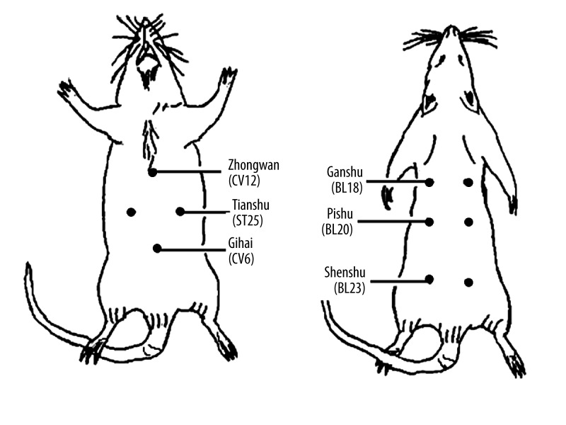
Rats were divided into 3 groups: OLETF rats with no acupuncture treatment (OLETF rats, 18 rats), OLETF rats treated with acupuncture (AcOLETF rats, 12 rats), and the control LETO rats (10 rats). Acupuncture treatment used a single light touch to pass a 15-mm No. 24 stainless steel needle through the skin adjacent to the surface of the muscle. Animals were held in 1 hand, with either the dorsal side or ventral side up, and the acupuncture was performed with the other hand, without anesthetic treatment. It required 5–10 s to apply a single needle at each acupoint. Acupuncture stimulation was applied to the following 6 points: the zhongwan (CV12), tianshu (ST25), qihai (CV6), ganshu (BL18), pishu (BL20), and shenshu points (BL23); the first 3 points are distributed on the ventral side and the latter 3 points distributed on the dorsal side of the body [9,14] (Figure 1). Zhongwan and qihai points were single points, while the others were bilateral paired points. Acupuncture stimulations were applied 2 times a week (Monday and Thursday), from 6 weeks of age to up to 39 weeks of age.
Figure 1.
Distribution of acupoints in the rat. In 6 acupoints, 3 points distributed on the ventral side (zhongwan, tianshu, and qihai points) are shown in the left-hand side, and 3 points distributed on the dorsal side (ganshu, pishu, and shenshu points) are shown in the right-hand side (each point is shown by a filled-in circle).
Every 4 weeks (sometimes, 9 weeks), rats were fasted for 20 h. Each rat was then immobilized by pushing it into a small cage made of metal mesh and the tail vein was cut for bleeding. The blood glucose concentration in the fasting state was measured using a simple blood glucose measurement kit (Rosh Diagnostic Co. Ltd., Switzerland), utilizing the glucose dehydrogenase method [15]. Blood glucose concentrations are expressed as mg/dl blood. The body weights of rat were also measured weekly, using a conventional electric balance (FP-12K, A & D, Co. Ltd., Tokyo).
Statistics
The measured values are expressed as the mean ± standard error of the mean (SEM). Statistical analysis of data was performed using 1-way analysis of variance (ANOVA), and post-hoc comparisons were made using the Bonferroni test. Statistical significance for the comparison was determined by the probability of less than 5% (P<0.05).
Results
Effects of acupuncture treatment on blood glucose concentration
The fasted blood glucose concentration of each rat was measured at 4-week intervals (some at 9-week intervals), by taking blood from the tail vein. At the start of the investigation, when the rats were 6 weeks old, the mean value of the blood glucose concentration was 63.2±5.6 mg/dl (n=18) for OLETF rats, 76.0±10.2 mg/dl (n=12) for AcOLETF rats, and 61.7±12.3 mg/dl (n=10) for LETO rats. These values were not significantly different from each other (P>0.05).
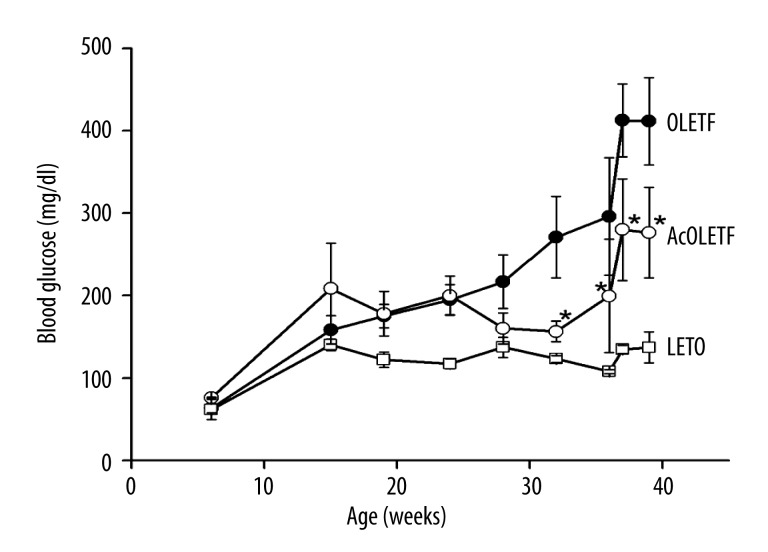
The change in blood glucose concentrations measured between 6 and 39 weeks of age (Figure 2) showed that it increased progressively with age, with a significant elevation compared to the initial value occurring at 16 weeks old, in each group of rats. In OLETF rats, the blood glucose concentration was further elevated with age, reaching a nearly maximum value of about 430 mg/dl at 37 weeks of age. In AcOLETF rats, the concentration of blood glucose was significantly elevated at 16 weeks old, but remained stable at around 200 mg/dl in the period between 16 and 36 weeks of age, and then began to rise again to reach the nearly maximum value of about 280 mg/dl at 37 weeks of age. Comparison of the blood glucose concentrations measured at 32–39 weeks of age indicated that each weekly value was significantly lower in AcOLETF rats than in OLETF rats (P<0.05). The concentration of blood glucose in LETO rats reached the maximum value of around 120~140 mg/dl at 16 weeks of age, and remained stable for up to 39 weeks of age. The blood glucose level in LETO rats were significantly lower than either OLETF or AcOLETF rats at any given week between 16 and 39 weeks of age (P<0.05).
Figure 2.
Changes in blood glucose concentrations in rats. Changes in blood glucose concentrations (mg/dl) are shown as a function of the age (weeks) of rats. The values are shown by the mean ±SEM (n=10–18). ●, OLETF rat; ○, AcOLETF rat; □, LETO rat. *, significant from OLETF rats (P<0.05).
These results indicate that the blood glucose concentration was significantly elevated in OLETF rats compared to LETO rats, and that the application of acupuncture treatment to the AcOLETF rats significantly delayed the elevation of blood glucose concentration with age, and significantly reduced the maximum level of blood glucose concentration in OLETF rats. However, the results also indicated that the acupuncture treatment did not reduce the level of blood glucose concentration of OLETF rats to that of LETO rats. Thus, acupuncture treatment was found to be effective in preventing the elevation of blood glucose concentration observed in OLETF rats.
Changes in body weight
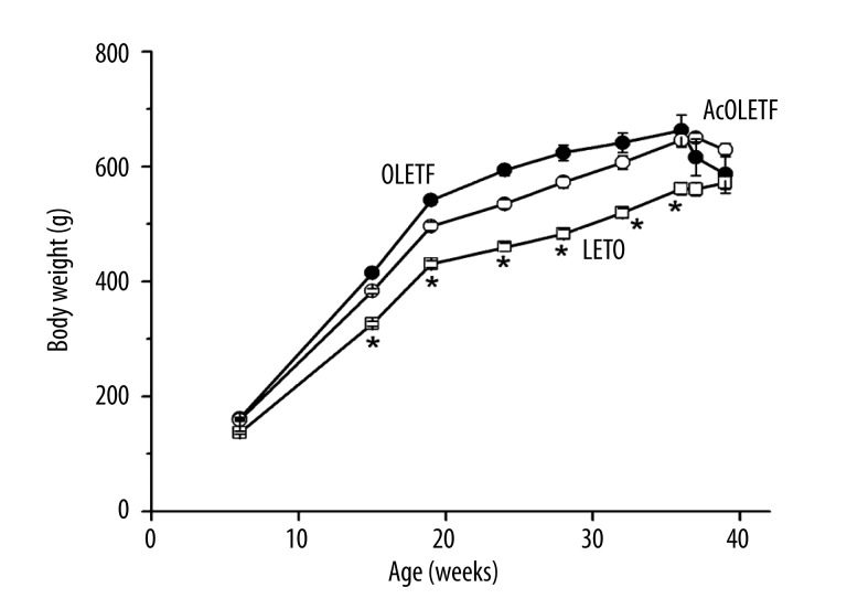
Body weights were measured every week, in all rats aged between 6 and 39 weeks old, and age-dependent changes in the mean value are summarized in Figure 3. At 6 weeks old, the average weight of each group of rats was 161.8±1.5 g (n=18) for OLETF rats, 159.6±2.8 g (n=12) for AcOLETF rats, and 137.1±2.4 g (n=10) for LETO rats. These values were not significantly different from each other (P>0.05). The body weight increased with age in all groups of rat, at a similar rate, and reached the maximum value of about 600~650 g at around 32 weeks old. The mean value of the body weight at 32 weeks old was 641.4±17.0 g (n=15) for OLETF rats, 607.2±11.6 g (n=11) for AcOLETF rats, and 519.3±7.9 g (n=10) for LETO rats. Thus, the body weight increased by more than 5 times during 30 weeks in all groups of rats. There was no significant difference in body weight between them at any given age (P>0.05). The body weight was significantly lower in LETO rats than in OLETF rats at all stages between 15 and 36 weeks of age.
Figure 3.
Changes in body weight of rats. Changes in body weight (g) are shown against the age (weeks) of rats. The values are shown by the mean ±SEM (n=10–18).●, OLETF rat; ○, AcOLETF rat; □, LETO rat. *, significant from both OLETF and AcOLETF rats (P<0.05).
Changes in physical conditions
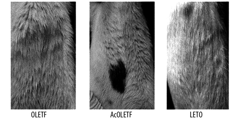
During the measurements of blood glucose concentration and body weight, there were some differences in the condition of the fur between the 3 groups of rats. During the experiments, the fur of LETO rats had a healthy condition and was shiny and well arranged. The fur of OLETF rats, however, was rough and lacked sheen. In acupuncture-treated AcOLETF rats, their fur was much brighter than in OLETF rats, but it was still less than that of LETO rats. A typical example of the abdominal fur obtained from rats at 32 weeks old is shown in Figure 4. Although these observations were not evaluated by scores, the difference between rat groups became more evident with age.
Figure 4.
Condition of the abdominal fur. Typical views of the abdominal fur of rats (ventral view) are shown in OLETF (left panel), AcOLETF (central panel), and LETO (right panel) rats. All panels were obtained from rats aged at 32 weeks old.
A difference between LETO and OLETF rats was also noted in urine production, although no successful quantification was made – the amount of urine was significantly increased in OLETF rats compared to LETO rats after 25 weeks of age. The rate and amount of urination were both nearly constant for LETO rats up to 39 weeks old. No detectable difference in urination was found between OLETF and AcOLETF rats.
Discussion
The present experiments revealed that in OLETF rats, an animal model for the type-2 diabetes mellitus, acupuncture treatment effectively inhibited the elevation of blood glucose concentration and also reduced the maximum concentration of blood glucose. The increase in the body weight associated with aging, however, could not be prevented by the acupuncture treatment. Furthermore, application of acupuncture stimulation improved the brightness of their fur, which was badly damaged during the development of diabetes mellitus. The condition of their hair coat may be a good indicator of the metabolic condition of their bodies, and the successful maintenance of the brightness of the fur in AcOLETF rats compared to OLETF rats suggests that the acupuncture treatment is effective in preventing a reduction in metabolism during the development of diabetes mellitus. Thus, the results strongly suggest that the application of acupuncture treatment twice weekly is effective in preventing the development of type-2 diabetes mellitus.
Clinically, 5 acupoints are generally selected for the acupuncture therapy for diabetes mellitus: the zhongwan point (CV12), tianshu point (ST25), qihai point (CV6), ganshu point (BL18), and pishu point (BL20). Acupuncture stimulation to these points is effective in elevating the activation of integrative mechanisms in the whole body (Kurono methods) [14]. In the present experiments, the shenshu point (BL23), which is known to enhance the transportation of body energy to the damaged place [3], was added to the acupoints selected by the Kurono methods [14], and our results indicated that the acupuncture stimulation to these 6 points was indeed effective in preventing hyperglycemic development in OLETF rats.
The acupoints employed in the present experiment for the treatment of hyperglycemia have been considered to facilitate the removal of unhealthy elements from the body, and thus increasing recovery from the diseased condition [14]. The results indicated that these 6 acupoints were effective in reducing the elevation of the concentration of blood glucose in OLETF rats, although it remain unclear how these points had any causal relation to the reduction of blood glucose concentrations. Application of acupuncture treatment to these 6 points was effective in the reduction of the blood glucose concentrations in rats that had developed type-1 diabetes as a result of streptozotocin treatment [9]. It seems likely that stimulation of these 6 points is effective in reducing blood glucose concentrations in both type-1 and type-2 diabetic animals. However, it remains unclear how these 6 points are comparable to those used in treatment of the human body, since the size and physical condition are not directly comparable between OLETF rats and humans.
Experiments using the glucose clamp method indicated that in male OLETF rats electric acupuncture stimulation applied to appropriate regions of the ears that affect the vagus nerves, particularly those that innervate both the auricle and the back of the body, protected against the development of insulin-resistant diabetes mellitus [13]. The electrical stimulation of acupoints distributed in the ear for a long period of time could also increase insulin resistance in OLETF rats [7]. The effectiveness of electrical acupuncture to the abdomen was shown to have a hypoglycemic effect on streptozotocin-induced type-1 diabetic rats [16]. In the present experiments, acupuncture with rather light stimuli was applied to the skin and under-skin levels using fine pins, and these stimuli were considered to produce more prolonged and sustained effects in comparison with electrical stimulation [14], and this produced a contrast to the electrical acupuncture stimulation that produced rather strong and acute effects in the body. Considering that diabetes mellitus is a slow-developing disease [1,7,14], application of weak and light stimuli with minimum intensity to effective points for a long period of time may be important, since these treatments will reduce the clinical burden on patients.
Application of acupuncture stimulation to points that are considered to influence the activity of the entire body, such as the zhongwan, tianshu, shenshu, ganshu, pishu, quchi, sanyinjiao, zusanli, and feishu points, is effective in increasing the secretion of insulin [5,17–19], and some of these points or functionally similar points distributed were also included in the present experiments. Diabetes mellitus induces a disorder of many functions in the body [1,7,14], and it is also reasonable to consider application of acupuncture treatment to points distributed in the hands and feet, since stimulation of these points is experientially proven to elevate the activity of the whole body [14]. The number of acupoints used in the treatment of diabetes mellitus in animal models is much smaller than the number of acupoints applied to humans, and the life styles (e.g., active times and food habits) are different between rats and humans. These differences may be also involved in the weaker effectiveness of acupuncture treatment in experimental animals. In some experiments, acupuncture treatment was applied while the activity of the animal had been inhibited by anesthetics [16], and this was also considered to weaken the effectiveness of acupuncture treatment in diabetic animal models. Although the present experiments were carried out carefully in the absence of anesthesia, in general the results obtained in laboratory experiments may not be directly applicable to humans. However, the genetic mechanism of the development of hyperglycemia is comparable between OLETF rats and humans [10–12]. Therefore, the results obtained in the present experiments may have significant implications for considering the application of acupuncture therapy in the treatment of diabetes mellitus, as discussed elsewhere [20].
The activity of vagus nerves distributed in smooth-muscle tissue isolated from the stomach remains enhanced in AcOLETF rats compared to OLETF rats, even several weeks after termination of the acupuncture treatment [21], and these results may suggest that acupuncture stimulation produces a sustained enhancement of the functional activity of autonomic nerves. Significant attenuation of neuromuscular transmission and disorder of the pacemaker mechanism are also found in the stomach muscle of OLETF rats [22]. It is generally considered that acupuncture therapy enhances the activity of autonomic control system of the body, including the enhanced activity of parasympathetic nerves [1,7,23]. In the present experiments, it is considered reasonable that the application of acupuncture stimulation facilitates the local circulation system and enhances glucose consumption in peripheral tissues, thus leading to reduced glucose levels in serum, and these effects may be comparable to those produced as a result of physical exercise.
Conclusions
Application of acupuncture stimulation to OLETF rats significantly reduced blood glucose concentrations, and the results suggest that this was an effective treatment for type-2 diabetes mellitus. Although the elevation of body weight was not significantly inhibited, the brightness and fineness of their hair coat were greatly improved by acupuncture treatment in AcOLETF rats.
Acknowledgements
The authors are grateful to Dr. John R. McLean, a Senior Associate of the University Melbourne, Australia, for his critical reading and useful comments to the manuscript. Experimental model animals, OLETF and LETO rats, were a gift from the Otsuka Pharmaceutical Co. Ltd., Japan. A part of the present experiments was supported by a Grant-in-Aid for Scientific Research from the Ministry of Education, Science, Technology and Sports of Japan to H.S. (21590237)
Footnotes
Source of support: Grant-in-Aid for Scientific Research from the Ministry of Education, Science, Technology and Sports of Japan (21590237)
References
- 1.Dey L, Attele AS, Yuan C-S. Alternative therapies for type 2 diabetes. Alternative Med Rev. 2000;7:45–58. [PubMed] [Google Scholar]
- 2.Nakamura H, Kurono Y, Watari N. A case report concerning acupuncture therapy on a diabetic patient (1) J Jpn Soc Acupunct Moxibust. 1993;43:196–200. [in Japanese] [Google Scholar]
- 3.Nakamura H, Kinuta A, Kurono Y. A case of acupuncture therapy for diabetes: a patient with hypoglycemia due to insulin treatment. J Jpn Soc Acupunct Moxibust. 1999;49:305–31. [in Japanese] [Google Scholar]
- 4.Yamada A, Nakamura H, Mizuno T, Kurono Y. Acupuncture treatment in patients with diabetes mellitus. J Jpn Soc Acupunct Moxibust. 2001;55:170–74. [in Japanese] [Google Scholar]
- 5.Kiyofuji S, Kajiyama S, Egawa M, et al. The effect of acupuncture stimulation to the bilateral Quchi points on insulin secretion. J Jpn Soc Acupunct Moxibust. 1990;40:292–98. [in Japanese with English abstract] [Google Scholar]
- 6.Wakuda T, Tanno Y. Effects of acupuncture therapy on diabetes mellitus – variation of blood sugar levels of insulin-dependent patient. J Jpn Soc Acupunct Moxibust. 2002;52:43–48. [in Japanese with English abstract] [Google Scholar]
- 7.Ishizaki N, Yano T, Yamamura Y. MIBYO-CHI: promotion of self-healing ability – application of acupuncture and moxibustion as prevention of diabetes mellitus. J Jpn Soc Acupunct Moxibust. 2003;53:159–66. [in Japanese] [Google Scholar]
- 8.Watari N, Kurono Y, Mabuchi Y. Effects of acupuncture treatment on the ultrastructure of pancreatic cells in alloxan-induced diabetic mice. Jpn J Endoclinol. 1978;34:425. [in Japanese] [Google Scholar]
- 9.Nakamura H, Kurono Y, Ishigami T, et al. Effects of acupuncture treatment on blood glucose concentrations and body weight in the streptozotocin-induced diabetic rats. J Jpn Soc Acupunct Moxibust. 1996;46:80–84. [in Japanese with English abstract] [Google Scholar]
- 10.Kawano K, Hirashima T, Mori S, et al. Spontaneous long term hyperglycemic rat with diabetic complications: Otsuka Long-Evans Tokushima Fatty (OLETF) strain. Diabetes. 1992;41:1422–28. doi: 10.2337/diab.41.11.1422. [DOI] [PubMed] [Google Scholar]
- 11.Mori Y, Yokoyama J, Nemoto M, et al. Characterization of a new genetically obese-hyperglycemic OLETF rat. Jikei Med J. 1992;39:349–59. [Google Scholar]
- 12.Kawano K, Hirashima T, Mori S, Natori T. OLETF (Otsuka Long-Evans Tokushima Fatty) rat: a new NIDDM rat strain. Diabetes Res Clin Pract. 1994;24:S317–20. doi: 10.1016/0168-8227(94)90269-0. [DOI] [PubMed] [Google Scholar]
- 13.Kato M, Fukuda F, Ishizaki N, et al. The effect of electro-acupuncture stimulation on insulin resistance in diabetic rats. J Orient Med. 1999;50:439–49. [in Japanese with English abstract] [Google Scholar]
- 14.Kurono Y. General textbook of acupuncture. Tokyo, Japan: FA Press; 1996. [Google Scholar]
- 15.Maguire GA, Price CP. Kinetic glucose dehydrogenase method for glucose measurement with a discrete kinetic analyzer overcomes interference by ascorbate. Clin Chem. 1984;30:157–58. [PubMed] [Google Scholar]
- 16.Shapira MY, Appelbaum EY, Hishberg B, et al. A sustained, non-insulin related, hypoglycaemic effect of electroacupuncture in diabetic Psammomys obesus. Diabetologia. 2000;43:809–13. doi: 10.1007/s001250051379. [DOI] [PubMed] [Google Scholar]
- 17.Hasegawa H, Terasawa S, Kumamoto Y, Odawara Y. Acupuncture therapy for diabetes mellitus: (3) Effect of acupuncture stimulus on the relationship between serum insulin and meridian point in normal subject. J Jpn Soc Acupunct Moxibust. 1981;31:55–59. [in Japanese with English abstract] [Google Scholar]
- 18.Hasegawa H, Terasawa S, Yuasa T, Yamagiwa Y, Odahara Y. Acupuncture therapy for diabetes mellitus: (2) Changes of serum insulin and blood sugar in patient with diabetes mellitus by acupuncture therapy. J Jpn Soc Acupunct Moxibust. 1981;30:147–52. (in Japanese with English abstract) [Google Scholar]
- 19.Kiyofuji S, Kajiyama S, Ishizaki N, et al. The effect of acupuncture stimulation on insulin secretion in rats using the bilateral Quchi (LI. 11) points. Meiji Acupunct Moxibust Med. 1990;7:43–48. [in Japanese with English summary] [Google Scholar]
- 20.Liang F, Kova D. Acupuncture: is it effective for treatment of insulin resistance? Diabetes Obes Metab. 2010;12:555–69. doi: 10.1111/j.1463-1326.2009.01192.x. [DOI] [PubMed] [Google Scholar]
- 21.Fukuta H, Koshita M, Nakamura E, et al. Acupuncture modulates mechanical responses of smooth muscle produced by transmural nerve stimulation in gastric antrum of genetically hyperglycemic rats. J Smooth Muscle Res. 2000;45:167–85. doi: 10.1540/jsmr.45.167. [DOI] [PubMed] [Google Scholar]
- 22.Takano H, Imaeda K, Koshita M, et al. Alteration of the properties of gastric smooth muscle in the genetically hyperglycemic OLETF rat. J Autonom Nerv Syst. 1998;70:180–88. doi: 10.1016/s0165-1838(98)00050-2. [DOI] [PubMed] [Google Scholar]
- 23.Kurono Y, Minagawa M, Ishigami T, et al. Acupuncture to Danzhong but not to Zhongting increases the cardiac vagal component of heart rate variability. Autonom Neurosci. 2011;161:116–20. doi: 10.1016/j.autneu.2010.12.003. [DOI] [PubMed] [Google Scholar]


