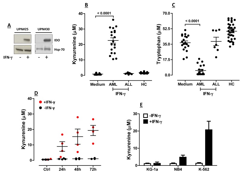
Figure 1. Expression and Function of IDO1 in BM Samples from Children with AML.
BM samples from children with AML obtained at diagnosis were cryopreserved. After thawing, BM mononuclear cells (MNC) were either stimulated with 100 ng/ml IFN-γ for up to 72 hours or were maintained in culture medium alone. Supernatants were collected and used for the measurement of kynurenine and tryptophan levels by RP-HPLC. (A) Up-regulation of IDO protein by IFN-γ (+) in 2 representative AML samples; UPN = Unique Patient Number; Hsp-70 = heat shock protein-70; (B) Release of kynurenine and (C) Consumption of tryptophan by AML blasts maintained in culture for 72 hours with or without exogenous IFN-γ. Blasts from a cohort of 7 children with acute lymphoblastic leukemia (ALL) were also challenged with IFN-γ to detect any IDO-mediated tryptophan breakdown. Comparisons between groups were performed with the Mann-Whitney U test for paired determinations. HC = healthy controls. Medium = blast cells maintained with complete culture medium alone; (D) Time-course experiments with AML blasts from 4 randomly selected BM samples that were either activated with IFN-γ (red dots) or left untouched (black dots). Bars depict the median and interquartile range; (E) Commercially available AML cell lines (see main text for details) were either stimulated with IFN-γ for 72 hours (black columns) or were maintained in culture medium alone (empty columns), prior to HPLC studies. Bars are representative of mean values and standard deviation recorded in 3 independent experiments run in duplicate.

