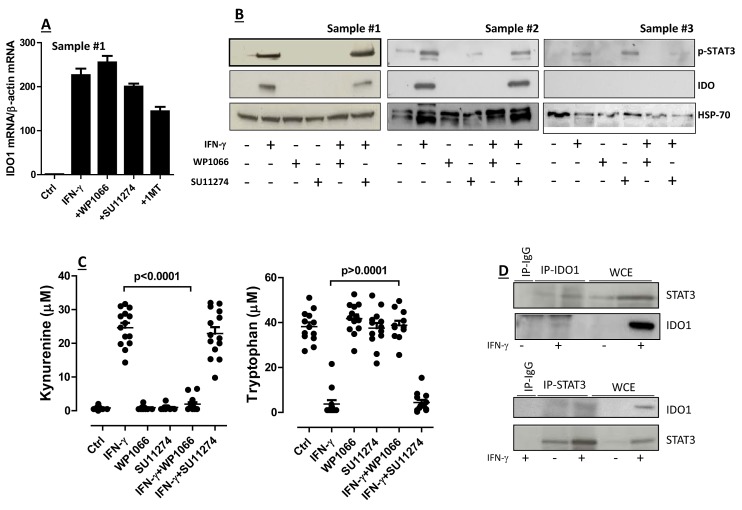
Figure 2. Regulation of IDO1 in AML Blasts.
(A) Quantitative RT-PCR and (B) Western blotting studies to detect IDO mRNA and protein in primary AML blasts. Samples were pre-treated for 2 hours with either of the following compounds: WP1066 or STAT3 Inhibitor III (28 μM final concentration), 1-methyl-tryptophan (1MT; 200 μM final concentration), SU11274 MET inhibitor (100 nM final concentration), followed by PCR or protein studies. Bars in panel A are representative of the mean ± standard deviation recorded in 3 independent experiments run in duplicate; (C) Release of kynurenine and depletion of tryptophan in supernatants of AML blasts that were pre-treated with either STAT3 inhibitors (WP1066) or MET inhibitors (SU11274) for 2 hours, and then activated with IFN-γ. Control cultures were established with either STAT3 inhibitors alone or with MET inhibitors alone. Comparisons were performed with the Mann-Whitney U test for paired determinations; (D) After challenge with IFN-γ (+) for 16 hours, leukemia cells were lysed, immunoprecipitated with anti-STAT3 antibodies and immunoblotted with anti-IDO1 antibodies (upper panel), or they were immunoprecipitated (IP) with anti-IDO1 antibodies and then immunoblotted with anti-STAT3 antibodies (lower panel). The specificity of each antibody used in these assays was confirmed using normal rabbit IgG as a negative control. Western blot runs were also performed with whole cell extracts (WCE).

