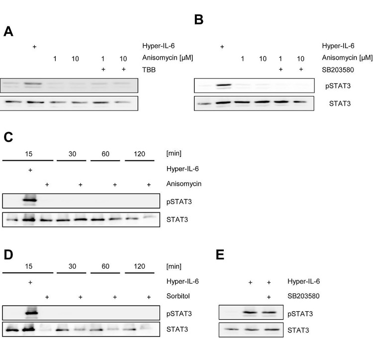
Fig 5. Activation of CK2 does not induce Jak/STAT signaling.

(A) HepG2 cells were serum starved for 3 h and stimulated with either 10 ng/ml Hyper-IL6 for 15 min, anisomycin for 30 min or left unstimulated. The CK2 inhibitor TBB was added 60 min prior to stimulation where indicated. (B) HepG2 cells were serum-starved for 3 h and stimulated with either 10 ng/ml Hyper-IL6 for 15 min, anisomycin for 30 min or left unstimulated. The p38 MAPK inhibitor SB203580 was added 60 min prior to stimulation where indicated. (C) HepG2 cells were serum-starved for 3 h and either left untreated, were stimulated with 10 ng/ml Hyper-IL-6 or 10 μM anisomycin for 15 min. Furthermore, either untreated or anisomycin-stimulated cells were harvested after 30, 60 or 120 min. (D) HepG2 cells were serum-starved for 3 h and either left untreated, were stimulated with 10 ng/ml Hyper-IL-6 or 500 mM sorbitol for 15 min. Furthermore, either untreated or sorbitol-stimulated cells were harvested after 30, 60 or 120 min. (E) HepG2 cells were serum-starved for 3 h and stimulated with either 10 ng/ml Hyper-IL-6 for 15 min or left unstimulated. The p38 MAPK inhibitor SB203580 was added 60 min prior to stimulation where indicated. Phosphorylation of STAT1 and STAT3 was assessed by Western blotting, and STAT1/STAT3 served as internal loading control, respectively. Shown is one representative Western blot from at least three independent experiments.
