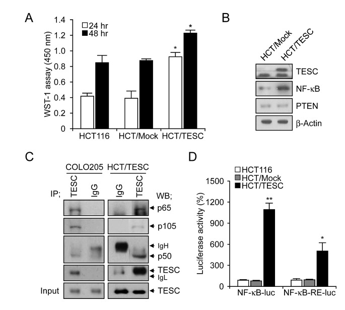
Figure 5. Interaction between TESC and NF-κB regulates cell proliferation in TESC-overexpressing HCT116 cells.
(A) Proliferation of HCT/TESC or HCT/Mock cells was measured using WST-1 reagent. *P < 0.01. (B) Western blot analysis of proteins involved in the NF-κB cell survival pathway. (C) TESC was immunoprecipitated using anti-TESC antibody, and precipitated proteins were analyzed by Western blotting using antibodies against NF-κB p65, NF-κB p50, or TESC. Total cell lysates (5% of input) were analyzed by Western blotting using anti-TESC antibody. Data are representative of three experiments. (D) NF-κB promoter activity was increased in HCT cells stably expressing NF-κB-luc and pcDNA-TESC plasmids (HCT/TESC-NF-κB-luc) compared with HCT cells expressing NF-κB-luc and pcDNA3.1 mock plasmids (HCT/Mock-NF-κB-luc), and in HCT cells stably expressing NF-κB-RE-luc and pcDNA-TESC plasmids (HCT/TESC-NF-κB-RE-luc) compared with HCT cells stably expressing NF-κB-RE-luc and pcDNA3.1 mock plasmids (HCT/Mock-NF-κB-RE-luc). Data are mean ± standard deviation from three independent experiments that were performed in triplicate. *P = 0.02; **P < 0.01.

