Abstract
Total talar extrusion without a soft tissue attachment is an extremely rare injury and is rarely reported. Appropriate treatment remains controversial. We describe the long-term outcomes of two patients who had complete talar extrusion without remaining soft tissue attachment treated with arthrodesis. Both of our patients had complications such as infection and progressive osteolysis. We suggest reimplantation of the extruded talus after thorough debridement as soon as possible as a reasonable option unless the talus is contaminated or missing, because an open wound may arise from inside to outside.
Keywords: Talus, Extrusion, Missing talus, Arthrodesis
A total talar extrusion as a result of a high energy injury is an unusual and rare injury.1,2,3,4,5,6,7,8,9,10) It is frequently associated with severe soft tissue injury, disruption of the talar blood supply, and fractures of surrounding bones. A completely extruded talus without remaining soft tissue attachments, a so-called missing talus, is extremely rare.1,2,4,5,6) The term "total extrusion of the talus" must be distinguished from total dislocation of the talus or a type 4 fracture dislocation of the talus. Total extrusion of the talus with severe contamination has a high possibility of avascular necrosis of the talus or infection and could have a different prognosis and treatment.
There is no consensus about appropriate treatment for an extruded talus. Some authors have advocated reimplantation of the extruded talus as the most appropriate treatment option.1,2,7,8,9) However tibiocalcaneal fusion is often frequently unavoidable after reimplantation due to complications such as deep infection, avascular necrosis, and resorption of bone.3,5) In the present article, we describe our experience with the long-term outcomes of two patients who had total talar extrusion, treated finally with delayed tibiocalcaneal fusion.
CASE REPORT
Case 1
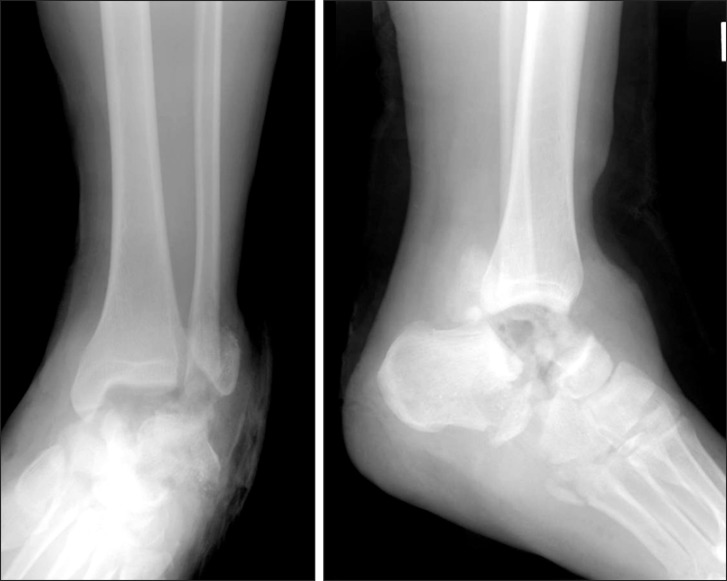
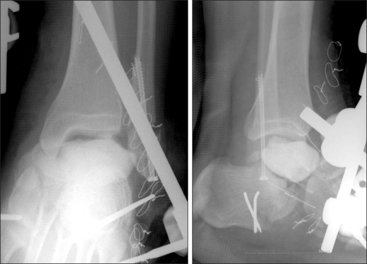
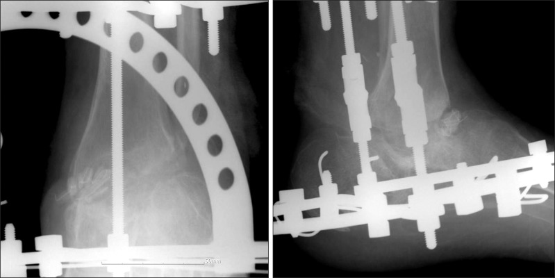
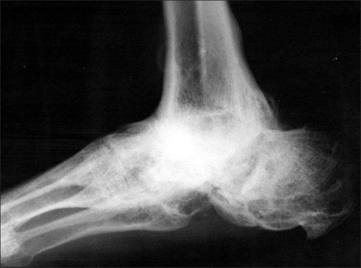
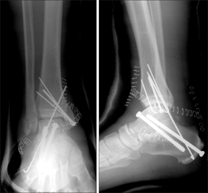
A 19-year-old man injured his left foot in a motorcycle accident. During the fall, the foot was forcibly twisted. He was brought to the emergency room 40 minutes after the accident. A clinical examination showed that the foot was supinated and severely contaminated on the anterolateral side. A Doppler examination did not show any vascular deficits. Initial radiographs of the ankle revealed complete absence of the talus and fractures of the lateral malleolus and anterior process of the calcaneus (Fig. 1). The patient was shifted to the operating room, and wound irrigation and debridement were performed, followed by internal fixation of the lateral malleolus and calcaneus. Because of the void created by the absent talus, a temporary antibiotic impregnated polymethyl-methacrylate cement spacer was inserted in the talar space, and a triplanar external fixator was placed across the ankle joint (Fig. 2). A few hour after the accident, the extruded talus was found in a trash basket near the site of the accident and was transferred to the hospital. It was heavily contaminated and scratched with soil and rubbish. We thoroughly cleaned the talus by pulsatile lavage in the operating room, and it was kept frozen. A reverse fasciocutaneous flap from the calf muscle was used for coverage of a palm-sized soft tissue defect of the anterolateral aspect of the ankle. The open wound was completely covered with additional split skin grafts. The cement spacer was removed 4 months later, and tibiocalcaneal fusion with bone grafts was performed instead of reimplantation of the frozen talus. The triplanar external fixator was converted to an Ilizarov external fixator. We packed the defect tightly with a cancellous bone graft from the anterior superior iliac spine and a frozen allograft of the femoral head. The wound was infected with purulent pus drainage 2 weeks after the surgery. Antibiotic-impregnated cement beads were inserted after removing the bone grafts and aggressive curettage of the talar space. Repetitive debridement and bead changes were done. We were able to control the infection 15 months after the injury. Compressive tibiocalcaneal fusion using an Ilizarov external fixator and an additional autogenous iliac bone graft were reperformed (Fig. 3). We achieved fusion 21 months after the injury followed by proximal tibial lengthening with the Ilizarov external fixator for the 4 cm leg length discrepancy. The patient is walking with full weight-bearing without any aids for regular activities of daily living 8 years after the injury. Solid fusion between the tibia, calcaneus, and navicular was observed on a lateral radiograph (Fig. 4).
Fig. 1.
Initial radiographs of the ankle, showing complete absence of the talus and fractures of the lateral malleolus and anterior process of the calcaneus.
Fig. 2.
Postoperative radiographs after inserting a cement spacer and stabilizing the ankle with an external fixator.
Fig. 3.
Postoperative radiographs after compressive tibiocalcaneal arthrodesis using an Ilizarov fixator.
Fig. 4.
Lateral radiograph of the ankle 8 years after injury, showing solid fusion.
Case 2
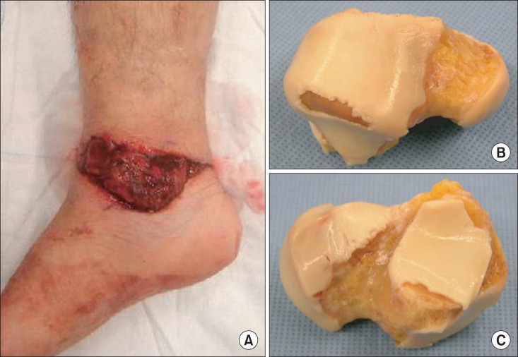
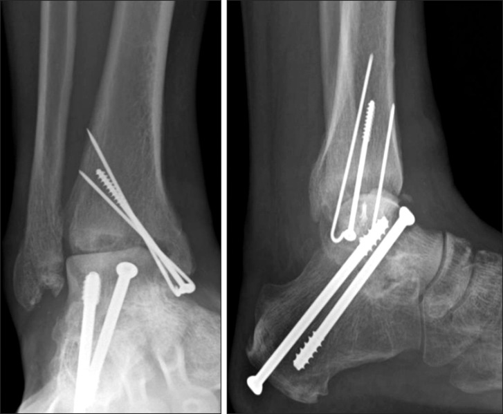
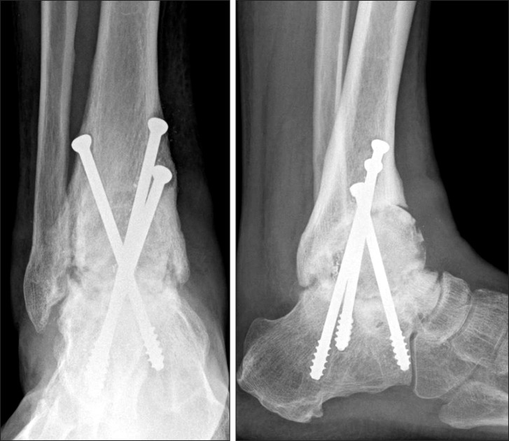
A 45-year-old man sustained a fall from 5 m while rock-climbing. He was transferred to the Emergency Department 1 hour after the accident. The patient sustained a pantalar dislocation with a > 10 cm sized transverse open wound on the medial aspect of the ankle (Fig. 5). He had no other major injuries. No distal neurovascular injury was detected. Initial radiographs showed complete absence of the talus and a tip fracture of both malleoli. The totally extruded talus was found on site but was severely contaminated with gravel, soil, and mud. It was immediately brought to the operating room. We had anticipated an inevitable avascular necrosis of the talus, as it was completely deprived of a blood supply; therefore, we planned to perform an initial subtalar fusion expecting revascularization from the calcaneus followed by ankle arthroplasty. The extruded talus was thoroughly washed and deep frozen. The talus was temporarily fixed using multiple Kirschner wires after massive wound debridement and irrigation because of the high risk for infection. Daily debridement and irrigation of the open wound was done for 3 days. A primary subtalar fusion was performed 4 days later, with reimplantation of the extruded talus and internal fixation with cannulated screws. Additional reduction and fixation of the fractured medial malleolus was performed (Fig. 6). Radiographs taken 6 months after the trauma, showed osteolysis on the distal tibial articular surface and loosening of the screws (Fig. 7). With suspicion of infection, curettage of the talar body was performed and an antibiotic impregnated cement spacer was inserted. However, the biopsy result revealed granulation tissue without significant neutrophil infiltration, with necrotic bone and marrow fibrosis. The culture reported no growth, and no abnormal laboratory findings were found. No infection was observed on follow-up evaluations. The ankle was fused using multiple screws, and a femoral head allograft was used to fill the talar defect 12 months after the trauma. Two years after the injury, the preserved talonavicular joint made mid-foot motion possible and tibio-talar-calcaneal fusion was achieved (Fig. 8). The patient has had a good functional outcome with the ability to enjoy a 4 hour course of tracking twice per week.
Fig. 5.
Photographs of the ankle and the totally extruded talus. (A) Medial view of the ankle. (B) Distal-dorsal view of the talus. (C) Plantar view of the talus.
Fig. 6.
Postoperative radiographs after reimplantation of the talus and subtalar fusion with pin and screws.
Fig. 7.
Radiographs of the ankle 7 months after injury, showing osteolysis of the distal tibia and loosening of the screws.
Fig. 8.
Radiographs of the ankle 3 years after injury, showing solid fusion.
DISCUSSION
A total talar extrusion associated with a high energy injury is rare and is frequently complicated by soft tissue injuries and fractures. This type of injury is prone to infection, avascular necrosis, secondary arthritis, and poor functional outcome. There is also controversy about the initial surgical treatment. Both primary talectomy and salvage of the talus have been recommended, but a primary talectomy in cases in which the talus has been extruded through the wound and contaminated has a high likelihood of infection. Smith et al.9) and Assal and Stern1) reported that a totally extruded talus should be managed with thorough cleaning and debridement of the wound followed by reimplantation. If the extruded talus is available and replaced, the survival of the talus probably depends on avoiding infection and avascular necrosis, and the existence of residual soft tissue attachment retaining a blood supply to the talus. Some authors have reported that reimplantation of an extruded talus with some retaining soft tissue attachments has relatively good clinical results.7,8,9) However, in cases without any soft tissue attachments, the result of reimplantation of a likely avascular and contaminated talus, which may act as a sequestrum, is questionable. Initial reimplantation of an extruded talus cannot restore vascular continuity. In contrast, reimplantation and wound closure can make it difficult to perform appropriate wound care that may require debridement and cleaning. Only nine cases in six reports have documented complete extrusion of the talus without any soft tissue attachments.1,2,4,5,6,9) Reimplantations were performed in five of the nine cases, and no subsequent infection or avascular necrosis were detected in the replaced talus. Only three cases developed osteoarthritis (Table 1). These results seem to be contradictory to our conception and are unusually excellent, even compared with those after fractures of the talar body or neck. More anatomical or physiological studies about a reimplanted talus might be needed. Our results were much worse than those of these studies. The causes of the poorer results were considered failure of infection control in case 1 and progressive bone resorption in case 2. Palomo-Traver et al.7) reported that the talus should be replaced unless it is totally extruded or grossly contaminated. It was suggested that impaired circulation related to trauma of the surrounding vascular and lymphatic structures leads to a nonhealing condition and subsequent infection; thus, predisposing the patient to acute and chronic bone infection.4,10) The cause of infection even after talectomy in case 1 seemed to be the unrepairable, contaminated open wound and the dead space created by the absent talus that made it difficult to control the infection. In case 1, we were worried about infection, and the talus was not available for reimplantation, whereas the talus in case 2 was contaminated but was available, and we replaced it after a thorough wash. We expected that avascular necrosis of the talus was inevitable and planned a primary subtalar fusion and revision ankle arthroplasty. We achieved the subtalar fusion, but had to perform massive debridement for unclearly progressive osteolysis around the ankle joint and then conducted a tibio-talar fusion with an allograft. We presumed that the repeated debridement and cleaning of the wound prevented the infection, but the cause of osteolysis may have been due to poor viability of the deep frozen talus.
Table 1.
Review of Cases Involving Open Total Extrusion of the Talus without Any Soft Tissue Attachments
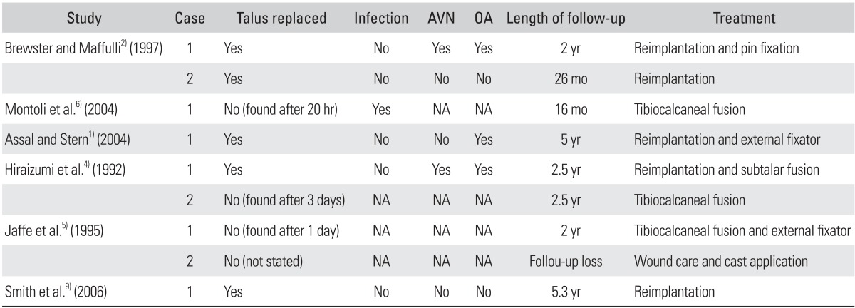
AVN: avascular necrosis of talus, OA: osteoarthritis, NA: not applicable.
Both of our patients were treated without initial reimplantation to prevent infection. Nevertheless, they had complications such as infection and unclearly progressive osteolysis and a long period of disability before achieving fusion. It is a reasonable option to replace an extruded talus as soon as possible after thorough debridement unless the talus is contaminated, because an open wound usually occurs from inside to outside.
Footnotes
No potential conflict of interest relevant to this article was reported.
References
- 1.Assal M, Stern R. Total extrusion of the talus: a case report. J Bone Joint Surg Am. 2004;86(12):2726–2731. doi: 10.2106/00004623-200412000-00021. [DOI] [PubMed] [Google Scholar]
- 2.Brewster NT, Maffulli N. Reimplantation of the totally extruded talus. J Orthop Trauma. 1997;11(1):42–45. doi: 10.1097/00005131-199701000-00011. [DOI] [PubMed] [Google Scholar]
- 3.Detenbeck LC, Kelly PJ. Total dislocation of the talus. J Bone Joint Surg Am. 1969;51(2):283–288. [PubMed] [Google Scholar]
- 4.Hiraizumi Y, Hara T, Takahashi M, Mayehiyo S. Open total dislocation of the talus with extrusion (missing talus): report of two cases. Foot Ankle. 1992;13(8):473–477. doi: 10.1177/107110079201300808. [DOI] [PubMed] [Google Scholar]
- 5.Jaffe KA, Conlan TK, Sardis L, Meyer RD. Traumatic talectomy without fracture: four case reports and review of the literature. Foot Ankle Int. 1995;16(9):583–587. doi: 10.1177/107110079501600913. [DOI] [PubMed] [Google Scholar]
- 6.Montoli C, De Pietri M, Barbieri S, D'Angelo F. Total extrusion of the talus: a case report. J Foot Ankle Surg. 2004;43(5):321–326. doi: 10.1053/j.jfas.2004.07.008. [DOI] [PubMed] [Google Scholar]
- 7.Palomo-Traver JM, Cruz-Renovell E, Granell-Beltran V, Monzonis-Garcia J. Open total talus dislocation: case report and review of the literature. J Orthop Trauma. 1997;11(1):45–49. doi: 10.1097/00005131-199701000-00014. [DOI] [PubMed] [Google Scholar]
- 8.Ries M, Healy WA., Jr Total dislocation of the talus: case report with a 13-year follow up and review of the literature. Orthop Rev. 1988;17(1):76–80. [PubMed] [Google Scholar]
- 9.Smith CS, Nork SE, Sangeorzan BJ. The extruded talus: results of reimplantation. J Bone Joint Surg Am. 2006;88(11):2418–2424. doi: 10.2106/JBJS.E.00471. [DOI] [PubMed] [Google Scholar]
- 10.Maffulli N, Francobandiera C, Lepore L, Cifarelli V. Total dislocation of the talus. J Foot Surg. 1989;28(3):208–212. [PubMed] [Google Scholar]


