Figure 1.

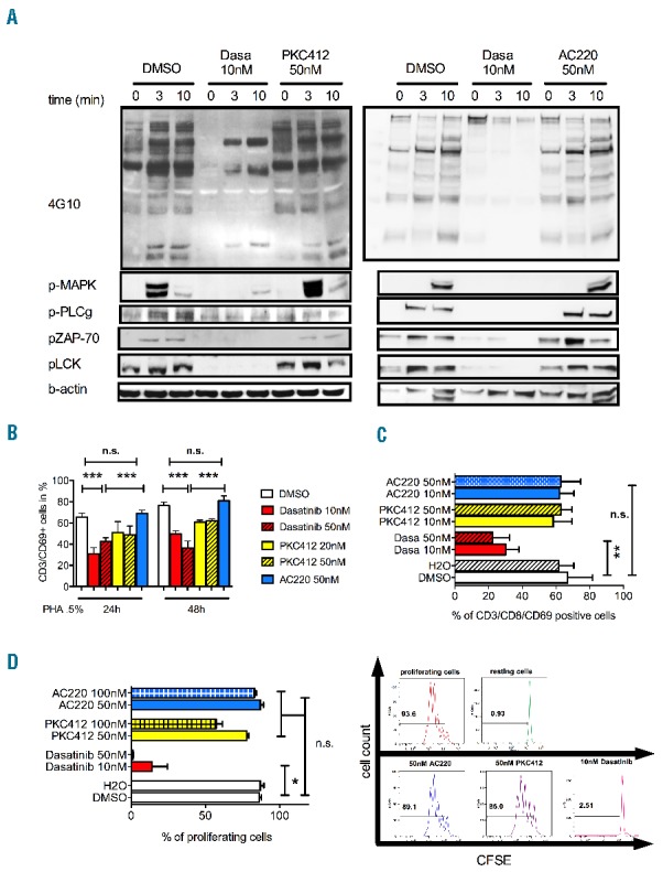
(A) Western blot analysis of T-cell receptor signaling upon inhibitor treatment. Cells were treated with the respective TKI for 48 h prior to short-term TCR stimulation. In detail, primary T cells from healthy donors (HD-TC) were treated with indicated concentrations of the respective kinase inhibitors (TKI) for 48 h and then stimulated with PHA (0.5%). Whole protein lysates were prepared immediately and after 3 and 10 min of PHA 0.5% stimulation. Midostaurin (PKC412, left panel) and quizartinib (AC220, right panel) did not reduce global tyrosine phosphorylation or activation of downstream signaling molecules (ZAP70, MAPK, LCK, PLCG1). Dasatinib treatment led to global reduction in global tyrosine phosphorylation and inhibition of downstream signaling pathways. Shown are 2 representative blots out of 6 healthy donors. (B, C) CD69 expression was determined by flow cytometry (n>5 per group). HD-TCs were stimulated with PHA 0.5% (B) or CD3/CD28-beads (for 48 h at a bead-to-cell ratio of 1:1; Dynabeads® Human T-Activator; Life Technologies) (C) and treated with indicated concentrations of the respective TKI. In both analyses all concentrations of midostaurin and quizartinib applied left CD69 expression unaffected. Dasatinib significantly reduced CD69 expression. (D) T-cell proliferation was assessed by CFSE labeling (n=4). HD-TCs stimulated for 24 h with 0.5% PHA/IL2 and co-incubated with kinase inhibitors dasatinib, midostaurin or quizartinib or DMSO. CFSE fluorescence was measured by flow cytometry on Day 5 post stimulation. 10nM dasatinib abrogated the proliferation activity (right). The FLT3-inhibitors midostaurin and quizartinib did not impair T-cell proliferation at concentrations applied.
