Abstract
Calcium-dependent protein kinases (CDPKs) are one of the largest protein kinases in plants and participate in different physiological processes through regulating downstream components of calcium signaling pathways. In this study, 41 CDPK genes, from GrCPK1 to GrCPK41, were identified in the genome of the diploid cotton, Gossypium raimondii. The phylogenetic analysis indicated that all these genes were divided into four subgroups and members within the same subgroup shared conserved exon-intron structures. The expansion of GrCPKs family in G. raimondii was due to the segmental duplication events, and the analysis of Ka/Ks ratios implied that the duplicated GrCPKs had mainly undergone strong purifying selection pressure with limited functional divergence. The cold-responsive elements in promoter regions were detected in the majority of GrCPKs. The expression analysis of 11 selected genes showed that GrCPKs exhibited tissue-specific expression patterns and the expression of GrCPKs were induced or repressed by cold treatment. These observations would lay an important foundation for functional and evolutionary analysis of CDPK gene family in Gossypium species.
Introduction
The plant growth and crop production are adversely affected by common stress conditions, such as drought, low temperature and high salinity [1]. Adaptation of plants to these environmental stresses includes the perception of stress signals and subsequent signal transduction, leading to the activation of various physiological and metabolic responses [2], [3], [4]. As a ubiquitous second messenger in cells, calcium (Ca2+) plays an important role in the signal transduction pathways. The transient changes of cytoplasmic Ca2+ concentration in response to various stresses were sensed and decoded by several Ca2+ sensors or Ca2+ binding proteins which relayed the signals into downstream response processes such as regulation of gene expression and phosphorylation cascades [5], [6].
There are four major classes of Ca2+ binding proteins characterized in plants, including calmodulins (CaM), calmodulin-like proteins (CaML), calcineurin B-like proteins (CBL) and calcium-dependent protein kinases (CDPK) [7], [8], [9]. Among them, CDPKs are the best characterized and are of particular interest, which constitute a large multigene family and are reported to be found throughout the plant kingdom from algae to angiosperms [10]. The CDPK protein possesses four characterized domains, a variable N-terminal domain, a catalytic Ser/Thr protein kinase domain, an autoinhibitory region, and a calmodulin-like domain [9], [11], [12], [13]. The N-terminal domain is highly variable and contains myristoylation or palmotylation sites which may contribute to membrane localization [12], [14]. And the calmodulin-like domain contains EF-hands structure for binding of Ca2+ [11], [12].
It was confirmed that CDPK genes were involved in regulating plant response to various stimuli, including abiotic and biotic stresses and hormones. Overexpression of the rice CDPK gene OsCDPK7 enhanced the tolerance of the transgenic rice plants to cold, salt, and drought stress [15], [16], and OsCPK12 overexpression plants increased the tolerance to salt stress but to blast fungus [17]. The low temperatures induced ZmCDPK1 expression in maize [18], and ZmCPK11 participated in touch- and wound-induced pathways [19]. In tobacco, NtCPK4 expression was increased in response to the treatment of gibberellin or NaCl [20]. In Arabidopsis, AtCPK10 was reported to participate in ABA- and Ca2+-medicated stomatal regulation in response to drought stress [21], and AtCPK3 and AtCPK6 were also shown as positive transducers of stomatal ABA signaling in guard cells [22]. AtCPK23 was demonstrated to function in response to drought and salt stresses [23]. In addition, some CDPK genes were also involved in pollen tube growth [24], root development [25], cell division and differentiation, and cell death [26], [27].
So far, many CDPK genes have been identified from numerous plant species. In Arabidopsis, 34 CDPK genes were revealed [9], [12]. And similarly, 31 CDPK genes in rice genome has identified by a genome-wide analysis [28]. In wheat, 20 CDPK genes including 14 full-length cDNA sequences were comprehensively studied [29]. And in poplar, 30 CDPK genes were identified [30], and 35 CDPK genes were initially revealed in maize genome [31]. Other higher plants such as tomato [32], potato [33], [34], tobacco [35], [36] and grapevine [37] also have multiple CDPK genes characterized in recent years. However, compared to the extensive studies of CDPK genes in many other plant species, little is known about this gene family in cotton. Till now, only one CDPK gene, GhCPK1, has been identified from upland cotton. And its transcripts accumulated primarily in the elongating fiber, which suggested that GhCPK1 might play a vital role in the calcium signaling events associated with fiber elongation [38], [39].
Cotton is the major source of natural fibers used in the textile industry and is cultivated worldwide. Cotton, which belongs to the genus of Gossypium, includes approximately 45 diploid and five tetraploid species and serves as an excellent model system for evolutionary studies of polyploidy plants [40]. With completion of the genome sequencing of the diploid cotton Gossypium raimondii [41], genome-wide analysis of all the genes belonging to specific gene families have been realized. Here, 41 CDPK genes from G. raimondii were identified by database searches and classified according to phylogenetic analysis. Furthermore, the expression profiles of CDPK genes in response to low temperature, which affects seed germination,plant development, and final yield in cotton, were investigated. The identification and comprehensive study for CDPK genes from G. raimondii will provide valuable information for breeding stress-resistant cotton and further studying of the biological function and evolutionary relationship of this family in cotton.
Materials and Methods
Gene retrieval and annotation
The G. raimondii genome database (release v2.1) [41] was downloaded from Phytozome (http://www.phytozome.net/cotton.php). The published CDPK protein information for Arabidopsis [12] and rice [28] were obtained from the Arabidopsis Information Resource (TAIR release 10, http://www.arabidopsis.org) and the Rice Genome Annotation Project Database (RGAP release 7, http://rice.plantbiology.msu.edu/index.shtml), respectively. To identify potential CDPK genes in G. raimondii, the Arabidopsis and rice CDPK proteins were used as queries by searching against the G. raimondii genome database using BlastP and tBlastN programs with default parameters. Subsequently, all hits were verified with the InterProScan program (http://www.ebi.ac.uk/Tools/pfa/iprscan/) [42] to confirm the presence of the protein kinase domain. Finally, the Pfam (http://pfam.sanger.ac.uk/) [43] and SMART (http://smart.embl-heidelberg.de/) [44] database were applied to manually determine each candidate member of the CDPK family. The EF hand was predicted by ScanProsite tool (http://prosite.expasy.org/scanprosite/) [45]. The N-myristoylation motif and the palmitoylation site were predicted by Myristoylator (http://web.expasy.org/myristoylator/) [46] and CSS-Plam program [47], respectively. The molecular weight (MW) of the full-length protein was calculated by Compute pI/Mw tool (http://web.expasy.org/cgi-bin/compute_pi/pi_tool) [48].
Phylogenetic analysis and Gene structure prediction
Multiple alignments of the full-length protein sequences were performed using Clustal X version 2.0 program [49] with default parameters. Phylogenetic trees were constructed using the method of Neighbor Joining MEGA 5.2 [50] with pairwise deletion option and poisson correction model. For statistical reliability, bootstrap tests were carried out with 1000 replicates.
The gene structures were obtained through comparing the genomic sequences and their predicted coding sequences of GSDS(http://gsds.cbi.pku.edu.cn/) [51].
Chromosomal locations and gene duplications
All CDPK genes were mapped on the G. raimondii chromosomes according to their starting positions given in the genome annotation document. The chromosome location image was generated by MapInspect software [52].
Gene duplication events of CDPK genes in G. raimondii were also investigated. The gene duplication was defined according to (1) the length of aligned sequence cover >80% of the longer gene, (2) the identity of the aligned regions >80%, and (3) only one duplication event was counted for tightly-linked genes [53], [54]. With the chromosomal locations of CDPK genes, two types of gene duplications were recognized, i.e., tandem duplication and segmental duplication.
Estimating Ka/Ks ratios for duplicated gene pairs
The CDPK duplicated gene pairs of G.raimondii were firstly aligned by Clustal X version 2.0 program [49]. Then Ks (synonymous substitution rate) and Ka (non-synonymous substitution rate) were calculated using the DnaSP v5.0 software (DNA polymorphism analysis) [55]. Finally, the Ka/Ks ratio was analyzed to assess the selection pressure for each gene pair.
Cis-element analysis
For promoter analysis, 2000 bp genomic DNA sequences upstream of the initiation codon (ATG) were retrieved from the genome sequence. These sequences were subjected to search in the PLACE database (http://www.dna.affrc.go.jp/PLACE/signalscan.html) [56] to identify putative cis-elements in promoter regions.
Plant materials and low temperature stress treatment
All the plants of G. raimondii were grown in a temperature-controlled chamber at 28°C with a photoperiod of 16 hours light and 8 hours dark. After ten days, the leaves, stems, roots, and cotyledons of some seedlings were collected to analyze tissue-specific expression, and the rest seedlings were used to examine the expression patterns of CDPK genes under low-temperature stress. Plant leaves of the seedlings grown in the temperature-controlled chamber treated at 10°C were harvested at 0, 3, 6, and 12 hours, which represented normal plants, slight stress, moderate stress, and severe stress, respectively. All collected samples were immediately frozen in liquid nitrogen and stored at −80°C.
RNA isolation and quantitative real-time PCR (qRT-PCR)
Total RNA was extracted from all samples using EASYspin Plus RNAprep Kit, and the first-strand cDNAs were synthesized with PrimerScript 1st Strand cDNA synthesis kit (TaKaRa). All protocols followed the manufacturer's protocol. For quantitative real-time PCR (qRT-PCR) assay, gene-specific primers were designed for the 11 selected CDPK genes according to their CDSs (Table S1). The qRT-PCR was performed with SYBR premix Ex taq (TakaRa) and CFX96 Realtime System (BioRad) by strictly following the manufacturer's instructions. Each reaction was done in a final volume of 20 µl containing 10 µl of SYBR premix Ex taq, 1.0 µl of cDNA sample, and 0.5 µl of each gene-specific primer. The qRT-PCR cycles were conducted with 40 cycles and an annealing temperature of 60°C, the amplification programs were as follows: 95°C for 30 seconds, 40 cycles of 94°C for 10 seconds, 60°C for 10 seconds, and 72°C for 15 seconds. The cotton UBQ7 gene was used as internal reference for all the qRT–PCR analyses. Each cDNA sample was tested in three replicates. The relative expression levels were calculated according to the 2−△△Ct method [57]. The expression profiles were clustered using the Cluster 3.0 software [58].
Results and Discussion
Identification and annotation of CDPK genes in G. raimondii
The availability of the G. raimondii genome sequences [41] makes it possible to identify all CDPK gene family members in the diploid cotton. BLASTP and TBLASTN programs were performed to search the candidate CDPK genes from the G. raimondii genome with the query sequences of Arabidopsis and rice CDPK genes. Then Pfam and SMART analyses were used to verify the retrieved sequences, and finally a total of 41 non-redundant CDPK genes were confirmed and described (Table1). According to the proposed nomenclature for CDPK genes [59], we designated these 41 CDPK genes as GrCPKs, from GrCPK1 to GrCPK41. The numbering of these GrCPKs was based on their position from top to bottom on corresponding chromosomes, from chromosome 1 to chromosome 13. Through analyzing the transcriptome sequencing data downloaded from the NCBI Sequence Read Archive (SRA), it was found that at least 37 GrCPKs were actively expressed in G. raimondii (Figure S1). The total number of CDPK genes identified from G. raimondii was greater than that in Arabidopsis [12] and rice [28]. The length and molecular weights (MW) of 41 GrCPK proteins were deduced from their predicted protein sequences. All identified GrCPK genes encoded proteins with amino acid numbers varying from 487 to 873 and molecular weight range between 54.6 kDa and 97.1 kDa, which were comparable with CDPK genes from other plant species [12], [28], [30], [31].
Table 1. The information of 41 CDPK genes from G. raimondii.
| Gene symbol | ID | Chromosome | Alternative splicing | CDS | Amino acid | MW (kDa) | No. of EF hands | N-terminal aa | N-Myristoylation | N-palmitoylation |
| GrCPK1 | Gorai.001G135000 | 1 | _ | 1542 | 513 | 57.3 | 4 | MGNCCSRG | Yes | Yes |
| GrCPK2 | Gorai.001G138000 | 1 | 2 | 1590 | 529 | 59.5 | 2 | MGNCCATT | No | Yes |
| GrCPK3 | Gorai.002G088800 | 2 | 4 | 1614 | 537 | 60.5 | 4 | MGSCLTKS | Yes | Yes |
| GrCPK4 | Gorai.002G153600 | 2 | 2 | 1776 | 591 | 66.3 | 4 | MGNSCAKS | Yes | Yes |
| GrCPK5 | Gorai.003G009500 | 3 | 8 | 1626 | 541 | 61.3 | 4 | MGACLSAT | Yes | Yes |
| GrCPK6 | Gorai.003G084000 | 3 | _ | 1527 | 508 | 57.5 | 3 | MGNCNGLP | No | Yes |
| GrCPK7 | Gorai.003G092900 | 3 | 3 | 1608 | 535 | 60.6 | 3 | MGNCCATP | Yes | Yes |
| GrCPK8 | Gorai.004G015100 | 4 | _ | 1617 | 538 | 60.5 | 4 | MGNCCSRG | Yes | Yes |
| GrCPK9 | Gorai.005G019800 | 5 | _ | 1599 | 532 | 60.7 | 4 | MGSCVARP | Yes | Yes |
| GrCPK10 | Gorai.005G074300 | 5 | _ | 1527 | 508 | 56.9 | 4 | MNNQSSSI | No | No |
| GrCPK11 | Gorai.005G216500 | 5 | 6 | 1707 | 568 | 63.6 | 4 | MGNTCRGS | No | Yes |
| GrCPK12 | Gorai.006G124800 | 6 | _ | 1575 | 524 | 58.6 | 4 | MGNCCSCG | Yes | Yes |
| GrCPK13 | Gorai.006G128200 | 6 | 3 | 1596 | 531 | 59.8 | 3 | MGNCCATP | No | Yes |
| GrCPK14 | Gorai.006G137800 | 6 | _ | 1596 | 531 | 60.2 | 3 | MGNCCVTS | No | Yes |
| GrCPK15 | Gorai.006G147600 | 6 | _ | 1836 | 611 | 68.4 | 4 | MGNNCFKT | No | Yes |
| GrCPK16 | Gorai.007G025000 | 7 | 5 | 1593 | 530 | 60.4 | 4 | MGNCNRPP | No | Yes |
| GrCPK17 | Gorai.007G035100 | 7 | 3 | 1653 | 550 | 62.3 | 4 | MGNCNACV | No | Yes |
| GrCPK18 | Gorai.007G194500 | 7 | 3 | 1665 | 554 | 62.8 | 4 | MGACLSTT | Yes | Yes |
| GrCPK19 | Gorai.007G378700 | 7 | _ | 1584 | 527 | 59.2 | 3 | MGNCCRSP | No | Yes |
| GrCPK20 | Gorai.008G013700 | 8 | _ | 1728 | 575 | 64.6 | 4 | MRLHYCMR | No | Yes |
| GrCPK21 | Gorai.008G251000 | 8 | 2 | 1605 | 534 | 60.2 | 4 | MGNCNSQP | Yes | Yes |
| GrCPK22 | Gorai.009G078000 | 9 | _ | 1599 | 532 | 59.5 | 4 | MGNLCSRS | Yes | Yes |
| GrCPK23 | Gorai.009G191500 | 9 | _ | 2622 | 873 | 97.1 | 4 | MGICQSLC | No | Yes |
| GrCPK24 | Gorai.009G290200 | 9 | 5 | 1617 | 538 | 60.4 | 4 | MNKKIAGS | No | No |
| GrCPK25 | Gorai.009G351200 | 9 | _ | 1605 | 534 | 60.7 | 4 | MGSCISAP | Yes | Yes |
| GrCPK26 | Gorai.009G394700 | 9 | _ | 1947 | 648 | 71.8 | 4 | MGNVCATL | No | Yes |
| GrCPK27 | Gorai.009G395400 | 9 | 5 | 1719 | 572 | 63.6 | 4 | MGNACAGP | No | Yes |
| GrCPK28 | Gorai.009G438300 | 9 | _ | 1575 | 524 | 58.7 | 4 | MGGCLTKT | Yes | Yes |
| GrCPK29 | Gorai.010G001300 | 10 | _ | 1581 | 526 | 59.1 | 4 | MGLCQSLG | Yes | Yes |
| GrCPK30 | Gorai.010G252400 | 10 | _ | 1584 | 527 | 59.3 | 4 | MGCCSSKN | Yes | Yes |
| GrCPK31 | Gorai.011G014200 | 11 | _ | 1656 | 551 | 62.0 | 4 | MGCFSSKH | Yes | Yes |
| GrCPK32 | Gorai.011G098300 | 11 | _ | 1635 | 544 | 62.0 | 4 | MGICLSTT | Yes | Yes |
| GrCPK33 | Gorai.011G228500 | 11 | 5 | 1764 | 587 | 65.4 | 4 | MGNTCVGP | No | Yes |
| GrCPK34 | Gorai.012G045700 | 12 | 2 | 1494 | 497 | 55.9 | 4 | MSRTSSGT | No | No |
| GrCPK35 | Gorai.012G114600 | 12 | _ | 1584 | 527 | 59.4 | 3 | MGNCCRSP | No | Yes |
| GrCPK36 | Gorai.012G138900 | 12 | 2 | 1659 | 552 | 62.0 | 4 | MGNTCRGP | No | Yes |
| GrCPK37 | Gorai.013G003100 | 13 | 2 | 1740 | 579 | 64.6 | 4 | MGNTCVGP | No | Yes |
| GrCPK38 | Gorai.013G064400 | 13 | 5 | 1464 | 487 | 54.6 | 4 | MRRAIDHQ | No | No |
| GrCPK39 | Gorai.013G064500 | 13 | 2 | 1572 | 523 | 58.3 | 3 | MGNTCLGS | No | Yes |
| GrCPK40 | Gorai.013G159300 | 13 | 3 | 1611 | 536 | 60.4 | 4 | MGGCLTKN | Yes | Yes |
| GrCPK41 | Gorai.013G253100 | 13 | _ | 1584 | 527 | 58.8 | 4 | MGNCCTRG | No | Yes |
_ represents no alternative splice variants.
All 41 CDPKs identified in our study possessed the typical CDPK structure with four characterized domains, including a variable N-terminal domain, a catalytic Ser/Thr protein kinase domain, an autoinhibitory region, and a calmodulin-like domain. The N-terminal domain of several CDPK proteins contained myristoylation motif with a Gly residue at the second position, which was thought to be critical for mediating protein-protein and protein-membrane interactions [60]. It was reported that CDPK genes which had an N-myristoylation motif tended to localize in the plasma membranes [14], [21], [29], [61], [62], demonstrating that myristoylation was required for membrane binding. In addition, palmitoylation, as a second type of lipid modification, was also necessary to stabilize for membrane association [14]. Among 41 CDPKs in our study, 18 CDPKs were predicted to have myristoylation motifs at the N-terminus, and all of them possessed at least one palmitoylation site. However, the membrane association of CDPKs was complex and might be affected by other motifs. For example, TaCPK3 and TaCPK15 lacking myristoylation motifs could also be associated with the membranes [29], while ZmCPK1 which was predicted to have an N-myristoylation motif was found to localize into the cytoplasm and nucleus [63]. Therefore, the subcellular localizations of these CDPKs were still needed to be further characterized experimentally.
The calmodulin-like domain contained Ca2+ binding EF hand structure that allow CDPK proteins to function as a Ca2+ sensor. In G. raimondii, 33 CDPKs contained four Ca2+ binding EF hands, seven CDPKs, including GrCPK6, GrCPK7, GrCPK13, GrCPK14, GrCPK19, GrCPK35, and GrCPK39, had three EF hand motifs each, whereas GrCPK2 had two EF hand motifs only. This difference in EF hand was also found in the CDPK family of other plants [9], [30], [31], [64]. Studies have demonstrated that the number and position of EF hands might be important for determining the Ca2+ regulation of CDPK activity [65], [66], and compared to C-terminal EF3 and EF4 motifs, N-terminal EF1 and EF2 motifs with lower Ca2+-binding affinities were more important for activating the kinases [67], [68].
Phylogenetic analysis of the CDPK gene family
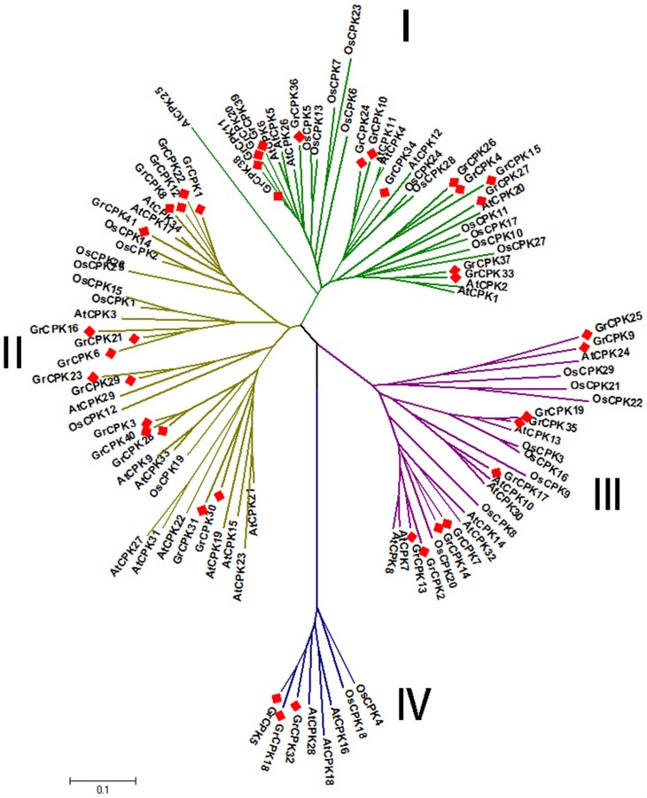
To detect the evolutionary relationships of CDPK genes, an unrooted phylogenetic tree was generated from alignments of the full-length sequences of G. raimondii, Arabidopsis and rice CDPK proteins. All the CDPK proteins were divided into four subgroups (Figure 1): CDPK I, CDPK II, CDPK III and CDPK IV. CDPK I contained 14 CDPKs from G. raimondii, 11 from rice, and ten from Arabidopsis. CDPK II contained 15 CDPKs from G. raimondii, eight from rice, and 13 from Arabidopsis. The CDPKs numbers in CDPK I and CDPK II from G. raimondii were greater than that from Arabidopsis and rice, mainly due to more CDPK genes in G. raimondii. In CDPK III, there were nine CDPKs from G. raimondii, eight from rice, and eight from Arabidopsis, while CDPK IV genes consisted of the smallest subfamilies in all of the three species, which contained three CDPKs from G. raimondii, two from rice, and three from Arabidopsis. And the number of genes in CDPK III and CDPK IV were approximately identical across these three species. Interestingly, among the four subfamilies, CDPK I in rice comprised the most members, while CDPK II in G. raimondii and Arabidopsis was the largest subfamily. Recent study indicated that poplar had total 30 CDPK genes with 11 genes in CDPK I, eight genes in CDPK II, nine genes in CDPK III and two genes in CDPK IV [30]. These results implied that the difference in the number of CDPK genes was mainly due to the occurrence of gene gain or loss in CDPK I or CDPK II subfamilies independently among the different organisms.
Figure 1. Unrooted phylogenetic tree of CDPK genes from G. raimondii, Arabidopsis and rice.
Four subfamilies are labeled as I, II, III, and IV. And the branches of each subfamily are indicated in a specific color.
Phylogenetic analysis also showed that GrCPKs were more closely allied to AtCPKs than to OsCPKs, consistent with the evolutionary relationships among G. raimondii, Arabidopsis, and rice. Moreover, all the CDPK genes from the three species sorted into four distinct clades, implying that these four subfamilies existed before the divergence of monocots and dicots, which also supported the hypothesis that CDPK genes radiated into four subfamilies before algae and land plants split [69].
Structural analysis and chromosomal localization of GrCPKs
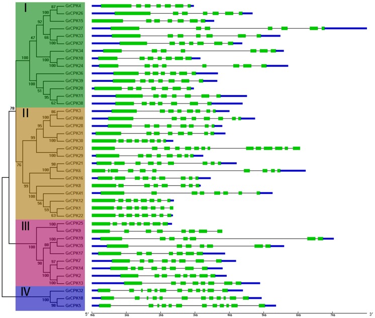
To get further insight into the possible structural evolution of GrCPKs, a separate unrooted phylogenetic tree was constructed using the protein sequences of all the CDPK genes from G. raimondii, and then the diverse exon-intron organizations of GrCPKs were compared. As shown in Figure 2, the GrCPKs clustered in the same subfamily shared very similar exon-intron structures. Most members in Group I possessed seven exons, except for GrCPK24, which contained eight exons. Most genes in Group II had eight exons, but GrCPK16 and GrCPK21 had nine exons each, GrCPK6 contained ten exons, and GrCPK23 contained 14 exons. Six genes in group III had eight exons, whereas three genes contained seven exons each. All genes in Group IV contained 12 exons. These conserved gene structure within each group supported their close evolutionary relationship.
Figure 2. Phylogenetic relationship and gene structure of CDPK genes from G. raimondii.
Four subfamilies labeled as I, II, III, and IV are marked with different color backgrounds. Exons are represented by green boxes and introns by black lines. The untranslated regions (UTRs) are indicated by thick blue lines.
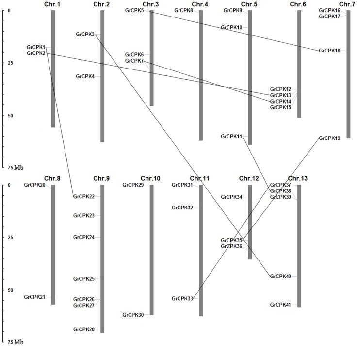
The 41 nonredundant CDPK genes were mapped on the 13 G. raimondii chromosomes (Figure 3). They were found to be distributed unevenly among 13 chromosomes and the number of CDPK genes on each chromosome varied widely. Chromosome 9 which contained 7 CDPK genes was the highest one in gene numbers, followed by chromosome 13 on which five genes were found. Chromosome 6 and 7 both had four genes, and chromosome 3, 5, 11, and 12 contained three genes each. Chromosome 1, 2, 8, and 10 had two genes each only, whereas only single CDPK gene was localized on chromosome 4.
Figure 3. Distributions of CDPK genes from G. raimondii in 13 chromosomes.
Chromosome numbers are indicated above each vertical bar. The scale represents megabases (Mb). The duplicated gene pairs are connected with black lines.
CDPK gene duplications and functional divergence
To elucidate the expanded mechanism of the CDPK gene family in G. raimondii, gene duplication events, including tandem and segmental duplications, were investigated, which were thought to play a significant role in the amplification of gene family members in the genome [70], [71]. A total of eight duplication events, GrCPK2/GrCPK13, GrCPK7/GrCPK14, GrCPK18/GrCPK5, GrCPK19/GrCPK35, GrCPK22/GrCPK1, GrCPK37/GrCPK33, GrCPK38/GrCPK11, and GrCPK40/GrCPK3, were found in the G. raimondii genome and all of them were segmental duplication events based on the chromosomal distribution of the CDPK genes (Figure 3). This result suggested that the CDPK gene family expansion in G. raimondii was mainly attributed to segmental duplication events.
The duplicated gene pairs might experience three alternative outcomes during the process of evolution, i.e., (i) one copy may simply become silenced and lost original functions (nonfunctionalization); (ii) one copy may acquire a novel, beneficial function, with the other copy retaining the original function (neofunctionalization); or (iii) both copies may become partition of original functions (subfunctionalization) [72]. We subsequently calculated the non-synonymous to synonymous substitution ratio (Ka/Ks) for each pair of duplicated CDPK genes, which showed the selective force acting on the protein, to reveal whether Darwinian positive selection was associated with functional divergence after gene duplications. Generally, Ka/Ks >1 indicates positive selection, Ka/Ks = 1 indicates neutral selection, while Ka/Ks <1 indicates negative or purifying selection. In this study, the Ka/Ks ratios for eight duplicated CDPK gene pairs were no larger than 0.2 (Table 2), which demonstrated that the CDPK genes from G. raimondii had mainly experienced strong purifying selection pressure with limited functional divergence after segmental duplications. These results suggested that functions of the duplicated CDPK genes did not diverge much during subsequent evolution.
Table 2. Ka/Ks analysis for the duplicated gene pairs.
| Duplicated gene 1 | Duplicated gene 2 | Ka | Ks | Ka/Ks | Purifying selection | Duplicate type |
| GrCPK2 | GrCPK13 | 0.068 | 0.422 | 0.161 | Yes | Segmental |
| GrCPK14 | GrCPK7 | 0.083 | 0.484 | 0.171 | Yes | Segmental |
| GrCPK18 | GrCPK5 | 0.057 | 0.589 | 0.096 | Yes | Segmental |
| GrCPK19 | GrCPK35 | 0.028 | 0.399 | 0.071 | Yes | Segmental |
| GrCPK22 | GrCPK1 | 0.080 | 0.683 | 0.118 | Yes | Segmental |
| GrCPK37 | GrCPK33 | 0.062 | 0.511 | 0.121 | Yes | Segmental |
| GrCPK38 | GrCPK11 | 0.034 | 0.515 | 0.065 | Yes | Segmental |
| GrCPK40 | GrCPK3 | 0.038 | 0.528 | 0.071 | Yes | Segmental |
Expression analysis of the CDPK genes in G. raimondii
Cold stress is one of the serious environmental stresses that most land plants might encounter during the process of their growth. And so far, numerous of CDPK genes identified in various plant species have been proven to play crucial roles in cold stress response. Cotton is a subtropical crop and its cultivation has been extended from tropical and subtropical to colder regions. Low temperature (<15°C ) can adversely affect plant development, resulting in poor germination, infection by fungi and other disease-causing organisms, and yield loss [73]. There is little information about the functions of CDPK genes in the cotton response to low temperature.
The cis-element analysis in promoter regions might provide some indirect evidence for the functional dissection of CDPK genes in stress responses [74]. To investigate the potential functions of GrCPKs in cotton under cold stress, the possible presence of low temperature-responsive element (LTRE) in the promoter regions of all the GrCPKs was detected by searching against the PLACE database (Table S2). The results revealed that the majority, 27 out of 41, of GrCPKs contained the LTRE in promoter sequences, indicating that these GrCPKs might participate in the signal transduction of the plant response to cold stress. In order to verify the expression patterns of these genes, eight GrCPKs which had the LTRE in promoter regions were selected for qRT-PCR analysis. In addition, as the gene expression was a complex process, the existence of some cis-elements did not always correlate with the gene function [74], therefore, three GrCPKs, GrCPK2, GrCPK11, and GrCPK27, which had no LTRE in promoter regions were also selected in this expression analysis.
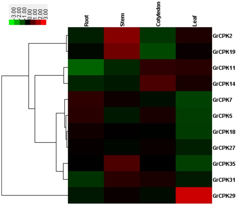
Firstly, qRT-PCR analysis was performed to examine expression patterns of 11 selected GrCPKs in roots, stems, cotyledons, and leaves of 10-day-old seedlings. As shown in Figure 4, GrCPK5 and GrCPK7 were expressed at high levels in roots, while GrCPK2, GrCPK19, and GrCPK35 shared high expressions in young stems. In cotyledons, GrCPK14 exhibited relatively high transcript abundance, whereas GrCPK29 were predominantly expressed in leaves, which showed about 6-fold higher expression than all other tissues. The results demonstrated that most of these 11 GrCPKs had specific spatial expression patterns, which was similar with CDPK genes in maize [54] and rice [74] that also exhibited tissue-specific expression.
Figure 4. Expression patterns of 11 GrCPKs in four representative tissues of G. raimondii seedlings.
The color bar represents the relative signal intensity values.
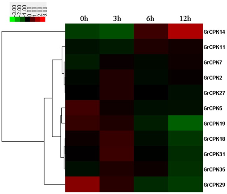
To further confirm the cold stress response of GrCPKs, the expression patterns of 11 GrCPKs in 10-day-old leaves under low temperature stress of 10°C were investigated. The expression levels of all the 11 CDPK genes responsive to slight cold stress (3 hours), moderate cold stress (6 hours), and severe cold stress (12 hours) were shown in Figure 5, comparing with normal plants. The result showed that the expression of all the 11 GrCPKs were induced or repressed by clod treatment. Among them, GrCPK2, GrCPK7, GrCPK11, GrCPK14, GrCPK18, GrCPK27, GrCPK31, and GrCPK35 were positively regulated response to cold stress according to their transcription levels. For example, GrCPK14 showed a significantly increase in its expression after long time cold stress treatment. Whereas, the transcription levels of the other three genes, GrCPK5, GrCPK19, and GrCPK29, were down-regulated in response to cold stress.
Figure 5. Expression profiling of 11 GrCPKs in leaves under 10°C treatment.
The color bar represents the relative signal intensity values.
Conclusions
A complete set of 41 CDPK genes were identified in the G. raimondii genome, which were categorized into four subgroups and mapped on the 13 chromosomes. Segmental duplication contributed to the expansion of GrCPKs, and the duplicated genes mainly undergone strong purifying selection pressure with limited functional divergence. The majority of GrCPKs contained the low temperature-responsive elements in their promoter sequences. The expression of GrCPKs were also induced or repressed by the clod stress treatment. The results would be helpful for better understanding of the biological functions of the CDPK genes in cotton and the evolutionary relationship of this family in Gossypium species.
Supporting Information
Expression analysis of GrCPKs using the transcriptome sequencing data. The transcriptome sequencing datasets of G. raimondii for three tissue samples (mature leaves, 0DPA ovules, and 3DPA ovules) were downloaded from the NCBI Sequence Read Archive (SRA) with accession numbers SRX111367, SRX111365 and SRX111366. Then sequenced reads of these three datasets were mapped to the sequences of GrCPKs, respectively. And matches were converted to RPKM to estimate gene expression levels. The expression profiles were clustered using the Cluster 3.0 software. The color bar represents the relative signal intensity values. DPA: Days Post Anthesis.
(TIF)
PCR primers used in this study.
(XLSX)
The LTRE in the promoter regions of GrCPKs .
(XLSX)
Acknowledgments
We are grateful to Lei Mei, Ning Zhu, Cheng Li, Yue Chen, Yingchao Sun and Jingjing Pan (Zhejiang University, China) for their support in this study.
Data Availability
The authors confirm that all data underlying the findings are fully available without restriction. The genome and gene sequences used in the manuscript are available from Phytozome (http://www.phytozome.net/cotton.php) with the accession IDs in Table 1.
Funding Statement
The works are support by the National Basic Research Program (973 program, No: 2010CB126006) and the National High Technology Research and Development Program of China (2011AA10A102, 2013AA102601). The funders had no role in study design, data collection and analysis, decision to publish, or preparation of the manuscript.
References
- 1. Xiong L, Schumaker KS, Zhu JK (2002) Cell signaling during cold, drought, and salt stress. Plant Cell 14 Suppl: S165–S183 [DOI] [PMC free article] [PubMed] [Google Scholar]
- 2. Yamaguchi-Shinozaki K, Shinozaki K (2006) Transcriptional regulatory networks in cellular responses and tolerance to dehydration and cold stresses. Annu Rev Plant Biol 57: 781–803. [DOI] [PubMed] [Google Scholar]
- 3. Valliyodan B, Nguyen HT (2006) Understanding regulatory networks and engineering for enhanced drought tolerance in plants. Curr Opin Plant Biol 9: 189–195. [DOI] [PubMed] [Google Scholar]
- 4. Tran LS, Nakashima K, Shinozaki K, Yamaguchi-Shinozaki K (2007) Plant gene networks in osmotic stress response: from genes to regulatory networks. Methods Enzymol 428: 109–128. [DOI] [PubMed] [Google Scholar]
- 5. Evans NH, McAinsh MR, Hetherington AM (2001) Calcium oscillations in higher plants. Curr Opin Plant Biol 4: 415–420. [DOI] [PubMed] [Google Scholar]
- 6. Tuteja N, Mahajan S (2007) Calcium signaling network in plants: an overview. Plant Signal Behav 2: 79–85. [DOI] [PMC free article] [PubMed] [Google Scholar]
- 7. McCormack E, Braam J (2003) Calmodulins and related potential calcium sensors of Arabidopsis. New Phytologist 159: 585–598. [DOI] [PubMed] [Google Scholar]
- 8. Kolukisaoglu U, Weinl S, Blazevic D, Batistic O, Kudla J (2004) Calcium sensors and their interacting protein kinases: genomics of the Arabidopsis and rice CBL-CIPK signaling networks. Plant Physiol 134: 43–58. [DOI] [PMC free article] [PubMed] [Google Scholar]
- 9. Cheng SH, Willmann MR, Chen HC, Sheen J (2002) Calcium signaling through protein kinases. The Arabidopsis calcium-dependent protein kinase gene family. Plant Physiol 129: 469–485. [DOI] [PMC free article] [PubMed] [Google Scholar]
- 10. Harmon AC, Gribskov M, Gubrium E, Harper JF (2001) The CDPK superfamily of protein kinases. New Phytologist 151: 175–183. [DOI] [PubMed] [Google Scholar]
- 11. Harper JF, Sussman MR, Schaller GE, Putnam-Evans C, Charbonneau H, et al. (1991) A calcium-dependent protein kinase with a regulatory domain similar to calmodulin. Science 252: 951–954. [DOI] [PubMed] [Google Scholar]
- 12. Hrabak EM, Chan CW, Gribskov M, Harper JF, Choi JH, et al. (2003) The Arabidopsis CDPK-SnRK superfamily of protein kinases. Plant Physiol 132: 666–680. [DOI] [PMC free article] [PubMed] [Google Scholar]
- 13. Klimecka M, Muszynska G (2007) Structure and functions of plant calcium-dependent protein kinases. Acta Biochim Pol 54: 219–233. [PubMed] [Google Scholar]
- 14. Martin ML, Busconi L (2000) Membrane localization of a rice calcium-dependent protein kinase (CDPK) is mediated by myristoylation and palmitoylation. Plant J 24: 429–435. [DOI] [PubMed] [Google Scholar]
- 15. Saijo Y, Hata S, Kyozuka J, Shimamoto K, Izui K (2000) Over-expression of a single Ca2+-dependent protein kinase confers both cold and salt/drought tolerance on rice plants. Plant J 23: 319–327. [DOI] [PubMed] [Google Scholar]
- 16. Saijo Y, Kinoshita N, Ishiyama K, Hata S, Kyozuka J, et al. (2001) A Ca2+-dependent protein kinase that endows rice plants with cold- and salt-stress tolerance functions in vascular bundles. Plant Cell Physiol 42: 1228–1233. [DOI] [PubMed] [Google Scholar]
- 17. Asano T, Hayashi N, Kobayashi M, Aoki N, Miyao A, et al. (2012) A rice calcium-dependent protein kinase OsCPK12 oppositely modulates salt-stress tolerance and blast disease resistance. Plant J 69: 26–36. [DOI] [PubMed] [Google Scholar]
- 18. Berberich T, Kusano T (1997) Cycloheximide induces a subset of low temperature-inducible genes in maize. Mol Gen Genet 254: 275–283. [DOI] [PubMed] [Google Scholar]
- 19. Szczegielniak J, Borkiewicz L, Szurmak B, Lewandowska-Gnatowska E, Statkiewicz M, et al. (2012) Maize calcium-dependent protein kinase (ZmCPK11): local and systemic response to wounding, regulation by touch and components of jasmonate signaling. Physiol Plant 146: 1–14. [DOI] [PubMed] [Google Scholar]
- 20. Zhang M, Liang S, Lu YT (2005) Cloning and functional characterization of NtCPK4, a new tobacco calcium-dependent protein kinase. Biochim Biophys Acta 1729: 174–185. [DOI] [PubMed] [Google Scholar]
- 21. Zou JJ, Wei FJ, Wang C, Wu JJ, Ratnasekera D, et al. (2010) Arabidopsis calcium-dependent protein kinase CPK10 functions in abscisic acid- and Ca2+-mediated stomatal regulation in response to drought stress. Plant Physiol 154: 1232–1243. [DOI] [PMC free article] [PubMed] [Google Scholar]
- 22. Mori IC, Murata Y, Yang Y, Munemasa S, Wang YF, et al. (2006) CDPKs CPK6 and CPK3 function in ABA regulation of guard cell S-type anion- and Ca2+-permeable channels and stomatal closure. PLoS Biol 4: e327. [DOI] [PMC free article] [PubMed] [Google Scholar]
- 23. Ma SY, Wu WH (2007) AtCPK23 functions in Arabidopsis responses to drought and salt stresses. Plant Mol Biol 65: 511–518. [DOI] [PubMed] [Google Scholar]
- 24. Estruch JJ, Kadwell S, Merlin E, Crossland L (1994) Cloning and characterization of a maize pollen-specific calcium-dependent calmodulin-independent protein kinase. Proc Natl Acad Sci U S A 91: 8837–8841. [DOI] [PMC free article] [PubMed] [Google Scholar]
- 25. Ivashuta S, Liu J, Liu J, Lohar DP, Haridas S, et al. (2005) RNA interference identifies a calcium-dependent protein kinase involved in Medicago truncatula root development. Plant Cell 17: 2911–2921. [DOI] [PMC free article] [PubMed] [Google Scholar]
- 26. Yoon GM, Cho HS, Ha HJ, Liu JR, Lee HS (1999) Characterization of NtCDPK1, a calcium-dependent protein kinase gene in Nicotiana tabacum, and the activity of its encoded protein. Plant Mol Biol 39: 991–1001. [DOI] [PubMed] [Google Scholar]
- 27. Lee SS, Cho HS, Yoon GM, Ahn JW, Kim HH, et al. (2003) Interaction of NtCDPK1 calcium-dependent protein kinase with NtRpn3 regulatory subunit of the 26S proteasome in Nicotiana tabacum . Plant J 33: 825–840. [DOI] [PubMed] [Google Scholar]
- 28. Ray S, Agarwal P, Arora R, Kapoor S, Tyagi AK (2007) Expression analysis of calcium-dependent protein kinase gene family during reproductive development and abiotic stress conditions in rice (Oryza sativa L. ssp. indica). Mol Genet Genomics 278: 493–505. [DOI] [PubMed] [Google Scholar]
- 29. Li AL, Zhu YF, Tan XM, Wang X, Wei B, et al. (2008) Evolutionary and functional study of the CDPK gene family in wheat (Triticum aestivum L.). Plant Mol Biol 66: 429–443. [DOI] [PubMed] [Google Scholar]
- 30. Zuo R, Hu R, Chai G, Xu M, Qi G, et al. (2013) Genome-wide identification, classification, and expression analysis of CDPK and its closely related gene families in poplar (Populus trichocarpa). Mol Biol Rep 40: 2645–2662. [DOI] [PubMed] [Google Scholar]
- 31. Ma P, Liu J, Yang X, Ma R (2013) Genome-wide identification of the maize calcium-dependent protein kinase gene family. Appl Biochem Biotechnol 169: 2111–2125. [DOI] [PubMed] [Google Scholar]
- 32. Chang WJ, Su HS, Li WJ, Zhang ZL (2009) Expression profiling of a novel calcium-dependent protein kinase gene, LeCPK2, from tomato (Solanum lycopersicum) under heat and pathogen-related hormones. Biosci Biotechnol Biochem 73: 2427–2431. [DOI] [PubMed] [Google Scholar]
- 33. Gargantini PR, Giammaria V, Grandellis C, Feingold SE, Maldonado S, et al. (2009) Genomic and functional characterization of StCDPK1. Plant Mol Biol 70: 153–172. [DOI] [PubMed] [Google Scholar]
- 34. Giammaria V, Grandellis C, Bachmann S, Gargantini PR, Feingold SE, et al. (2011) StCDPK2 expression and activity reveal a highly responsive potato calcium-dependent protein kinase involved in light signalling. Planta 233: 593–609. [DOI] [PubMed] [Google Scholar]
- 35. Witte CP, Keinath N, Dubiella U, Demouliere R, Seal A, et al. (2010) Tobacco calcium-dependent protein kinases are differentially phosphorylated in vivo as part of a kinase cascade that regulates stress response. J Biol Chem 285: 9740–9748. [DOI] [PMC free article] [PubMed] [Google Scholar]
- 36. Yang DH, Hettenhausen C, Baldwin IT, Wu J (2012) Silencing Nicotiana attenuata calcium-dependent protein kinases, CDPK4 and CDPK5, strongly up-regulates wound- and herbivory-induced jasmonic acid accumulations. Plant Physiol 159: 1591–1607. [DOI] [PMC free article] [PubMed] [Google Scholar]
- 37. Dubrovina AS, Kiselev KV, Khristenko VS (2013) Expression of calcium-dependent protein kinase (CDPK) genes under abiotic stress conditions in wild-growing grapevine Vitis amurensis . J Plant Physiol 170: 1491–1500. [DOI] [PubMed] [Google Scholar]
- 38. Huang QS, Wang HY, Gao P, Wang GY, Xia GX (2008) Cloning and characterization of a calcium dependent protein kinase gene associated with cotton fiber development. Plant Cell Rep 27: 1869–1875. [DOI] [PubMed] [Google Scholar]
- 39. Wang H, Mei W, Qin Y, Zhu Y (2011) 1-Aminocyclopropane-1-carboxylic acid synthase 2 is phosphorylated by calcium-dependent protein kinase 1 during cotton fiber elongation. Acta Biochim Biophys Sin (Shanghai) 43: 654–661. [DOI] [PubMed] [Google Scholar]
- 40. Chen ZJ, Scheffler BE, Dennis E, Triplett BA, Zhang T, et al. (2007) Toward sequencing cotton (Gossypium) genomes. Plant Physiol 145: 1303–1310. [DOI] [PMC free article] [PubMed] [Google Scholar]
- 41. Paterson AH, Wendel JF, Gundlach H, Guo H, Jenkins J, et al. (2012) Repeated polyploidization of Gossypium genomes and the evolution of spinnable cotton fibres. Nature 492: 423–427. [DOI] [PubMed] [Google Scholar]
- 42. Quevillon E, Silventoinen V, Pillai S, Harte N, Mulder N, et al. (2005) InterProScan: protein domains identifier. Nucleic Acids Res 33: W116–W120. [DOI] [PMC free article] [PubMed] [Google Scholar]
- 43. Punta M, Coggill PC, Eberhardt RY, Mistry J, Tate J, et al. (2012) The Pfam protein families database. Nucleic Acids Res 40: D290–D301. [DOI] [PMC free article] [PubMed] [Google Scholar]
- 44. Letunic I, Doerks T, Bork P (2012) SMART 7: recent updates to the protein domain annotation resource. Nucleic Acids Res 40: D302–D305. [DOI] [PMC free article] [PubMed] [Google Scholar]
- 45. de Castro E, Sigrist CJ, Gattiker A, Bulliard V, Langendijk-Genevaux PS, et al. (2006) ScanProsite: detection of PROSITE signature matches and ProRule-associated functional and structural residues in proteins. Nucleic Acids Res 34: W362–W365. [DOI] [PMC free article] [PubMed] [Google Scholar]
- 46. Bologna G, Yvon C, Duvaud S, Veuthey AL (2004) N-Terminal myristoylation predictions by ensembles of neural networks. Proteomics 4: 1626–1632. [DOI] [PubMed] [Google Scholar]
- 47. Ren J, Wen L, Gao X, Jin C, Xue Y, et al. (2008) CSS-Palm 2.0: an updated software for palmitoylation sites prediction. Protein Eng Des Sel 21: 639–644. [DOI] [PMC free article] [PubMed] [Google Scholar]
- 48. Bjellqvist B, Basse B, Olsen E, Celis JE (1994) Reference points for comparisons of two-dimensional maps of proteins from different human cell types defined in a pH scale where isoelectric points correlate with polypeptide compositions. Electrophoresis 15: 529–539. [DOI] [PubMed] [Google Scholar]
- 49. Larkin MA, Blackshields G, Brown NP, Chenna R, McGettigan PA, et al. (2007) Clustal W and Clustal X version 2.0. Bioinformatics 23: 2947–2948. [DOI] [PubMed] [Google Scholar]
- 50. Tamura K, Peterson D, Peterson N, Stecher G, Nei M, et al. (2011) MEGA5: molecular evolutionary genetics analysis using maximum likelihood, evolutionary distance, and maximum parsimony methods. Mol Biol Evol 28: 2731–2739. [DOI] [PMC free article] [PubMed] [Google Scholar]
- 51. Guo AY, Zhu QH, Chen X, Luo JC (2007) [GSDS: a gene structure display server]. Yi Chuan 29: 1023–1026. [PubMed] [Google Scholar]
- 52. Zhao Y, Zhou Y, Jiang H, Li X, Gan D, et al. (2011) Systematic analysis of sequences and expression patterns of drought-responsive members of the HD-Zip gene family in maize. PLoS One 6: e28488. [DOI] [PMC free article] [PubMed] [Google Scholar]
- 53. Wei H, Li W, Sun X, Zhu S, Zhu J (2013) Systematic analysis and comparison of nucleotide-binding site disease resistance genes in a diploid cotton Gossypium raimondii . PLoS One 8: e68435. [DOI] [PMC free article] [PubMed] [Google Scholar]
- 54. Kong X, Lv W, Jiang S, Zhang D, Cai G, et al. (2013) Genome-wide identification and expression analysis of calcium-dependent protein kinase in maize. BMC Genomics 14: 433. [DOI] [PMC free article] [PubMed] [Google Scholar]
- 55. Librado P, Rozas J (2009) DnaSP v5: a software for comprehensive analysis of DNA polymorphism data. Bioinformatics 25: 1451–1452. [DOI] [PubMed] [Google Scholar]
- 56. Higo K, Ugawa Y, Iwamoto M, Korenaga T (1999) Plant cis-acting regulatory DNA elements (PLACE) database: 1999. Nucleic Acids Res 27: 297–300. [DOI] [PMC free article] [PubMed] [Google Scholar]
- 57. Livak KJ, Schmittgen TD (2001) Analysis of relative gene expression data using real-time quantitative PCR and the 2−△△CT Method. Methods 25: 402–408. [DOI] [PubMed] [Google Scholar]
- 58. de Hoon MJ, Imoto S, Nolan J, Miyano S (2004) Open source clustering software. Bioinformatics 20: 1453–1454. [DOI] [PubMed] [Google Scholar]
- 59. Hrabak EM, Dickmann LJ, Satterlee JS, Sussman MR (1996) Characterization of eight new members of the calmodulin-like domain protein kinase gene family from Arabidopsis thaliana . Plant Mol Biol 31: 405–412. [DOI] [PubMed] [Google Scholar]
- 60. Johnson DR, Bhatnagar RS, Knoll LJ, Gordon JI (1994) Genetic and biochemical studies of protein N-myristoylation. Annu Rev Biochem 63: 869–914. [DOI] [PubMed] [Google Scholar]
- 61.Mehlmer N, Wurzinger B, Stael S, Hofmann-Rodrigues D, Csaszar E, et al. (2010) The Ca2+-dependent protein kinase CPK3 is required for MAPK-independent salt-stress acclimation in Arabidopsis. Plant J. [DOI] [PMC free article] [PubMed]
- 62. Lu SX, Hrabak EM (2013) The myristoylated amino-terminus of an Arabidopsis calcium-dependent protein kinase mediates plasma membrane localization. Plant Mol Biol 82: 267–278. [DOI] [PMC free article] [PubMed] [Google Scholar]
- 63. Wang C, Shao J (2013) Characterization of the ZmCK1 gene encoding a calcium-dependent protein kinase responsive to multiple abiotic stresses in maize. Plant Molecular Biology Reporter 31: 222–230. [Google Scholar]
- 64. Asano T, Tanaka N, Yang G, Hayashi N, Komatsu S (2005) Genome-wide identification of the rice calcium-dependent protein kinase and its closely related kinase gene families: comprehensive analysis of the CDPKs gene family in rice. Plant Cell Physiol 46: 356–366. [DOI] [PubMed] [Google Scholar]
- 65. Hong Y, Takano M, Liu C, Gasch A, Chye M, et al. (1996) Expression of three members of the calcium-dependent protein kinase gene family in Arabidopsis thaliana . Plant molecular biology 30: 1259–1275. [DOI] [PubMed] [Google Scholar]
- 66. Zhao Y, Pokutta S, Maurer P, Lindt M, Franklin RM, et al. (1994) Calcium-binding properties of a calcium-dependent protein kinase from Plasmodium falciparum and the significance of individual calcium-binding sites for kinase activation. Biochemistry 33: 3714–3721. [DOI] [PubMed] [Google Scholar]
- 67. Christodoulou J, Malmendal A, Harper JF, Chazin WJ (2004) Evidence for differing roles for each lobe of the calmodulin-like domain in a calcium-dependent protein kinase. J Biol Chem 279: 29092–29100. [DOI] [PubMed] [Google Scholar]
- 68. Franz S, Ehlert B, Liese A, Kurth J, Cazale AC, et al. (2011) Calcium-dependent protein kinase CPK21 functions in abiotic stress response in Arabidopsis thaliana . Mol Plant 4: 83–96. [DOI] [PubMed] [Google Scholar]
- 69. Chen F, Fasoli M, Tornielli GB, Dal Santo S, Pezzotti M, et al. (2013) The evolutionary history and diverse physiological roles of the grapevine calcium-dependent protein kinase gene family. PLoS One 8: e80818. [DOI] [PMC free article] [PubMed] [Google Scholar]
- 70. Cannon SB, Mitra A, Baumgarten A, Young ND, May G (2004) The roles of segmental and tandem gene duplication in the evolution of large gene families in Arabidopsis thaliana . BMC Plant Biol 4: 10. [DOI] [PMC free article] [PubMed] [Google Scholar]
- 71. Maere S, De Bodt S, Raes J, Casneuf T, Van Montagu M, et al. (2005) Modeling gene and genome duplications in eukaryotes. Proc Natl Acad Sci U S A 102: 5454–5459. [DOI] [PMC free article] [PubMed] [Google Scholar]
- 72. Lynch M, Conery JS (2000) The evolutionary fate and consequences of duplicate genes. Science 290: 1151–1155. [DOI] [PubMed] [Google Scholar]
- 73. Kargiotidou A, Deli D, Galanopoulou D, Tsaftaris A, Farmaki T (2008) Low temperature and light regulate delta 12 fatty acid desaturases (FAD2) at a transcriptional level in cotton (Gossypium hirsutum). J Exp Bot 59: 2043–2056. [DOI] [PMC free article] [PubMed] [Google Scholar]
- 74. Wan B, Lin Y, Mou T (2007) Expression of rice Ca2+-dependent protein kinases (CDPKs) genes under different environmental stresses. FEBS Lett 581: 1179–1189. [DOI] [PubMed] [Google Scholar]
Associated Data
This section collects any data citations, data availability statements, or supplementary materials included in this article.
Supplementary Materials
Expression analysis of GrCPKs using the transcriptome sequencing data. The transcriptome sequencing datasets of G. raimondii for three tissue samples (mature leaves, 0DPA ovules, and 3DPA ovules) were downloaded from the NCBI Sequence Read Archive (SRA) with accession numbers SRX111367, SRX111365 and SRX111366. Then sequenced reads of these three datasets were mapped to the sequences of GrCPKs, respectively. And matches were converted to RPKM to estimate gene expression levels. The expression profiles were clustered using the Cluster 3.0 software. The color bar represents the relative signal intensity values. DPA: Days Post Anthesis.
(TIF)
PCR primers used in this study.
(XLSX)
The LTRE in the promoter regions of GrCPKs .
(XLSX)
Data Availability Statement
The authors confirm that all data underlying the findings are fully available without restriction. The genome and gene sequences used in the manuscript are available from Phytozome (http://www.phytozome.net/cotton.php) with the accession IDs in Table 1.


