18F-FDG PET/CT has been widely validated in recent years for detection and follow-up of differentiated carcinoma of the thyroid and can have a complementary role in patients with high levels of serum thyroglobulin and a negative 131I whole body scan [1, 2] . A 68-year-old woman, who had undergone thyroidectomy 7 years before for papillary carcinoma of the thyroid, came under our observation during follow-up. Serum thyroglobulin was 524 ng/ml (normal <3). A 131I whole body scan showed only a pathological uptake in the left laterocervical region.
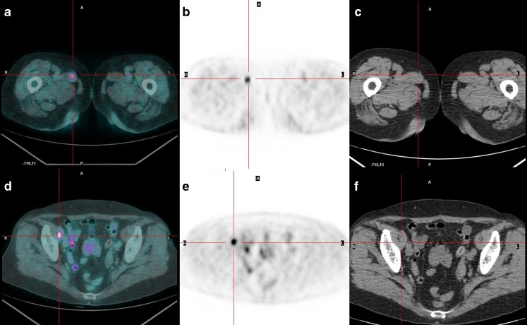
An 18F-FDG PET/CT showed two muscular distant lesions, involving the right adductor longus and right iliopsoas muscles. The lesions were confirmed as metastases from papillary carcinoma by biopsy (Fig. 1).
Fig. 1.
18F-FDG PET/CT confirms the presence of the lesion revealed by 131I scan and shows also two muscular distant lesions, involving the right adductor longus muscle (a PET/CT fused transaxial images, b PET transaxial images, c CT transaxial images) and right iliopsoas muscle (d PET/CT fused transaxial images, e PET transaxial images, f CT transaxial images). The lesions were confirmed as metastases from papillary carcinoma by biopsy. To the best of our knowledge, this is also the first described case of a double distant muscle metastasis imaged with 18F-FDG PET/CT
Although extrathyroidal extension to the soft tissues of the neck may occur, distant metastases are rare in patients affected by papillary carcinoma of the thyroid [3]. Skeletal muscle metastases from a differentiated thyroid carcinoma are extremely rare, and only a few cases are reported in the literature [4, 5]. To the best of our knowledge, this is also the first described case of a double distant muscle metastasis imaged with 18F-FDG PET/CT.
Acknowledgments
Conflict of interest
None.
References
- 1.Joensuu H, Ahonen A. Imaging of metastases of thyroid carcinoma with fluorine-18Fluorodeoxyglucose. J Nucl Med. 1987;28:910–4. [PubMed] [Google Scholar]
- 2.Hooft L, Hoekstra OS, Devillé W, Lips P, Teule GJ, Boers M, et al. Diagnostic accuracy of F-18-fluorodeoxyglucose positron emission tomography in the follow-up of papillary and follicular thyroid cancer. J Clin Endocrinol Metab. 2001;86:3779–86. doi: 10.1210/jc.86.8.3779. [DOI] [PubMed] [Google Scholar]
- 3.Panoussopoulos D, Theodoropoulos G, Vlahos K, Lazaris AC, Papadimitrou K. Distant solitary skeletal muscle metastasis from papillary thyroid carcinoma. Int Surg. 2007;92:226–9. [PubMed] [Google Scholar]
- 4.Qiu ZL, Luo QY. Erector spinae metastases from differentiated thyroid cancer identified by I-131 SPECT/CT. Clin Nucl Med. 2009;34:137–40. doi: 10.1097/RLU.0b013e31819675b6. [DOI] [PubMed] [Google Scholar]
- 5.Zhao L, Li L, Li F, Zhao Z. Rectus abdominis muscle metastasis from papillary thyroid cancer identified by I-131 SPECT/CT. Clin Nucl Med. 2010;35:360–1. doi: 10.1097/RLU.0b013e3181d6265b. [DOI] [PubMed] [Google Scholar]


