Abstract
The effect of aging on natural killer cell homeostasis is not well studied in humans or in animal models. We compared natural killer (NK) cells from young and aged mice to investigate age-related defects in NK cell distribution, and development. Our findings indicate aged mice have reduced NK cells in most peripheral tissues, but not in bone marrow. Reduction of NK cells in periphery was attributed to a reduction of the most mature CD11b+ CD27− NK cells. Apoptosis was not found to explain this specific reduction of mature NK cells. Analysis of NK cell development in bone marrow revealed that aged NK cells progress normally through early stages of development, but a smaller percentage of aged NK cells achieved terminal maturation. Less mature NK cells in aged bone marrow correlated with reduced proliferation of immature NK cells. We propose advanced age impairs bone marrow maturation of NK cells, possibly affecting homeostasis of NK cells in peripheral tissues. These alterations in NK cell maturational status have critical consequences for NK cell function in advanced age: reduction of the mature circulating NK cells in peripheral tissues of aged mice affects their overall capacity to patrol and eliminate cancerous and viral infected cells.
1. Introduction
Studies on immunosenescence have primarily focused on the impairment of adaptive immunity in part because of the reduced responsiveness of elderly people to vaccination (Gardner et al., 2001). It is well accepted that lymphocytes of adaptive immunity exhibit reduced function and altered composition with aging, but less is known about the lymphocytes of innate immunity, natural killer (NK) cells. NK cells are known as innate cells based on their spontaneous killing of tumor cells and their antiviral properties. The increased incidence of infectious diseases and cancer among the elderly, suggests NK cell responses are impaired in advanced ages. Because NK cells consist of various subsets with different functions, reduced function with advanced age may be the result of altered homeostasis. To date, there is an incomplete understanding of how aging affects NK cell homeostasis. In this study we examined NK cell phenotype, tissue distribution and development in a model of naturally aged C57BL/6J mice.
Our current understanding of NK cell development is that NK cells are produced in the bone marrow and seed the peripheral tissues during their last stages of maturation. Although immature NK cells can be found in liver, thymus, spleen and lymph nodes, the bone marrow is considered the primary site for NK cell development (Di Santo, 2008; Yokoyama et al., 2004). In the bone marrow, NK cell precursors (NKPs) undergo several stages of differentiation that can be tracked by the coordinated expression of cell surface markers (Kim et al., 2002). Immature NK cells that have acquired Ly49 receptors undergo functional maturation during a developmental stage that corresponds with an increase expression of maturation markers, and a significant expansion of their numbers in the bone marrow (Kim et al., 2002). It is proposed that NK cells acquire function after they express high levels of CD11b and CD43 (Kim et al., 2002). During these late developmental stages and after their release to the periphery, a reduction of CD27 and an increase of KLRG1 on NK cell surface is observed, making the CD11b+ CD27− KLRG1+ NK cells the most differentiated NK cell subset (Huntington et al., 2007). CD11b+ CD27− NK cells generally compose the majority of NK cells circulating in peripheral blood (up to 90%) and in non-lymphoid tissues. This NK cell subset is the major producer of IFN-γ and cytotoxic function upon activation (Di Santo, 2008; Yokoyama et al., 2004).
Our laboratory has previously shown that influenza infection is more severe in the absence of NK cells (Nogusa et al., 2008) and that aged mice have reduced NK cells infiltrating in the lungs during the early days of influenza infection (Beli et al., 2011; Nogusa et al., 2008). We also have shown that aged NK cells had reduced ability to produce IFN-γ in response to influenza infection and to various stimulants in vitro which was correlated with significantly reduced numbers and percentages of mature, CD11b+ CD27− NK cells in aged mice (Beli et al., 2011). In this manuscript, we show that aged mice have reduced NK cells in most peripheral tissues but not in the bone marrow. Reduction of total NK cells is attributed to a specific reduction of the mature, CD11b+ CD27− NK cell subset. Analysis of the developmental stages of NK cells in the bone marrow revealed that aged mice had similar NK cells belonging to the early stages of development but reduced NK cells in the terminal maturation stage, suggesting a block in their terminal maturation. We attribute the reduction of mature circulation of NK cells to reduced proliferation of NK cells in the bone marrow, as evidence for increased death in the peripheral tissues was not observed.
2. Materials and Methods
Mice
Male, C57BL/6J, young adult (6 month- from now on referred as young) and aged (22 month) mice were purchased from National Institute on Aging colony (Charles River Laboratories, Wilmington, MA, USA). Mice were acclimated for at least one week, housed in micro-isolator cages in a biosafety level 2 room at the Michigan State Research Containment Facility, an Association for Assessment and Accreditation of Laboratory Animal Care International certified facility. All animal procedures were approved by the Michigan State Institutional Animal Care and Use Committee and were in accordance with National Research Council guidelines.
Cell Preparation
Cells isolated from spleen and lungs were prepared according previously published procedures (Beli et al., 2011) with minor modifications. Collected lungs were dissociated using the GentleMACS™ Dissociator (Miltenyi Biotec Inc, Auburn, CA, USA). The homogenates were incubated at 37°C for 1 hour in a 5% CO2 incubator with digestion buffer [1mg/mL Collagenase D (Sigma-Aldrich) and 80 Kuntz units/mL (Roche) in RPMI-1640 media (Simga-Aldrich) with 5% FBS (Atlanta Biologicals) The digested tissue was grinded through a 40μm cell strainer and single cell suspensions washed 2 times with 5% FBS- RPMI media. Red blood cell lysis was performed with ACK lysis buffer. Spleens were grounded with a Dounce homogenizer, washed and directly lysed with ACK lysis buffer to remove red blood cells. Bone marrow cells were flushed from femurs according previously published methods (Hayakawa et al., 2010) and red blood cells were lysed directly with ACK lysis buffer Livers were homogenized using the GentleMACS™ Dissociator and lymphocytes were isolated using 37.5% Percoll solution according to previously published methods (Hayakawa et al., 2010). Blood was collected in heparin tubes, and mononuclear cells were isolated using 1083 Histopaque (Sigma-Aldrich, St. Louis, MO, USA) according to manufacturer instructions. For the lymph nodes, we collected inguinal and brachial lymph nodes. Single cell suspensions of lymph nodes were prepared by forcing through a 70 μm filter and washed with RPMI. The number of cells isolated from each tissue was determined by counting live cells using a hematocytometer and trypan blue staining.
Flow Cytometry
For cell surface staining, one million, and for intracellular staining, two million cells were stained with the appropriate antibodies on ice for 30 min according to previously published methods (Yokoyama and Kim, 2008). Before cell surface staining, cells were treated with Fc-Block (antibody to CD16/32) except during staining for CD16 on NK cells. A combination of 7 or 8 of the following monoclonal antibodies conjugated to FITC or Alexa Fluor-488, PE, PE-Cy7, PerCP-Cy5.5, Alexa Fluor-700, APC or efluor-647, APC-Cy7, Pacific Blue or efluor-450 and biotin was used: NK1.1 (PK136), NKp46 (29A1.4), CD3 (145-2C11), CD19 (1D3), CD11b (M1/70), CD27 (LG.3A10), KLRG1 (2F1), CD122 (TMb1), CD49b (DX5), CD43 (S7), NKG2D (CX5), NKG2ACE (20d5), NKG2A (16a11), CD94 (18d3), CD16/32 (93), 2B4 (eBio244F4), CD62L (MEL-14), CD11a (2D7), B220 (RA3-6B2), CD90.2 (53-2.1), CD69 (H1.2F3), CD127 (A7R34), Ly6C (AL-21), CXCR3 (CXCR3-173), CCR7 (4B12), CXCR6 (221002), CD160 (13-1601-80), PD-1 (J43), Ki67 (B56), Bim (Ham 151-149), Bcl-2 (3F11), Caspase-3 (C92-605), Annexin V and 7-AAD. Antibodies were purchased from (San Diego BD Pharmingen (La Jolla, CA, USA), ebiosciences, CA, USA), Biolegend (San Diego, CA, USA) or R&D Systems (Minneapolis, MN, USA). Streptavidin PE, PE-Cy7, or, APC-Cy7 (BD and ebiosciences) were used as secondary reagents for biotinylated antibodies. For intracellular staining, cells were first surface-stained followed by fixation/permeabilization using BD Bioscience Fix/Perm and Perm/Wash buffer according to manufacturer’s instructions. Subsequently, cells were incubated with antibodies for intracellular markers, washed, and analyzed by flow cytometry. Data were acquired with FACS-Canto II or LSR II (BD) and analyzed with FlowJo software (Treestar, Ashland, OR, USA). The lymphocyte population was gated based on forward and side scatter and NK cells were identified as CD3/CD19 negative and NK1.1/NKp46 positive cells within the lymphocyte gate. Total numbers of NK cells were calculated by multiplying the percentage of NK cells from the live cell gate by the total number of cells determined by hematocytometer counts for each tissue.
Determination of apoptosis
Freshly isolated single cell suspension of spleen and bone marrow cells were directly stained intracellularly with Bcl-2, Bim and caspace-3 according to the intracellular protocol described above. For annexin V staining we incubated splenocytes and bone marrow cells (2×10⁷ cells/mL) at 37°C for 10 hours in a 5% CO2 incubator in serum free media. Cells were collected and stained for cell surface markers and Annexin V according to manufacturer’s instructions (BD Biosciences).
BrdU incorporation assay
Mice were injected i.p. every 12 hours with 100uL of 1mg/mL BrdU for a total of 96 hours. Mice were euthanized, tissues processed, and cells were surfaced stained as described above. Subsequently, cells were fixed, permeabilized, and stained with anti-BrdU according to manufacture instructions (BD Biosciences).
Statistical Analysis
All statistics were generated using Prism 6. Two-tailed student’s t-test was used to compare adult and aged NK cells with a significance level of p<0.05, Welch’s correction was used on data with unequal variance which is specified in figure legends.
3. Results
NK cell phenotype is altered in aged mice
Initially we examined a plethora of markers associated with NK cells to characterize the phenotype of aged NK cells at resting conditions (Figure 1). Resting NK cells from the spleens of aged mice had similar expression of NKp46, CD16, 2B4, NKG2D, CD94, integrins CD11a and DX5, chemokine receptors CCR7 and CXCR6, activation marker CD69 and exhaustion/senescent molecules CD160 and PD-1. However, aged NK cells had reduced expression of markers associated with mature NK cells including CD43, CD11b, KLRG1, CD62L, and Ly6C and they had increased expression of markers found mainly on immature NK cells, such as NKG2AEC, CD27, CXCR3, CD127, CD90.2 and B220. Altogether our phenotypic characterization indicated that in aged mice NK cells some maturational defects.
Figure 1. Phenotypic characterization of NK cells from spleens of young and aged mice.
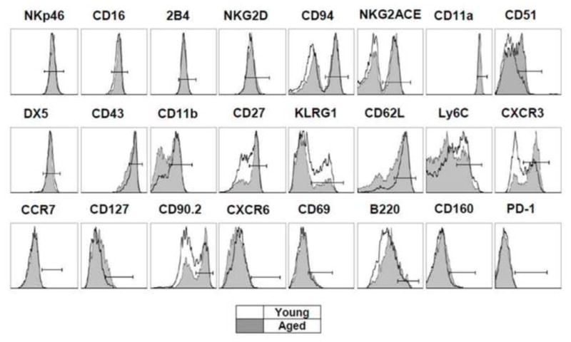
Histograms are representative of the average values for each group, n = 5–15, depending on the marker, but n is equal in young and aged NK cells for each marker. Empty histograms represent NK cells from young mice, while shaded histograms are NK cells from aged mice.
Aged mice have reduced total and mature NK cells in most peripheral tissues
Based on previous data indicating reduced NK cells in aged mice during influenza infection (Beli et al., 2011), we determined whether the distribution of NK cells in various tissues differs between adult and aged mice at resting conditions. Aged mice had significantly reduced percentage of NK cells in spleen, liver, lung, and blood (Figure 2A) and total numbers in spleen, liver and blood (Figure 2B). When we examined the distribution of CD11b/CD27 NK cells, we observed that aging resulted in a reduction of the relative frequency of terminal mature, CD11b+ CD27− NK cells in most of the peripheral tissues (Figure 2C). Reduced relative frequency of mature NK cells appeared to be due to a selective reduction of the absolute number of mature NK cells and not due to an increase of the immature NK cells (Figure 2D). We further examined NK cells for the distribution of KLRG1/CD27 subsets, another set of markers used to characterize NK subsets based on their maturation status. Immature NK cells express CD27 and lack KLRG1, while mature NK cells lack CD27 and express KLRG1 (Huntington et al., 2007). Consistent with what we observed for aged NK cell subsets based on CD11b and CD27, aged mice had increased percent of immature (KLRG1− CD27+) NK cells and reduced percent of mature (KLRG1+ CD27−) NK cells with aging (Figure 2E). Furthermore, the reduced frequency of KLRG1+ CD27− also appeared to be due to a selective reduction of the absolute number of mature NK cell and not due to an increase of the immature NK cells (data not shown). Altogether, we show that aged mice have reduced numbers of NK cells in the peripheral tissues but not in the bone marrow, where aged mice show similar if not higher NK cell numbers.
Figure 2. NK cell distribution in various peripheral tissues of young and aged mice.
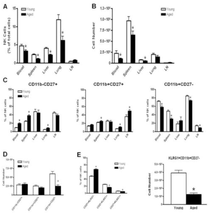
NK cells gated as NK1.1+, NKp46+, CD3ε−, CD19− cells. (A) NK cells as percentage of lymphocytes gated based on their FSC/SSC profile. (B) Numbers of NK cells in spleen (n=15), lungs (n=15), blood (NK cells/PBMCs per mL of blood) (n=15), liver (n=5) and lymph nodes (n= 5). (C) NK cell subsets based on CD11b/CD27 markers as a percentage of lymphocytes. (D) Numbers of CD11b/CD27 NK cell subsets in spleen. (E) NK cell subsets based on KLRG1/CD27 markers as percentage of lymphocytes. Asterisks (*) indicate statistically significant difference between young and aged groups, t-test, p < 0.05.
Aged mice have increased percentage of bone marrow NK cells that accumulate in stage IV of development
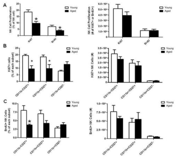
Having shown that aged NK cells have maturational defects that are detectable in peripheral tissues, we hypothesized that aging affects the development of NK cells in the bone marrow. First we compared the frequency of NK cells in the bone marrow; surprisingly, the frequency of NK cells in the bone marrow was increased in aged mice (Figure 3A). Total bone marrow NK cells were not different, albeit slightly higher in aged mice (Figure 3B). To examine whether NK cell maturation is blocked in the bone marrow of aged mice, we compared the stages of NK cell maturation (Di Santo, 2006; Yokoyama et al., 2004) following a gating strategy shown in Figure 3C. We observed that approximately the same percentage of NK cells were classified as being in early stages of development, Stages I–IV but less aged NK cells belonged to the subsequent stages of phenotypic maturation, Stage V (Figure 3D). As expected the majority of bone marrow NK cells are immature, CD11b− CD27+ NK cells that could belong to any developmental stage between I–IV. In aged mice, the immature (CD11b− CD27+) NK cell percent was significantly increased while the terminally mature (CD11b+ CD27−) NK cell percent was almost 2 fold less compared to young mice (Figure 3E), while there was no difference on the absolute numbers (data not shown). Because developing NK cells undergo expansion after they acquire their Ly49 receptors (Stage IV) and before they become mature (Stage V) (REF, Kim 2002), we examined whether proliferation of aged NK cells was reduced in the bone marrow, resulting in less mature NK cells. We used both Ki67 and the BrdU incorporation assay as indicators of proliferating cells. While we did not observe any differences in the spleen, a smaller percentage of NK cells from the bone marrow of aged mice were Ki67 and BrdU positive (Figure 4B). We then compared the ability of each CD11b/CD27 subset to proliferate in the bone marrow. We observed that a reduced percentage of immature CD11b− CD27+ were Ki67+ (Figure 4C) and BrdU+ (Figure 4D) in the bone marrow of aged mice compared to young. Therefore, less immature NK cells were proliferating in the aged bone marrow microenvironment. Comparisons of the absolute numbers of NK cells from each subset that had proliferated did not reflect the reduction in the percentages because aged mice have slightly higher NK cell total numbers. Overall these data indicate that in the aged bone marrow a smaller percentage of immature NK cells are proliferating. This reduction in the proliferation rates may indicate reduced turn over of NK cells or impaired generation of NK cells in general.
Figure 3. Bone marrow NK cell development.
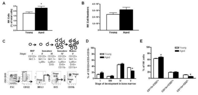
Bone marrow NK cells as a percent of lymphocytes (A) and total number (B) (n=10). (C) Scheme of NK cell development in the bone marrow {Kim, 2002 #40}. (D) NK cell subsets belonging to each stage of development as a percent of CD122+CD3−CD19− cells (n=5) (E) Maturation subsets of bone marrow NK cells as a percent of total NK cells (n=10).
Figure 4. NK cell proliferation in young and aged mice.
(A) Percentages and numbers of Ki67+ and BrdU+ NK cells in the bone marrow (n=8). (B) Percentages and numbers of Ki67+ cells for each CD11b/CD27 subset in the bone marrow (n=8). (C) Percentages and numbers of BrdU+ cells for each CD11b/CD27 subset in the bone marrow (n=8). Asterisks (*) indicate statistical differences between young and aged groups, t-test, p < 0.05.
NK cells from aged mice have similar survival as NK cells from young mice
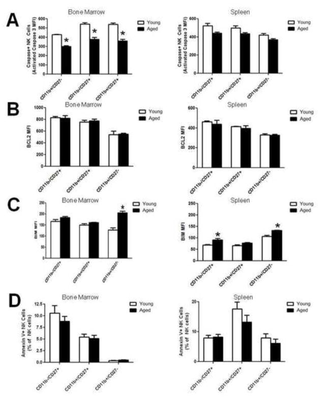
To understand the reasons of the reduced numbers of NK cells observed in the peripheral tissues we explored the apoptotic potential of NK cells in aged and young mice. To explore whether aged NK cells underwent higher degree of apoptotic death, we stained freshly isolated bone marrow and spleen cells for intracellular caspase -3, a downstream key player of apoptosis. As it is shown in figure 5A, aged NK cells have reduced caspace-3 staining in the bone marrow and similar to young in the spleen. These data suggest that aged NK cells are resistant to apoptosis. To confirm these data, we examined whether aged NK cells have increased expression of the anti-apoptotic molecule Bcl-2 and reduced expression of the proapoptotic molecule Bim. We observed that aged NK cells have similar levels of Bcl-2 as young NK cells but they had higher levels of Bim, which was only related to the most mature, CD11b+CD27− NK cells in the bone marrow and to some of the subsets in the spleen (Figure 5C). Finally, we incubated freshly isolated bone marrow and spleen single cell suspensions for 10 hours in-vitro in media that did not contain FBS or other growth factors and checked apoptosis induction by Annexin V staining. As it is shown in Figure 5D, neither NK cells from bone marrow nor from spleen of aged mice had higher Annexin- V staining under these conditions. Altogether, aged NK cells show resistance to apoptotic death through the pathways we examined and under homeostatic conditions other mechanisms contribute to the reduction of the numbers of NK cells in the peripheral tissues.
Figure 5. Analysis of the apoptotic potential of resting NK cells from young and aged mice.
(A) Freshly isolated bone marrow and spleen cells were stained intracellularly for caspace-3. Median Fluoresence Intensity (MFI) of expression for each NK cell subset, (n=5). (B) Freshly isolated bone marrow and spleen cells were stained intracellularly for Bcl-2. MFI of expression for each NK cell subset, (n=5). (C) Freshly isolated bone marrow and spleen cells were stained intracellularly for BIM. MFI of expression for each NK cell subset, (n=5). (D) Annexin staining for each NK subset for bone marrow and spleen cells incubated for 10 hrs without growth factors at 37°C, (n=10). Asterisks indicate statistical significant difference between the two age groups, t-test, p < 0.05.
4.5 Discussion
Previous studies from our laboratory implied that advanced age results in defects of NK cell function. During influenza infection aged mice had reduced infiltration of NK cells in their lungs and significant reduction of their function (Beli et al., 2011; Gardner, 2005; Nogusa et al., 2008). We also showed that aged mice had reduced mature NK cells in their spleen and lungs. Based on these data, we hypothesize that altered distribution of NK cell subsets with different maturational status results in an overall impairment of their function in aged mice. The studies presented herein examine NK cell distribution, maturation and development in aged mice and present novel data that support a blockage of NK cell maturation during their development in the bone marrow.
First, we observed that aged mice have reduced NK cells in major peripheral tissues but not in the bone marrow. There are previous reports that aged mice have reduced NK cells in their spleen (Beli et al., 2011; Dussault and Miller, 1994; Fang et al., 2010; Saxena et al., 1984) and increased NK cells in their bone marrow (King et al., 2009). Yet, because other studies report no age-related differences in NK cells (Dong et al., 2000; Koo et al., 1982; Nogusa et al., 2008; Plett and Murasko, 2000) and most human studies report increased numbers of circulating NK cells with aging (Almeida-Oliveira et al., 2011; Borrego et al., 1999; Facchini et al., 1987; Lutz et al., 2005; Sansoni et al., 1993; Vitale et al., 1992) the effect of aging on NK cell distribution required more attention. We identified NK cells by excluding B and T cells as well excluding cells that express NK cell markers such as NKT cells that are reported to be increased with aging (Plackett et al., 2004). Differences on the effects of aging in homeostasis of NK cells in humans and mice may be explained by the history of infections, vaccinations, and physiological changes, such as diet, occurring throughout the lifespan that shape the human immune system. Thus, what is observed in the elderly is not the result of aging alone. In contrast, in the aged mouse, housed in specific pathogen-free conditions and fed a standard diet, the immune system specifically reflects the effects of aging on immune cells. Finally, human studies are limited to peripheral blood samples and it is not known whether aging affects distribution of NK cells within other tissues. In our model, we observed that aging resulted in reduced NK cells in most peripheral tissues, with the exception of the bone marrow.
Reduced frequency of NK cells in peripheral tissues was attributed to a specific reduction of the mature CD11b+ CD27− NK cell subset. The suggestion that aged mice have less mature NK cells in their peripheral tissues is supported by very early studies comparing the lymphokine activated killer cell (LAK) activity in young and aged mice but it was not recognized and documented as a possible mechanism for alteration of the homeostasis and function of NK cells in aged individuals (Kawakami and Bloom, 1987). The fact that similar but delayed LAK activity was observed in splenocytes isolated from aged mice was attributed to a phenotypic difference between LAK precursors in aged mice from that of young mice. The majority of LAK precursors in young mice are considered to be mature NK cells while aged mice had reduced frequency of these precursors (Saxena et al., 1984). Additionally it was shown that LAK activity from bone marrow, which is comprised mostly from immature NK cells in both young and aged mice, was independent of age (Kawakami and Bloom, 1988). Indeed, LAK cells from bone marrow cells primarily consisted of CD90.2+ NK cells in both young and aged mice in contrast to spleens, where there is a preferential accumulation of CD90.2+ NK cells in aged mice (Kawakami and Bloom, 1988). Overall, past literature and our studies confirm that aged mice have reduced functionally mature NK cells in the peripheral tissues, but not in the bone marrow.
To understand why aged mice have reduced mature NK cells in the periphery, we examined the development of NK cells in the bone marrow. Our findings revealed that there was no age-related difference in the frequency of NK cells belonging at the early developmental stages when NK cells are still considered immature. Aged mice showed impairment only during the transition from Stage IV to Stage V before terminal maturation and upregulation of CD11b (Kim et al., 2002). At this stage, immature NK cells expand but at the bone marrow of aged mice, the level of expansion was reduced. Thus, we suggest that reduced proliferation in the bone marrow of aged mice may result in reduced production of NK cells. In contrast to bone marrow, proliferation of NK cells in the spleen was the same in both young and aged mice, similar to findings from peripheral blood NK cells in young and elderly humans (Lutz et al., 2011). Previous in vivo labeling experiments also showed NK cell production rates are decreased with aging in humans (Zhang et al., 2007) and in mice (Dussault and Miller, 1994). In humans, NK cells remain in the bone marrow for a significant time -almost 10 days - after their expansion and before they migrate to the peripheral blood to mature (Zhang et al., 2007). Thus, it is possible that bone marrow signals during that period affect NK cell maturation. Although impaired proliferation in the bone marrow can partially explain the reduced NK cell maturation, studies on the role of certain transcription factors regulating NK cell development and adoptive transfer experiments could give more insight into the specific mechanisms.
Reduced NK cells in peripheral tissues but not in bone marrow suggests impaired egress of mature NK cells and/or decreased migration to peripheral tissues in resting steady state conditions. Decreased terminal maturation of NK cells in the aged mice may impede the acquisition of the appropriate receptors and signals that would allow them to mature and egress from the bone marrow. However, in non-homeostatic conditions, mobilization of progenitor cells from the bone marrow of aged mice is not impaired but on the contrary GM-CSF induced mobilization was increased significantly in aged mice (Xing et al., 2006). The same authors showed adherence of hematopoietic stem cells to bone marrow stromal cells in aged mice was reduced because of altered integrin and adhesion molecule expression. Thus, it is possible that by altering expression of the appropriate adhesion molecules, aging affects the homing and homeostatic egress of NK cells from the bone marrow at steady state, while increases their mobilization during an inflammatory condition. Further research is required to shed light on the trafficking of NK cells in aged organisms. Such research can optimize conditions for cell therapies designed for the elderly.
Defects in the bone marrow of aged mice resulting in impaired generation of mature NK cells unable to seed the peripheral tissues can partly explain reduced NK cell homeostasis in the peripheral tissues. We therefore characterized the apoptotic potential of NK cells in aged mice. Our results indicate that the turn over of NK cells in aged mice is similar if not reduced compared to young mice. Caspace-3 staining on NK cells was reduced in the bone marrow of aged mice and similar to young mice in the spleen. Furthermore, no differences were observed in Bcl-2, an antiapoptotic molecule, neither with annexin V staining on NK cells that were incubated ex vivo for 10 hours in media without growth factors. The only indication that aged NK cells may undergo a stress was that some subsets had higher BIM expression possibly implicating the TGF-β induced apoptotic pathway (Ramesh et al., 2009). In contrast to what is observed in resting conditions, when we activated NK cells with YAC-1 cells at various effector to target ratio, aged NK cells showed increased Annexin V staining compared to young NK cells (data not shown). Similarly, it was shown that aged NK cells undergo higher apoptosis upon activation with IFN-α/β and YAC-1 cells, related to increased expression of IFNα/β receptor and CD95 in aged NK cells (Plett et al., 2000). However, higher activation induced death does not explain the reduced number of circulating NK cells in aged mice at resting conditions. Although we examined a variety of indicators of apoptosis it is possible that other molecules/executioners and pathways are implicating in the homeostatic turn over of NK cells in aged mice. Thus, we cannot exclude the possibility that aged NK cells do not survive very well in the aged circulation.
4.6 Conclusions
Overall, the data presented in this study are the first to characterize in detail the development of NK cells in aged mice and provide inside into their maturation. We propose a model shown in figure 5 where in aged mice, NK cells progress normally through the first stages of development, but during terminal maturation, the percentage of mature NK cells available to traffic to the periphery is significantly reduced. We propose that reduced proliferation at that stage may affect the generation of mature NK cells and their capacity to egress the bone marrow and seed the peripheral tissues. However, reduced generation of NK cells from the bone marrow can at least partially explain the reduced numbers of NK cells observed in the circulation. Nevertheless, the observed altered distribution of NK cell subsets with reduced maturation status and the reduced presence of NK cells in the circulation of aged mice can have severe implications in their immune responses to viral infections and cancers.
Figure 6. Proposed model for the effects of aging on NK cells.
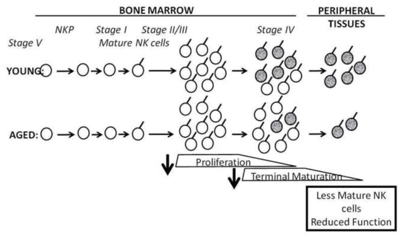
NK cells from aged mice proceed normally through the first stages of development, but because of reduced expansion during maturation, the percentage of mature NK cells ready to traffic to the periphery is significantly reduced. Overall altered distribution of NK cell subsets with reduced maturation status in the circulation of aged mice can have severe implications in their immune responses to viral infections and cancers (modified from (Di Santo, 2006; Yokoyama et al., 2004).
Highlights.
Aging modulates NK cell surface receptor expression
Aged mice have less CD11b+CD27− NK cells in peripheral tissues
Aged mice show a defect during late stages of maturation in the bone marrow, associated with reduced proliferation.
Acknowledgments
The research reported in this article was supported by NIA Grant R01AG034949.
Footnotes
The content is solely the responsibility of the authors and does not necessarily represent the official views of the NIA or NIH.
Publisher's Disclaimer: This is a PDF file of an unedited manuscript that has been accepted for publication. As a service to our customers we are providing this early version of the manuscript. The manuscript will undergo copyediting, typesetting, and review of the resulting proof before it is published in its final citable form. Please note that during the production process errors may be discovered which could affect the content, and all legal disclaimers that apply to the journal pertain.
References
- Almeida-Oliveira A, Smith-Carvalho M, Porto LC, Cardoso-Oliveira J, Ribeiro Ados S, Falcao RR, Abdelhay E, Bouzas LF, Thuler LC, Ornellas MH, Diamond HR. Age-related changes in natural killer cell receptors from childhood through old age. Hum Immunol. 2011;72:319–329. doi: 10.1016/j.humimm.2011.01.009. [DOI] [PubMed] [Google Scholar]
- Beli E, Clinthorne JF, Duriancik DM, Hwang I, Kim S, Gardner EM. Natural killer cell function is altered during the primary response of aged mice to influenza infection. Mech Ageing Dev. 2011;132:503–510. doi: 10.1016/j.mad.2011.08.005. [DOI] [PMC free article] [PubMed] [Google Scholar]
- Borrego F, Alonso MC, Galiani MD, Carracedo J, Ramirez R, Ostos B, Pena J, Solana R. NK phenotypic markers and IL2 response in NK cells from elderly people. Exp Gerontol. 1999;34:253–265. doi: 10.1016/s0531-5565(98)00076-x. [DOI] [PubMed] [Google Scholar]
- Di Santo JP. Natural killer cell developmental pathways: a question of balance. Annu Rev Immunol. 2006;24:257–286. doi: 10.1146/annurev.immunol.24.021605.090700. [DOI] [PubMed] [Google Scholar]
- Di Santo JP. Functionally distinct NK-cell subsets: developmental origins and biological implications. Eur J Immunol. 2008;38:2948–2951. doi: 10.1002/eji.200838830. [DOI] [PubMed] [Google Scholar]
- Dong L, Mori I, Hossain MJ, Kimura Y. The senescence-accelerated mouse shows aging-related defects in cellular but not humoral immunity against influenza virus infection. J Infect Dis. 2000;182:391–396. doi: 10.1086/315727. [DOI] [PubMed] [Google Scholar]
- Dussault I, Miller SC. Decline in natural killer cell-mediated immunosurveillance in aging mice--a consequence of reduced cell production and tumor binding capacity. Mech Ageing Dev. 1994;75:115–129. doi: 10.1016/0047-6374(94)90080-9. [DOI] [PubMed] [Google Scholar]
- Facchini A, Mariani E, Mariani AR, Papa S, Vitale M, Manzoli FA. Increased number of circulating Leu 11+ (CD 16) large granular lymphocytes and decreased NK activity during human ageing. Clin Exp Immunol. 1987;68:340–347. [PMC free article] [PubMed] [Google Scholar]
- Fang M, Roscoe F, Sigal LJ. Age-dependent susceptibility to a viral disease due to decreased natural killer cell numbers and trafficking. J Exp Med. 2010 doi: 10.1084/jem.20100282. [DOI] [PMC free article] [PubMed] [Google Scholar]
- Gardner EM. Caloric restriction decreases survival of aged mice in response to primary influenza infection. J Gerontol A Biol Sci Med Sci. 2005;60:688–694. doi: 10.1093/gerona/60.6.688. [DOI] [PubMed] [Google Scholar]
- Gardner EM, Bernstein ED, Dran S, Munk G, Gross P, Abrutyn E, Murasko DM. Characterization of antibody responses to annual influenza vaccination over four years in a healthy elderly population. Vaccine. 2001;19:4610–4617. doi: 10.1016/s0264-410x(01)00246-8. [DOI] [PubMed] [Google Scholar]
- Hayakawa Y, Andrews DM, Smyth MJ. Subset analysis of human and mouse mature NK cells. Methods Mol Biol. 2010;612:27–38. doi: 10.1007/978-1-60761-362-6_3. [DOI] [PubMed] [Google Scholar]
- Huntington ND, Tabarias H, Fairfax K, Brady J, Hayakawa Y, Degli-Esposti MA, Smyth MJ, Tarlinton DM, Nutt SL. NK cell maturation and peripheral homeostasis is associated with KLRG1 up-regulation. J Immunol. 2007;178:4764–4770. doi: 10.4049/jimmunol.178.8.4764. [DOI] [PubMed] [Google Scholar]
- Kawakami K, Bloom ET. Lymphokine-activated killer cells and aging in mice: significance for defining the precursor cell. Mech Ageing Dev. 1987;41:229–240. doi: 10.1016/0047-6374(87)90043-1. [DOI] [PubMed] [Google Scholar]
- Kawakami K, Bloom ET. Lymphokine-activated killer cells derived from murine bone marrow: age-associated difference in precursor cell populations demonstrated by response to interferon. Cell Immunol. 1988;116:163–171. doi: 10.1016/0008-8749(88)90218-3. [DOI] [PubMed] [Google Scholar]
- Kim S, Iizuka K, Kang HS, Dokun A, French AR, Greco S, Yokoyama WM. In vivo developmental stages in murine natural killer cell maturation. Nat Immunol. 2002;3:523–528. doi: 10.1038/ni796. [DOI] [PubMed] [Google Scholar]
- King AM, Keating P, Prabhu A, Blomberg BB, Riley RL. NK cells in the CD19− B220+ bone marrow fraction are increased in senescence and reduce E2A and surrogate light chain proteins in B cell precursors. Mech Ageing Dev. 2009;130:384–392. doi: 10.1016/j.mad.2009.03.002. [DOI] [PMC free article] [PubMed] [Google Scholar]
- Koo GC, Peppard JR, Hatzfeld A. Ontogeny of Nk-1+ natural killer cells. I. Promotion of Nk-1+ cells in fetal, baby, and old mice. J Immunol. 1982;129:867–871. [PubMed] [Google Scholar]
- Lutz CT, Karapetyan A, Al-Attar A, Shelton BJ, Holt KJ, Tucker JH, Presnell SR. Human NK cells proliferate and die in vivo more rapidly than T cells in healthy young and elderly adults. J Immunol. 2011;186:4590–4598. doi: 10.4049/jimmunol.1002732. [DOI] [PMC free article] [PubMed] [Google Scholar]
- Lutz CT, Moore MB, Bradley S, Shelton BJ, Lutgendorf SK. Reciprocal age related change in natural killer cell receptors for MHC class I. Mech Ageing Dev. 2005;126:722–731. doi: 10.1016/j.mad.2005.01.004. [DOI] [PMC free article] [PubMed] [Google Scholar]
- Nogusa S, Ritz BW, Kassim SH, Jennings SR, Gardner EM. Characterization of age-related changes in natural killer cells during primary influenza infection in mice. Mech Ageing Dev. 2008;129:223–230. doi: 10.1016/j.mad.2008.01.003. [DOI] [PubMed] [Google Scholar]
- Plackett TP, Boehmer ED, Faunce DE, Kovacs EJ. Aging and innate immune cells. J Leukoc Biol. 2004;76:291–299. doi: 10.1189/jlb.1103592. [DOI] [PubMed] [Google Scholar]
- Plett A, Murasko DM. Genetic differences in the age-associated decrease in inducibility of natural killer cells by interferon-alpha/beta. Mech Ageing Dev. 2000;112:197–215. doi: 10.1016/s0047-6374(99)00091-3. [DOI] [PubMed] [Google Scholar]
- Plett PA, Gardner EM, Murasko DM. Age-related changes in interferon-alpha/beta receptor expression, binding, and induction of apoptosis in natural killer cells from C57BL/6 mice. Mech Ageing Dev. 2000;118:129–144. doi: 10.1016/s0047-6374(00)00164-0. [DOI] [PubMed] [Google Scholar]
- Ramesh S, Wildey GM, Howe PH. Transforming growth factor beta (TGFbeta)-induced apoptosis: the rise & fall of Bim. Cell Cycle. 2009;8:11–17. doi: 10.4161/cc.8.1.7291. [DOI] [PMC free article] [PubMed] [Google Scholar]
- Sansoni P, Cossarizza A, Brianti V, Fagnoni F, Snelli G, Monti D, Marcato A, Passeri G, Ortolani C, Forti E, et al. Lymphocyte subsets and natural killer cell activity in healthy old people and centenarians. Blood. 1993;82:2767–2773. [PubMed] [Google Scholar]
- Saxena RK, Saxena QB, Adler WH. Interleukin-2-induced activation of natural killer activity in spleen cells from old and young mice. Immunology. 1984;51:719–726. [PMC free article] [PubMed] [Google Scholar]
- Vitale M, Zamai L, Neri LM, Galanzi A, Facchini A, Rana R, Cataldi A, Papa S. The impairment of natural killer function in the healthy aged is due to a postbinding deficient mechanism. Cell Immunol. 1992;145:1–10. doi: 10.1016/0008-8749(92)90307-b. [DOI] [PubMed] [Google Scholar]
- Xing Z, Ryan MA, Daria D, Nattamai KJ, Van Zant G, Wang L, Zheng Y, Geiger H. Increased hematopoietic stem cell mobilization in aged mice. Blood. 2006;108:2190–2197. doi: 10.1182/blood-2005-12-010272. [DOI] [PMC free article] [PubMed] [Google Scholar]
- Yokoyama WM, Kim S. Analysis of individual natural killer cell responses. Methods Mol Biol. 2008;415:179–196. doi: 10.1007/978-1-59745-570-1_11. [DOI] [PubMed] [Google Scholar]
- Yokoyama WM, Kim S, French AR. The dynamic life of natural killer cells. Annu Rev Immunol. 2004;22:405–429. doi: 10.1146/annurev.immunol.22.012703.104711. [DOI] [PubMed] [Google Scholar]
- Zhang Y, Wallace DL, de Lara CM, Ghattas H, Asquith B, Worth A, Griffin GE, Taylor GP, Tough DF, Beverley PC, Macallan DC. In vivo kinetics of human natural killer cells: the effects of ageing and acute and chronic viral infection. Immunology. 2007;121:258–265. doi: 10.1111/j.1365-2567.2007.02573.x. [DOI] [PMC free article] [PubMed] [Google Scholar]


