Abstract
Objective
The aim of this study was to evaluate the marginal adaptation of Biodentine in comparison with Mineral Trioxide Aggregate (MTA) and Intermediate Restorative Material (IRM), as a root end filling material, using Scanning Electron Microscopy (SEM).
Materials and Methods:
Thirty permanent maxillary central incisors were chemo-mechanically prepared and obturated. Three millimetres of the root end were resected and 3mm retro cavity preparation was done using ultrasonic retrotips. The samples were randomly divided into three groups (n=10) and were restored with root end filling materials: Group I – MTA, Group II – Biodentine, Group III – IRM. The root ends were sectioned transversely at 1mm and 2mm levels and evaluated for marginal adaptation using SEM. The gap between dentin and retro filling material was measured at four quadrants. The mean gap at 1mm level and 2mm level from the resected root tip and combined mean were calculated. The data were statistically analyzed, using one-way ANOVA and Tukey’s HSD post hoc test for intergroup analysis and paired t-test for intragroup analysis.
Results:
The overall results showed no statistically significant difference between MTA and IRM but both were superior when compared to Biodentine. At 1mm level there was no statistically significant difference among any of the tested materials. At 2mm level MTA was superior to both IRM and Biodentine.
Conclusion:
In overall comparison, MTA and IRM were significantly superior when compared to Biodentine in terms of marginal adaptation, when used as retrograde filling material.
Keywords: Marginal adaptation, Biodentine, MTA, IRM, Root end filling material, Scanning electron microscopy
INTRODUCTION
The success of periapical surgery is dictated by elimination of infected tissues and adequate apical seal [1]. Ideal apical seal prevents ingress of residual irritants into the periapical region and percolation of periapical tissue fluid in to the canal system [2]. Various root end filling materials have been tested for their sealing ability and newer materials are still under research. The root end filling material should possess ideal properties such as bio-compatibility, dimensional stability, radiopacity, ability to set in a wet environment, antibacterial properties, easy handling, adequate compressive strength and hardness, osteoinductive and osteoconductive properties and adherence to the canal walls to provide a good apical seal [3,4]. Among the various root end filling materials tested, Mineral Trioxide Aggregate (MTA) has shown good sealing ability and biocompatibility in previous in-vitro and in-vivo studies [5]. In recent years, various materials like Biodentine [6], CER (Cemento Endodontico Rapido/Fast endodontic cement) [7], ERRM (Endosequence Root Repair Material) [8] and Endocem (MTA-derived pozzolan cement) [9] have been introduced with the aim of overcoming some of the disadvantages of the MTA, such as the difficulties in handling and long setting time [10, 11, 12].
Biodentine is a relatively new material introduced as a dentine substitute. Biodentine powder is mainly composed of highly pure tricalcium silicate, which regulates the setting reaction. Other components are calcium carbonate (filler) and zirconium dioxide (radiopacifier). The liquid contains calcium chloride (setting accelerator), water reducing agent (super-plasticizer) and water. The super-plasticizer reduces the viscosity of the cement and improves handling [6]. The manufacturer claims that this material can be used for pulp capping, pulpotomy, apexification, root perforation, internal and external resorption and also as a root end filling material in periapical surgery. In the previous studies, Biodentine showed biocompatibility and the ability to induce odontoblast differentiation and mineralization in cultured pulp cells [13]. The main benefits of Biodentine over other calcium silicate based materials are the reduced setting time, better handling and mechanical properties [11]. The importance of marginal adaptation is that it may have an indirect correlation with the sealing ability of retro-filling materials. There is no previous study assessing the marginal adaptation of Biodentine when used as a root end filling material [14]. Hence, the aim of this study was to evaluate the marginal adaptation of Biodentine in comparison with MTA and IRM, as a root end filling material, using Scanning Electron Microscopy (SEM).
MATERIALS AND METHODS
Thirty freshly extracted maxillary central incisors with mature apices were selected for the study. All the teeth were cleaned, autoclaved and stored in 0.2% thymol solution until they were used. Access cavity preparation was done using a #2 round diamond point (NSK, Japan) and coronal preflaring was done using Gates-Glidden drills (MANI, Inc, Japan).
Size #10 K-file (Mani, Inc, Japan) was introduced into the root canal until it was visible at the apex and then 1mm was subtracted from that point to establish the working length. Biomechanical preparation was done using step-back technique with apical enlargement up to #60 size K-file (Mani Inc., Japan). Copious irrigation with 3% sodium hypochlorite (Vensons, India) was done all through the procedure. Final irrigation was done with 17% EDTA (Prime Dental Products, India) followed by 3% sodium hypochlorite for 1 minute each and rinsing with saline. The canals were dried using absorbent points and obturation was done with 2% gutta percha points (Dentsply Maillefer, China) and zinc oxide eugenol sealer (Bombay Burmah trading corp., Mumbai, India), using the lateral condensation technique. After 24 hrs. of obturation, the root ends were resected 3mm from the apex using a No.1557 fissure bur; retrograde cavity was prepared to a depth of 3mm coaxially using surgical ultrasonic retro-preparation tips (Satelec AS6D, France).
Then the teeth were randomly divided into 3 groups, with each group containing 10 teeth.
Group 1- MTA (Dentsply/Tulsa Dental, Tulsa, OK),
Group 2- Biodentine (Septodont, Saint Maurdes Fossés, France),
Group 3- IRM (Dentsply International Inc, U.S.A).
Each group received their corresponding root end filling material. All the materials were mixed according to the instructions given by the manufacturers.
All root end filling materials were placed incrementally, following which radiographs were taken labio-palatally and mesio-distally to confirm proper filling of the material. After root end filling, a moist cotton pellet was placed on MTA for setting of the material and all the samples were stored in relative humidity (95%) at 37°C for 5 days.
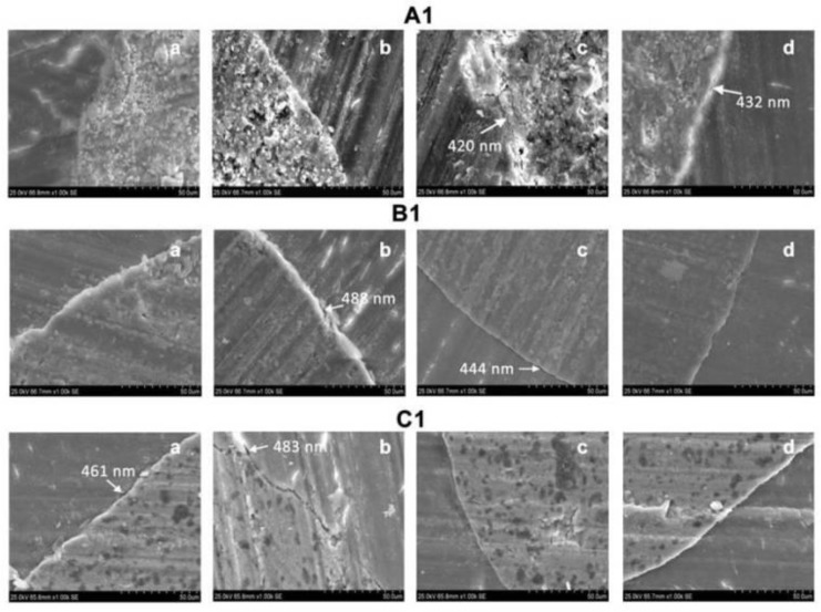
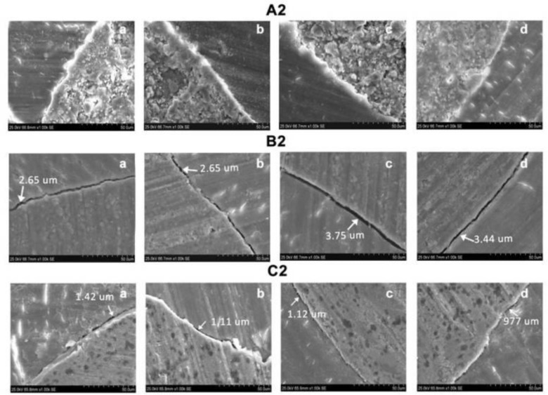
The samples were mounted in a resin block to create a platform for sectioning with a hard tissue microtome and sectioned apically at 1mm and 2mm levels from the apex. The samples were gold sputtered and viewed under scanning electron microscopy (1000X magnification) for evaluating the adaptation of the material to the canal walls. The largest gap present between the material and canal wall was measured in four quadrants (Fig 1 and Fig 2) and the mean gap was calculated for each sample.
Fig 1.
A1 (a–d), B1 (a–d), C1 (a–d) Marginal adaptation of MTA, Biodentine and IRM respectively at 1mm level under SEM at X 1000 magnification.
Fig 2.
A2 (a–d), B2 (a–d), C2 (a–d) Marginal adaptation of MTA, Biodentine and IRM respectively at 2mm level under SEM at X 1000 magnification.
Statistical Analysis:
The data obtained were recorded and analyzed. One-way ANOVA and Tukey’s HSD Post-hoc test were done for intergroup analysis to compare the 1mm, 2mm and overall values of the three groups; Paired t-test was used for intragroup analysis to compare the 1mm and 2mm values within each group, with the significance level of 0.05 using SPSS version 17.0 software.
RESULTS
The overall results showed that the mean gap at the dentin-retrograde filling material interface was maximum for Biodentine (1. 446 ± 0.367 μm), followed by IRM (0. 942 ± 0.353 μm) and MTA (0. 792 ± 0.201 μm).
The difference between MTA and IRM was not statistically significant (P>0.05). But both showed statistically significant difference when compared to Biodentine (P<0.05).
Intra-group and inter-group comparison at 1mm level and 2mm level are shown in Table 1. At 1mm level there was no significant difference among the groups.
Table 1.
Marginal gap at dentin-retrograde filling material interface at 1mm and 2mm levels.
| MTA Mean ± SD (μm) | Biodentine Mean ± SD (μm) | IRM Mean ± SD (μm) | P value | |
|---|---|---|---|---|
| Root Section at 1 mm | 0.847 ± 0.298 | 1.345 ± 0.717 | 0.689 ± 0.699 | 0.056 |
| Root Section at 2 mm | 0.738 ± 0.466 | 1.489 ± 0.459 | 1.362 ± 0.425 | 0.002 |
| p value | 0.623 | 0.632 | 0.029 |
At 2mm level, MTA was superior to both IRM and Biodentine.
DISCUSSION
A successful periapical surgery requires appropriate root-end resection, preparation and adequate apical seal. In root-end resection at least 3mm of the root-end must be eliminated to reduce 98% of the apical ramifications and 93% of the lateral canals, which might be responsible for endodontic failure.
Perpendicular resection minimizes the number of exposed dentinal tubules [15]. Hence 3mm of root-end was resected perpendicular to the long axis of the tooth in this study. Ultrasonic tips were reported to have better control and ability to stay centered in the canal and reduce perforation risk [16]. Since diamond coated ultrasonic tips reduce the chance for microcrack formation [17], the diamond coated ultrasonic tips were used to prepare a 3mm retrograde cavity coaxially.
Incremental placement of the retrograde filling material was done to minimize the voids and enhance the quality of the filling. Numerous materials are used as retrograde filling material namely MTA, GIC, IRM, super EBA and composite resins. Biodentine is a relatively new tricalcium silicate based material, which forms hydrated calcium silicate gel (CSH) and calcium hydroxide on hydration. Marginal adaptation is one of the desirable properties for a retrograde filling material. SEM aids in assessing the marginal adaptation at the filling material - tooth interface under higher magnification [18].
Torabinejad et al. claimed that the longitudinal type of sectioning might create false gaps in the interface between dentin and root end filling material thereby affecting the evaluation of marginal adaptation [19].
Transverse sections allow the visualization of the restoration-dentin interface throughout the circumference. A few previous studies have evaluated the marginal adaptation of the root end filling material to the canal wall using resin replicas. Orosco et al. stated that for evaluation of marginal adaptation of the retrofilling material, the samples can be directly viewed under SEM after gold sputtering and there is no need for creation of resin replicas, as direct SEM evaluation of the samples did not result in artificial gap formation [20].
Hence, we sectioned the samples transversely and examined the interface directly under SEM [21].
Marginal adaptation was evaluated only at 1mm and 2mm levels; 3mm level section was avoided, as it would encroach upon the junction between the retrograde filling material and the gutta-percha [22]. It is not clear why at 1mm level there was no difference in marginal adaptation among the three tested materials. But at 2mm level, MTA was superior to IRM and Biodentine and this superiority of MTA over IRM for marginal adaptation is in accordance with a previous report by Torabinejad et al [19]. Furthermore in the overall result, MTA and IRM were superior to Biodentine.
In a recent study, a fast setting MTA-derived pozzolan cement (Endocem), showed tight sealing with the mould comparable to MTA [9]. But in this study, Biodentine which is a fast setting tricalcium silicate based material showed inferior marginal adaptation when compared to MTA and IRM. In previous clinical trials, comparable surgical success rates were reported for MTA and IRM. In a randomized controlled trial on surgical success rate, Chong et al. concluded that MTA was not superior to IRM [23]. Lindeboom et al. showed that MTA and IRM had similar clinical results when used as a root end filling material [24]. Another clinical study by Zuolo et al. showed that the surgical success rate of IRM as a root end filling material was 91.2% clinically and radiographically, for a follow up period of 4 years [25]. The inadequacy in the marginal adaptation may influence the sealing ability and the clinical success rate.
A few properties such as biocompatibility and the ability to induce mineralization have been studied earlier.
Other properties such as washout resistance and dimensional stability have not yet been evaluated. These properties need to be investigated, since Biodentine has a very limited published literature supporting its use.
CONCLUSION
As far as this in-vitro study can discern, it may be concluded that:Marginal adaptation at the 1mm level was similar among MTA, IRM and Biodentine. At the 2mm level, MTA was superior to both IRM and Biodentine. In the overall comparison, the marginal adaptation of MTA and IRM were superior to Biodentine.
REFERENCES
- 1.Kim S, Kratchman S. Modern endodontic surgery concepts and practice: a review. J Endod. 2006 Jul;32(7):601–23. doi: 10.1016/j.joen.2005.12.010. [DOI] [PubMed] [Google Scholar]
- 2.Gutmann JL, Harrison JW. Surgical Endodontics. 1st ed. Boston: Blackwell Scientific Publications; 1991. [Google Scholar]
- 3.Johnson BR, Fayad MI, Witherspoon DE. Periradicular surgery. In: Hargreaves KM, Cohen S, editors. Cohen’s Pathways of the pulp. 10th ed. St. Louis: Mosby; 2011. pp. 720–76. [Google Scholar]
- 4.Chong BS, Ford TRP. Root-end filling materials: rationale and tissue response. Endodontic Topics. 2005;11(1):114–30. [Google Scholar]
- 5.Torabinejad M, Parirokh M. Mineral trioxide aggregate : A comprehensive literature review-part II : leakage and biocompatibility investigations. J Endod. 2010 Feb;36(2):190–202. doi: 10.1016/j.joen.2009.09.010. [DOI] [PubMed] [Google Scholar]
- 6.Goldberg M, Pradelle-Plasse N, Tran XV, Colon P, Laurent P, Aubut V, et al. Emerging trends in (bio) material research Physico – chemical properties of Biodentine. In: Goldberg M, editor. Biocompatibility or cytotoxic effects of dental composites. 1st ed. Oxford: Coxmoor publishing co; 2009. pp. 181–203. [Google Scholar]
- 7.Gomes-Filho JE, Rodrigues G, Watanabe S, Estrada Bernabé PF, Lodi CS, Gomes AC, et al. Evaluation of the tissue reaction to fast endodontic cement (CER) and Angelus MTA. J Endod. 2009 Oct;35(10):1377–80. doi: 10.1016/j.joen.2009.06.010. [DOI] [PubMed] [Google Scholar]
- 8.Ma J, Shen Y, Stojicic S, Haapasalo M. Biocompatibility of two novel root repair materials. J Endod. 2011 Jun;37(6):793–8. doi: 10.1016/j.joen.2011.02.029. [DOI] [PubMed] [Google Scholar]
- 9.Choi Y, Park S, Lee S, Hwang Y, Yu M, Min K. Biological effects and washout resistance of a newly developed fast-setting pozzolan cement. J Endod. 2013 Apr;39(4):467–72. doi: 10.1016/j.joen.2012.11.023. [DOI] [PubMed] [Google Scholar]
- 10.Asgary S, Shahabi S, Jafarzadeh T, Amini S, Kheirieh S. The properties of a new endodontic material. J Endod. 2008 Aug;34(8):990–3. doi: 10.1016/j.joen.2008.05.006. [DOI] [PubMed] [Google Scholar]
- 11.Santos AD, Moraes JCS, Araujo EB, Yukimitu K, Valerio Filho WV. Physico-chemical properties of MTA and a novel experimental cement. Int Endod J. 2005 Jul;38(7):443–7. doi: 10.1111/j.1365-2591.2005.00963.x. [DOI] [PubMed] [Google Scholar]
- 12.Ber BS, Hatton JF, Stewart GP. Chemical modification of ProRoot MTA to improve handling characteristics and decrease setting time. J Endod. 2007 Oct;33(10):1231–4. doi: 10.1016/j.joen.2007.06.012. [DOI] [PubMed] [Google Scholar]
- 13.Zanini M, Sautier JM, Berdal A, Simon S. Biodentine Induces Immortalized Murine Pulp Cell Differentiation into Odontoblast - like Cells and Stimulates Biomineralization. J Endod. 2012 Sep;38(9):1220–6. doi: 10.1016/j.joen.2012.04.018. [DOI] [PubMed] [Google Scholar]
- 14.Stabholz A, Shani J, Friedman S. Marginal adaptation of retrograde fillings and its correlation with sealability. J Endod. 1995 May;11(5):218–23. doi: 10.1016/s0099-2399(85)80063-7. [DOI] [PubMed] [Google Scholar]
- 15.Mjör IA, Smith MR, Ferrari M, Mannose F. The structure of dentine in the apical region of human teeth. Int Endod J. 2001 Jul;34(7):346–53. doi: 10.1046/j.1365-2591.2001.00393.x. [DOI] [PubMed] [Google Scholar]
- 16.Engel TK, Steiman HR. Preliminary investigation of ultrasonic root end preparation. J Endod. 1995 Sep;21(9):443–5. doi: 10.1016/S0099-2399(06)81524-4. [DOI] [PubMed] [Google Scholar]
- 17.Rodríguez-Martos R, Torres-Lagares D, Castellanos-Cosano L, Serrera-Figallo MA, Segura-Egea JJ, Gutierrez-Perez JL. Evaluation of apical preparations performed with ultrasonic diamond and stainless steel tips at different intensities using a scanning electron microscope in endodontic surgery. Med Oral Patol Oral Cir Bucal. 2012 Nov;17(6):e988–93. doi: 10.4317/medoral.17961. [DOI] [PMC free article] [PubMed] [Google Scholar]
- 18.Badr AE. Marginal Adaptation and Cytotoxicity of Bone Cement Compared with Amalgam and Mineral Trioxide Aggregate as Root-end Filling Materials. J Endod. 2010 Jun;36(6):1056–60. doi: 10.1016/j.joen.2010.02.018. [DOI] [PubMed] [Google Scholar]
- 19.Torabinejad M, Smith PW, Kettering JD, Pitt Ford TR. Comparative investigation of marginal adaptation of mineral trioxide aggregate and other commonly used root-end filling materials. J Endod. 1995 Jun;21(6):295–9. doi: 10.1016/S0099-2399(06)81004-6. [DOI] [PubMed] [Google Scholar]
- 20.Orosco FA, Bramante CM, Garcia RB, Bernardineli N, de Moraes IG. Sealing ability, marginal adaptation and their correla tion using three root-end filling materials as apical plugs. J Appl Oral Sci. 2010 Mar-Apr;18(2):127–34. doi: 10.1590/S1678-77572010000200006. [DOI] [PMC free article] [PubMed] [Google Scholar]
- 21.Costa AT, Post LK, Xavier CB, Weber JB, Gerhardt-Oliveira M. Marginal adaptation and microleakage of five root-end filling materials : an in vitro study. Minerva Stomatol. 2008 Jun;57(6):295–300. [PubMed] [Google Scholar]
- 22.Xavier CB, Weismann R, de Oliveira MG, Demarco FF, Pozza DH. Root-end filling materials: apical microleakage and marginal adaptation. J Endod. 2005 Jul;31(7):539–42. doi: 10.1097/01.don.0000152297.10249.5a. [DOI] [PubMed] [Google Scholar]
- 23.Chong BS, Pitt Ford TR, Hudson MB. A prospective clinical study of Mineral Trioxide Aggregate and IRM when used as root-end filling materials in endodontic surgery. Int Endod J. 2003 Aug;36(8):520–6. doi: 10.1046/j.1365-2591.2003.00682.x. [DOI] [PubMed] [Google Scholar]
- 24.Lindeboom JA, Frenken JW, Kroon FH, van den Akker HP. A comparative prospective randomized clinical study of MTA and IRM as root-end filling materials in single-rooted teeth in endodontic surgery. Oral Surg Oral Med Oral Pathol Oral Radiol Endod. 2005 Oct;100(4):495–500. doi: 10.1016/j.tripleo.2005.03.027. [DOI] [PubMed] [Google Scholar]
- 25.Zuolo ML, Ferreira MO, Gutmann JL. Prognosis in periradicular surgery: a clinical prospective study. Int Endod J. 2000 Mar;33(2):91–8. doi: 10.1046/j.1365-2591.2000.00263.x. [DOI] [PubMed] [Google Scholar]


