Abstract
Genetic analysis of Rickettsia prowazekii has been hindered by the lack of selectable markers and efficient mechanisms for generating rickettsial gene knockouts. We have addressed these problems by adapting a gene that codes for rifampin resistance for expression in R. prowazekii and by incorporating this selection into a transposon mutagenesis system suitable for generating rickettsial gene knockouts. The arr-2 gene codes for an enzyme that ADP-ribosylates rifampin, thereby destroying its antibacterial activity. Based on the published sequence, this gene was synthesized by PCR with overlapping primers that contained rickettsial codon usage base changes. This R. prowazekii-adapted arr-2 gene (Rparr-2) was placed downstream of the strong rickettsial rpsL promoter (rpsLP), and the entire construct was inserted into the Epicentre EZ::TN transposome system. A purified transposon containing rpsLP-Rparr-2 was combined with transposase, and the resulting DNA-protein complex (transposome) was electroporated into competent rickettsiae. Following selection with rifampin, rickettsiae with transposon insertions in the genome were identified by PCR and Southern blotting and the insertion sites were determined by rescue cloning and inverse PCR. Multiple insertions into widely spaced areas of the R. prowazekii genome were identified. Three insertions were identified within gene coding sequences. Transposomes provide a mechanism for generating random insertional mutations in R. prowazekii, thereby identifying nonessential rickettsial genes.
Rickettsia prowazekii, the causative agent of epidemic typhus, is an obligate, intracellular, parasitic bacterium that grows directly within the cytoplasm of its eukaryotic host cell, unbounded by a vacuolar membrane. R. prowazekii is exquisitely adapted to this cytoplasmic environment, as evidenced by its expression of specialized systems, such as those for the transport of high-energy compounds, that exploit this metabolically rich environment (24, 25). However, rickettsial obligate dependence on the intracytoplasmic milieu does not prevent this versatile pathogen from infecting organisms as diverse as humans, flying squirrels, and the arthropod vector of epidemic typhus, the human body louse. Rickettsial pathogenicity, in both the arthropod vector and the human host, is due to intracellular growth of the rickettsiae followed by the lysis of the host cell and the infection of additional cells. Thus, an understanding of rickettsial growth and metabolism within the eukaryotic cytoplasm is essential to understanding the basis of rickettsial pathogenicity.
Previous studies on the physiology of rickettsial intracellular growth are now complemented by the genome sequences of several rickettsial species, including that of R. prowazekii (2, 19). The R. prowazekii genome contains 834 open reading frames (ORFs), a number of pseudogenes, and a high proportion of noncoding regions (2). While some of the 834 gene products can be annotated confidently by their extensive identity to proteins of known function in other organisms, there are a large number of genes of questionable annotation or unknown function.
Analysis of gene function in the rickettsiae is hampered not only by the obvious difficulties associated with manipulating an obligate intracellular pathogen, but also by a paucity of genetic tools, including selectable markers and efficient mechanisms for generating gene knockouts. Rachek et al. demonstrated that DNA could be introduced into R. prowazekii by electroporation and that the transforming rickettsial DNA could be inserted into the chromosome by homologous recombination (21). For this transformation, the target gene was rpoB and a point mutation that imparted rifampin resistance served as the selectable marker. Subsequently, Rachek and coworkers employed the Escherichia coli ereB erythromycin resistance gene as a selectable marker in rickettsial transformation and demonstrated targeted insertion of the transforming plasmid into the gltA locus of R. prowazekii (20). Unfortunately, erythromycin selection for rickettsial transformants proved to be problematic. The number of rickettsiae per host cell can affect the ability of the antibiotic to inhibit sensitive rickettsiae, and during selection there is a background of high spontaneous erythromycin resistance (20). The use of green fluorescent protein (GFP) as a screenable marker in rickettsial transformations has also been reported for Rickettsia typhi and for Rickettsia conorii (17, 19). For these experiments, gfp was fused to the rpoB gene for homologous recombination into the genome. Although insertion was confirmed by PCR and a population of rickettsiae-infected cells was identified by flow cytometry, individually fluorescent rickettsiae could not be detected by fluorescent microscopy. These rickettsial transformation studies demonstrate the feasibility of generating rickettsial mutants by insertional inactivation. However, to our knowledge, such mutants have not been isolated. In this report, we describe an additional antibiotic selection, rifampin resistance generated by ribosylation of the antibiotic (8, 18, 23), that, when coupled with an efficient transposon mutagenesis system (10, 11), provides a functional mechanism for the insertional mutagenesis of R. prowazekii and the identification of nonessential rickettsial genes.
MATERIALS AND METHODS
Bacterial strains and culture conditions.
The bacterial strains and plasmids used in this study are listed in Table 1. Rickettsiae were purified from the yolk sacs of embryonated chicken eggs inoculated with R. prowazekii (passage 283) as described previously (25). Rickettsiae purified from yolk sacs were used in transformation experiments directly, after suspension in 0.25 M sucrose, or were frozen in a sucrose-phosphate-glutamate-magnesium solution (SPG-Mg; 0.218 M sucrose, 3.76 mM KH2PO4, 7.1 mM K2HPO4, 4.9 mM potassium glutamate, and 10 mM MgCl2) and subsequently used for infecting mouse fibroblast L929 cells. Rickettsia-infected L929 cells were grown in modified Eagle medium (Mediatech, Atlanta, Ga.) supplemented with 10% newborn-calf serum and 2 mM l-glutamine (Sigma, St. Louis, Mo.). The isolation of rickettsiae from infected L929 cells was carried out according to previously published procedures (21) with the following modification for releasing rickettsiae from infected cells. Rickettsiae-infected L929 cells (three 185-cm2 tissue culture flasks containing approximately 2 × 10⁷ cells/flask with 200 to 300 rickettsiae/cell) were disrupted with the use of minibeads and a minibead mixer (United Laboratory Plastics, St. Louis, Mo.). Briefly, the infected cells were collected by centrifugation at 4°C and suspended in 3 ml of cold SPG-Mg. A 1-ml aliquot of the suspended cells was placed into a 2-ml tube containing 1.2 g of 1-mm-diameter glass beads. The tube was shaken at 4,800 rpm for 10 s and cooled on ice, and the shaking was repeated twice. Rickettsiae were separated from host cell debris by centrifugation and suspended in SPG-Mg for storage or in 0.25 M sucrose for use in transformations (∼1 × 10¹⁰ rickettsiae/ml). Rickettsial electroporations were performed as previously described (21). With a field strength of 17 kV/cm, there was no loss of rickettsial viability (21). For selection, rifampin was added to supplemented medium at a final concentration of 200 ng/ml and the rifampin-containing medium was changed every 2 to 3 days. Rickettsial growth was monitored by microscopic examination of Gimenez-stained (9) infected cells on coverslips. Chromosomal DNA was isolated with the PUREGENE DNA purification kit (Gentra Systems, Inc., Minneapolis, Minn.), while plasmid DNA was isolated with a QIAprep spin miniprep kit (QIAGEN, Valencia, Calif.). PCR products were isolated by gel electrophoresis, excised from the gel, and purified with the QIAquick gel extraction kit (QIAGEN). DNA sequencing was performed by the DNA Sequencing and Synthesis Facility, Iowa State University, Ames. Probes used in Southern hybridizations (22) were 32P-labeled by using the Multiprime DNA labeling system (Amersham, Piscataway, N.J.) and [α-32P]dATP (ICN, Irvine, Calif.).
TABLE 1.
Bacterial strains and plasmids
| Strain or plasmid | Description | Source and/or referencea |
|---|---|---|
| Strains | ||
| R. prowazekii Madrid E | Rifs | 7 |
| E. coli XL1-Blue | recA1 endA1 gyrA96 thi-1 hsdR17 supE44 relA1 lac [F′ proAB lacIqZΔM15 Tn10 (Tetr)] | Stratagene (5) |
| E. coli TransforMax EC100D pir-116 | F−mcrA Δ(mrr-hsdRMS-mcrBC) Ω80dlacZΔM15 ΔlacX74 recA1 endA1 araD139 Δ(ara, leu) 7697 galU galK λ−rpsL nupG pir-116(DHFR) | Epicentre (16) |
| Plasmids | ||
| pBluescript II SK(+) | Ampr | Stratagene |
| pMOD-3 | Ampr | Epicentre |
| pMW1076 | Plasmid with R. prowazekii rpsL promoter and ribosomal binding site, Ampr | This study |
| pMW1405 | pBluescript with inserted synthesized Rparr-2 gene, Ampr Rifr | This study |
| pMW1406 | Plasmid containing the Rparr-2 gene under the control of the rpsL promoter and ribosomal binding site, Rifr | This study |
| pMW1407 | pBluescript with PCR fragment rpsLP-Rparr-2 amplified from pMW1406, Ampr Rifr | This study |
| pMW1409 | pMOD-3 with XbaI/HindIII fragment of pMW1407, Rifr Ampr | This study |
Where both a source and a reference are given, the reference is in parentheses.
Individual rickettsial clones were isolated by using limiting dilution. Briefly, L929 cell populations in which rickettsiae could be visualized microscopically in a low percentage of the population and that were positive by PCR for Rparr-2 were planted into 96-well tissue culture plates. The number of cells planted into each well (a minimum of 1,000 cells/well) was determined by the ratio of rickettsia-infected cells to uninfected cells. Cell mixtures were planted so that there would be a high probability that rickettsia-containing wells represented a clonal population. After 10 to 12 days of growth with medium changes every 2 to 3 days, rickettsia-positive microtiter dish wells were identified by PCR with primers targeted to a chromosomal gene (sdhA). Wells positive for the rickettsial sdhA gene were then analyzed for the Rparr-2 gene by using specific primers (DW603 and DW605). Cells from Rparr-2-positive wells were expanded and stored frozen in Gibco cell culture freezing medium (Invitrogen Corp., Carlsbad, Calif.).
E. coli strains were grown in Luria-Bertani medium (3). Where appropriate for the selection of E. coli transformants, ampicillin and rifampin were added to a final concentration of 50 μg/ml.
Gene synthesis and transposon construction.
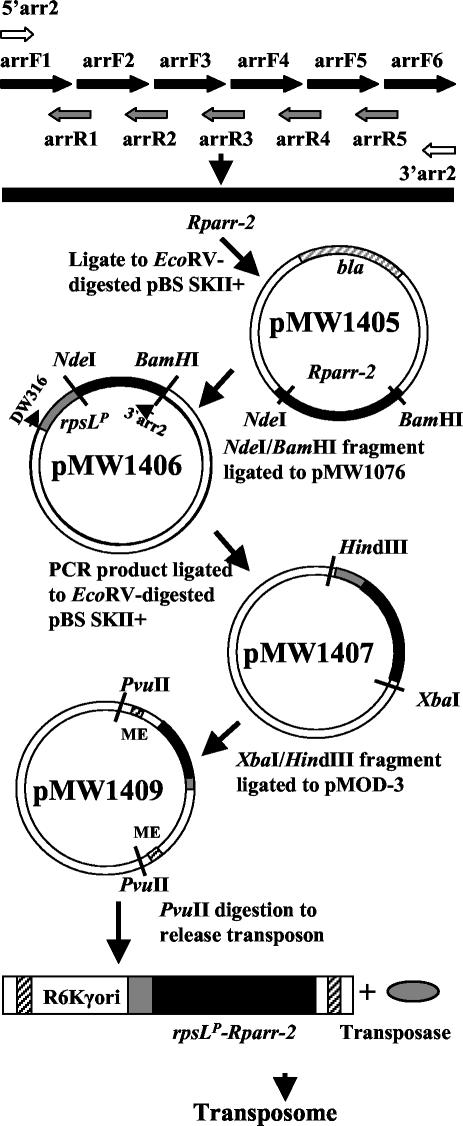
A rickettsial arr-2 rifampin resistance gene (Rparr-2) was synthesized on the basis of rickettsial codon usage preferences (Fig. 1). The Rparr-2 sequence was generated by a PCR-mediated gene synthesis strategy involving the assembly of oligonucleotides (1, 12). Oligonucleotides used in the synthesis of Rparr-2 are listed in Table 2. Oligonucleotides of 80 bases spanning the sequence of Rparr-2 on one strand were mixed with 40-base oligonucleotides corresponding to the opposite strand and spanning the junctions of the 80-base oligonucleotides (Fig. 1). In addition, two outer primers (5′arr2 and 3′arr2) used to generate the full-length double strands were also included. These two primers contained selected restriction endonuclease cleavage sites (NdeI and BamHI) as well as a ribosomal binding site within the 5′ primer. The 100-μl reaction mixture contained 4 pmol of each synthesis oligonucleotide, 100 pmol of each of the outer primers, 200 μM of each deoxynucleoside triphosphate, 2 U of Vent DNA polymerase, and 10 μl of 10× ThermoPol reaction buffer (New England Biolabs, Beverly, Mass.). The PCR conditions consisted of 25 cycles of 95°C for 1 min, 50°C for 1 min, and 72°C for 1 min. After this initial reaction, 5 μl of the reaction mixture was used as a template in a subsequent PCR for which the reaction mixture contained only the two outer primers. The conditions for the secondary PCR were identical to those for the primary amplification with the exception that the annealing temperature was increased to 60°C. Amplified DNA was separated by gel electrophoresis, revealing a single band of the predicted size. The band was excised from the gel and purified. The synthesized gene was subsequently placed under the control of the R. prowazekii rpsL promoter and ribosomal binding site.
FIG. 1.
Schematic outlining the synthesis of Rparr-2 and the construction of the rpsLP-Rparr-2 transposon. Arrows at the top of the figure represent oligonucleotides, while the solid black bar represents the synthesized Rparr-2 gene.
TABLE 2.
Oligonucleotides
| Oligonucleo- tide name | Sequence (5′ to 3′) |
|---|---|
| arrF1 | TAAGGAGGTATCATATGGTAAAAGATTGGATTCCTATTTCTCATGATAATTATAAACAAGTACAAGGACCATTTTATCAT |
| arrF2 | GGAACTAAAGCTAATTTAGCAATTGGTGATTTACTAACTACAGGATTTATTTCTCATTTTGAAGATGGTCGAATTCTTAA |
| arrF3 | ACATATTTATTTTTCAGCTTTAATGGAACCAGCAGTTTGGGGAGCTGAACTTGCTATGTCACTATCTGGTCTTGAAGGTC |
| arrF4 | GTGGTTATATATATATAGTTGAACCAACAGGACCATTTGAAGATGATCCAAATCTTACAAATAAAAAATTTCCTGGTAAT |
| arrF5 | CCAACACAATCTTATAGAACTTGTGAACCTTTAAGAATTGTTGGTGTTGTTGAAGATTGGGAAGGACATCCTGTTGAATT |
| arrF6 | AATAAGAGGAATGTTAGATTCATTAGAAGATTTAAAACGTCGTGGTTTACATGTTATTGAAGATTAAGGATCCTCTAGAC |
| arrR1 | CTAAATTAGCTTTAGTTCCATGATAAAATGGTCCTTGTAC |
| arrR2 | AAGCTGAAAAATAAATATGTTTAAGAATTCGACCATCTTC |
| arrR3 | ACTATATATATATAACCACGACCTTCAAGACCAGATAGTG |
| arrR4 | CTATAAGATTGTGTTGGATTACCAGGAAATTTTTTATTTG |
| arrR5 | AATCTAACATTCCTCTTATTAATTCAACAGGATGTCCTTC |
| 5′arr2 | TAAGGAGGTATCATATGGTAAAAG |
| 3′arr2 | GTCTAGAGGATCCTTAATCTTC |
| DW316 | AACATACTTGCTTTTATAGG |
| DW603 | CTAAACCCTCATGGCTAACG |
| DW605 | CTGCTGGTTCCATTAAAGCTG |
| DW615 | CTATACTGTTTCCTATGAGAGACG |
| DW616 | CTTACCTAAATCAAATAACGCAGC |
A schematic outlining the construction of the Rparr-2 transposon is presented in Fig. 1. The purified synthetic Rparr-2 gene was ligated into the EcoRV site of the pBluescript II SK(+) vector, generating plasmid pMW1405. This plasmid conferred an ampicillin- and rifampin-resistant phenotype when introduced into E. coli. A BamHI-NdeI fragment containing Rparr-2 was cleaved from pMW1405 and cloned into the similarly cut plasmid pMW1076. This plasmid contains the PCR-amplified R. prowazekii rpsL promoter region and ribosomal binding site cloned into the EcoRV site of pBluescript II SK(+). The resulting construct, pMW1406, places the Rparr-2 gene under the control of the rickettsial rpsL promoter (rpsLP), with the start codon of the Rparr-2 gene appropriately placed downstream of the rpsL ribosomal binding site. To generate the final transposon vector, the rpsLP-Rparr-2 gene cassette was amplified by using an rpsL-specific primer (DW316) in conjunction with the 3′arr2 reverse primer. The amplified fragment was inserted into pBluescript II SK(+) to generate pMW1407. This construct was sequenced to confirm the predicted sequence of the rpsLP-Rparr-2 cassette. The sequence of this cassette is available in GenBank under accession number AY396719. Plasmid pMW1407 was digested with HindIII and XbaI, and the resulting Rparr-2-containing fragment was cloned into the similarly digested EZ:TN pMOD-3〈R6Kγori/MCS〉 transposon construction vector (Epicentre, Madison, Wis.), generating pMW1409. A transposon containing the Rparr-2 gene was released from pMW1409 by digestion with PvuII and purified for transformation experiments.
Rescue cloning and inverse PCR.
Identification of transposon insertion sites was accomplished by rescue cloning. Chromosomal DNA (2 to 8 μg), isolated from rifampin-resistant rickettsiae that exhibited a positive PCR result for the R. prowazekii sdhA gene and the Rparr-2 gene, was digested with BclI and then self-ligated in a 500- or 1,000-μl reaction volume with 20 or 40 U of T4 DNA ligase (5 U/μl; Invitrogen), respectively, overnight at 16°C. Large-volume ligations were performed to favor intramolecular interactions. Following ligation, the DNA was precipitated with 95% ethanol and the precipitated DNA was washed twice with 70% ethanol, dried, and suspended in 6 μl of water at a final concentration of 200 ng/μl. This DNA preparation was used to transform electrocompetent E. coli TransforMax EC100D pir-116 cells (Epicentre). Transformants were selected on Luria-Bertani plates containing rifampin. Transformant colonies containing the Rparr-2 gene were identified by colony hybridization by using a portion of the transposon containing rpsLP-Rparr-2 as a 32P-labeled probe. Positive Rparr-2 transformants were colony purified, and plasmids were isolated with a mini-prep kit (QIAGEN). PCR was used to confirm that the purified plasmid contained Rparr-2, and the plasmid DNA was sequenced. DNA sequencing of the rescued plasmid was performed by using transposon primers SqFP and SqRP (Epicentre). As an alternative method for identifying insert sites, ligated DNA preparations described above were used as templates in an inverse PCR with the SqFP and SqRP primers. DNA fragments obtained by this method were directly sequenced by using the SqFP and SqRP primers.
RESULTS
Transposome mutagenesis of R. prowazekii and identification of transposon insertions.
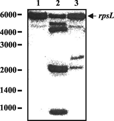
Plasmid pMW1409 was digested with PvuII, releasing the Rparr-2 transposon. The purified Rparr-2 transposon was mixed with transposase to generate the transforming transposome according to the manufacturer's recommendation (Epicentre). Transposome (60 ng) was mixed with competent rickettsiae and electroporated. Following electroporation, the rickettsiae were allowed to infect L929 cells (∼1.4 × 10⁸ cells) which were planted into fifteen 185-cm2 SoLo tissue culture flasks (Nalge Nunc, Naperville, Ill.). At 24 h after infection, rifampin (200 ng/ml) was added for selection. Growth under rifampin selection was continued for 10 to 15 days, at which time rickettsiae could be visualized microscopically. Rifampin-resistant rickettsiae were isolated, and DNA was extracted for use as template DNA in PCRs and for Southern blotting. The presence of the Rparr-2 gene was detected by PCR in DNA isolated from the rifampin-resistant rickettsiae (data not shown). Four out of seven independent transformation experiments yielded rickettsial populations that were positive by PCR for the presence of Rparr-2. This result was confirmed by Southern blotting, which revealed the presence of multiple Rparr-2 hybridizing bands within the rickettsial DNA isolated from two of the independent rickettsial transformations (Fig. 2). These results suggest the presence of populations of rickettsiae with different insertions. Subsequent characterization of the T-12 population (see below) revealed that it contained two independent insertions. The presence of four hybridizing bands (Fig. 2, lane 2) results from the fact that each of the inserts is cleaved by HindIII.
FIG. 2.
Hybridization of the R. prowazekii rpsLP-Rparr-2 gene cassette to R. prowazekii chromosomal DNA digested with HindIII. Lane 1, DNA isolated from the Madrid E strain; lane 2, DNA isolated from rifampin-resistant rickettsiae of transformation T-12; lane 3, DNA isolated from rifampin-resistant rickettsiae of transformation T-15. Molecular size markers are indicated, as is the 5.9-kb rpsL HindIII fragment.
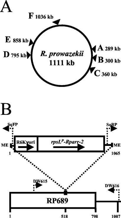
To identify specific insertion sites, rescue cloning was employed. The transposon used in these studies contains the R6Kγ origin. By digesting the chromosomal DNA with a restriction enzyme that does not cut within the transposon (BclI), it is possible to generate fragments containing the transposon and attached rickettsial sequences that can be self-ligated and transformed into the pir+ strain, which is able to support replication of plasmids containing the R6Kγ origin. Colonies positive for the presence of Rparr-2 were identified by colony hybridization or by PCR. Plasmids isolated from rifampin-resistant colonies were screened by PCR to confirm the presence of Rparr-2 and sequenced to identify the rickettsial DNA sequence linked to the transposon. One or two separate insertions were identified for each experiment (Table 3). The sites of these insertions within the R. prowazekii chromosome are shown in Fig. 3A.
TABLE 3.
Transposon insertion sites identified by rescue cloning
| Site | 9-bp repeat (5′-3′) | Genomic position | Disrupted ORF
|
Description | ||
|---|---|---|---|---|---|---|
| ORF | Size (bp) | Insertion site | ||||
| A | GTTATCAAT | 289923 | Between RP229 and RP230 | |||
| B | TTGCAGCAC | 300565 | RP243 | 1,053 | 289 | ermA |
| C | CCCTAAAGC | 360084 | RP294 | 1,413 | 254 | gppA |
| D | ATTACATAC | 795091 | Between RP623 and RP624 | |||
| E | GTTATATCC | 858752 | RP689 | 798 | 518 | Unknown function |
| F | AGGTAAAAG | 1036224 | RP823 | 2,235 | 2211 | ftsK |
FIG. 3.
(A) Schematic of the R. prowazekii chromosome showing the locations of Rparr-2 transposon insertions. (B) Gene map showing the insertion of the Rparr-2 transposon into the R. prowazekii RP689 ORF. Dotted arrows indicate the positions and directions of synthesis of primer sites. Numbers indicate size in base pairs. ME, mosaic ends; rpsLP, rpsL promoter.
A transposon insertion was also identified by inverse PCR. Chromosomal DNA digested and ligated as described above was used as a template in inverse PCR with transposon-specific primers. A PCR product obtained by this method was sequenced and revealed one of the same insertions identified by the rescue cloning technique.
Isolation of R. prowazekii knockout strains.
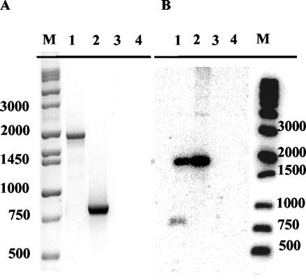
In order to demonstrate the existence of a stable R. prowazekii insertional mutant, a specific clone was isolated by limiting dilution from a population of L929 cells infected with rifampin-resistant rickettsiae of the T-12 experiment. By the rescue cloning and inverse PCR techniques described above, the transposon insertion site was identified within the RP689 ORF (Fig. 3B). When DNA isolated from this clone was used as a template for PCR with primers that would amplify the wild-type RP689 gene, a single product of the predicted size was obtained, confirming the insertion of the transposon within this gene (Fig. 4A, lane 1). Sequencing of the PCR product confirmed the insertion of the transposon at a site 518 bp downstream of the start codon. Based on the Poisson distribution and the fact that the smaller wild-type product was not amplified, it can be assumed that a pure clone of the RP689 insertional knockout was isolated. This assumption was confirmed by Southern blotting with Rparr-2 as a probe. By use of only the Rparr-2 portion of the transposon as the probe rather than the rpsLP-Rparr-2 fragment used in the hybridizations shown in Fig. 2, only a single HindIII fragment is targeted. A single hybridizing band is observed for T-12B1 (Fig. 4B, lane 2) demonstrating that T-12B1 is a pure clone. In the original T-12 population (Fig. 4B, lane 1), two hybridizing bands are observed. By the cloning techniques described above, the additional band was found to correspond to an emrA insertion. In the case of the RP689 clone, no phenotypic differences from wild-type R. prowazekii have been observed.
FIG. 4.
(A) PCR analysis of the RP689 insertion. Chromosomal primers that amplify a portion of the RP689 gene (DW615 and DW616) (Fig. 3B) were used in PCRs with template DNAs from R. prowazekii. Lane 1, T-12B1 RP689 insertion clone DNA; lane 2, Madrid E DNA; lane 3, L-929 cell DNA; lane 4, water. Size markers (M) are indicated in base pairs. (B) Southern blot analysis. Lane 1, T-12 DNA; lane 2, T-12B1 DNA; lane 3, Madrid E DNA; lane 4, L-929 cell DNA.
DISCUSSION
The sequence of the R. prowazekii genome revealed the gene repertoire available for this bacterium to enable it to grow and survive in eukaryotic cell cytoplasm. The next step is to understand the function of each of the 834 identified coding regions and characterize their environmental and temporal expression patterns. Anticipated advances in microarray analysis and proteomics will certainly advance our knowledge of the expression patterns of rickettsial genes; however, gene knockouts would provide definitive proof that a particular gene is or is not required for rickettsial survival and intracellular growth under a specific environmental condition (e.g., growth in lice versus that in humans). While several reports have documented the feasibility of rickettsial transformation by electroporation and have demonstrated that recombination of DNA into the chromosome does occur (17, 19-21), the isolation of rickettsial gene knockouts has not been reported. In this study, we have identified an additional gene for the selection of rickettsial transformants and demonstrated the feasibility of using a commercially available transposome system to generate rickettsial mutants.
R. prowazekii is sensitive to rifampin, the first antibiotic used, in a study by Rachek et al. (21), to demonstrate rickettsial transformation. However, in that study, the rifampin resistance was due to a point mutation in the rpoB gene, a large, essential gene coding for the β subunit of RNA polymerase, a gene unsuitable for general genetic selections. Fortunately, the arr-2 gene provides another mechanism for rifampin selection. It codes for a transferase enzyme that inactivates the antibiotic and, due to its small size, was easily synthesized for expression in R. prowazekii. While it is unknown whether the change in arr-2 codon usage was necessary, it was decided that every effort would be made to ensure the optimal expression of arr-2 in R. prowazekii. This approach also improves upon the erythromycin selection described for R. prowazekii (20). While successful, the ereB erythromycin resistance gene used in previous rickettsial transformations was a much larger gene that retained its original codon usage pattern and ribosome binding site. Combined with the fact that erythromycin was less effective than rifampin at eliminating nontransformed rickettsiae, the use of rifampin for rickettsial selection provides a more effective tool for genetic manipulations.
The use of the transposome system also provided an efficient mechanism for generating random rickettsial chromosomal insertions. The fact that the recipient rickettsiae did not have to express the transposase was considered a major advantage. While both the E. coli ereB gene and gfp have been expressed in rickettsiae (17, 19, 20), in vivo expression of a transposase could have presented problems. Thus, with active transposase attached to the transposon DNA, the only barrier for transposition was entrance into the cell, a process accomplished by electroporation.
It was interesting that in each of the successful transformation experiments (four out of seven), only a few insertion sites, one or two per experiment, were identified by Southern blotting. This result may be due to a limited number of nonessential sites being available for transposon insertion, although the fact that independent experiments produced different insertion patterns argues against this hypothesis. A more likely possibility is that this outcome was due to less-than-optimal experimental design and the inherent inefficiency associated with rickettsial transformant isolation. With our standard rickettsial transformation protocol, an experiment requires approximately 2 weeks of rickettsial growth under antibiotic selection before a population of resistant bacteria is visualized. Additional weeks of growth may then be required to obtain sufficient populations for analysis and cloning. Since we use microscopic visualization of a rifampin-resistant population as the positive indicator of a successful transformation, individual transformants must compete not only with other transformants but also with spontaneous, rifampin-resistant mutants that arise in every experiment. Thus, any individual transposon insertion mutant may be a minor part of the entire population obtained after extended growth. Also, if a mutant has a growth disadvantage, it may be difficult to detect it within the larger population. Of course, growth-inhibited mutants are just the mutants that would be most exciting to isolate and study. To overcome this problem, the electroporated rickettsiae will need to be cloned by limiting dilution techniques earlier in the experimental protocol. Identification of mutated genes by cloning into E. coli may also select for a limited subset of mutations, since some rickettsial fragments may be unstable in E. coli or lethal to the host cell.
Of the six insertions identified in this study, two were in intervening regions. Considering that the R. prowazekii genome contains a high percentage of noncoding DNA (24%), the proportion of insertion sites in intervening regions was not unexpected. Another insertion was located at the extreme 3′ end of the ftsK gene (22 bases upstream of the stop codon). Since this gene codes for FtsK, a protein involved in cell division and chromosome segregation (4, 6, 14), it is assumed that this insertion does not generate an inactive protein. Three insertions (RP243, RP294, and RP689) are located deep within the coding sequence of each gene, generating mutants that most likely would not retain their function. These genes represent the first rickettsial genes to be experimentally placed in the category of nonessential genes, at least for R. prowazekii growing in mouse fibroblast L929 cells. Whether the gene products of these genes are required when rickettsiae are growing in other environments is under investigation.
The three insertional mutants identified raise some intriguing questions concerning rickettsial growth. The RP243 insertional knockout is especially interesting, since it has been designated emrA. This gene codes for a product with homology to the EmrA multidrug resistance protein A. In other bacterial systems, this protein, in conjunction with EmrB, forms a multidrug-resistant pump (15). An EmrB homolog is also present in R. prowazekii. Thus, this particular mutant may be more sensitive to environments containing toxic compounds and antibiotics. RP294 is designated gppA, which has been associated with stress responses. In E. coli, the enzyme encoded by gppA hydrolyzes the 5′-γ-phosphate of pppGpp to form ppGpp (13). RP689 codes for a protein of unknown function. Such genes account for approximately 38% of the 834 protein-coding genes. Interestingly, the upstream RP688 gene product is highly homologous to RP689. Although RP688 has an additional 50 residues at the amino terminus and RP689 extends 14 residues past the overlap at the carboxy terminus, within the 200-residue overlap, the proteins are 87% identical. Studies are under way to determine whether the RP688 gene can also be disrupted without affecting rickettsial growth and whether it will be possible to isolate a double mutant of both genes.
In summary, rifampin resistance encoded by a rickettsial adapted arr-2 gene provides effective selection for rickettsial transformants. With the transposome system, it is now possible to generate random insertional mutations of rickettsial genes. In addition, this selection can also be used in targeted gene knockouts generated by homologous recombination. These methods provide effective tools for identifying nonessential genes of R. prowazekii and for incorporating this information into hypotheses concerning the requirements for rickettsial obligate intracellular growth.
Acknowledgments
We thank Herbert Winkler for helpful discussions and for critical reading of the manuscript.
This work was supported by Public Health Service grant AI20384 from the National Institute of Allergy and Infectious Diseases.
REFERENCES
- 1.Alexeyev, M. F., and H. H. Winkler. 1999. Gene synthesis, bacterial expression and purification of the Rickettsia prowazekii ATP/ADP translocase. Biochim. Biophys. Acta 1419:299-306. [DOI] [PubMed] [Google Scholar]
- 2.Andersson, S. G. E., A. Zomorodipour, J. O. Andersson, T. Sicheritz-Ponten, U. C. M. Alsmark, R. M. Podowski, A. K. Nslund, A. S. Eriksson, H. H. Winkler, and C. G. Kurland. 1998. The genome sequence of Rickettsia prowazekii and the origin of mitochondria. Nature 396:133-143. [DOI] [PubMed] [Google Scholar]
- 3.Ausubel, F., R. Brent, R. E. Kingston, D. D. Moore, J. G. Seidman, J. A. Smith, and K. Struhl. 1997. Current protocols in molecular biology, vols. 1, 2, and 3. John Wiley & Sons, Inc., New York, N.Y.
- 4.Begg, K. J., S. J. Dewar, and W. D. Donachie. 1995. A new Escherichia coli cell division gene, ftsK. J. Bacteriol. 177:6211-6222. [DOI] [PMC free article] [PubMed] [Google Scholar]
- 5.Bullock, W. O., J. M. Fernandez, and J. M. Short. 1987. A high efficiency plasmid transforming recA Escherichia coli strain with beta-galactosidase selection. BioTechniques 5:376-379. [Google Scholar]
- 6.Capiaux, H., C. Lesterlin, K. Perals, J. M. Louarn, and F. Cornet. 2002. A dual role for the FtsK protein in Escherichia coli chromosome segregation. EMBO Rep. 3:532-536. [DOI] [PMC free article] [PubMed] [Google Scholar]
- 7.Clavero, G., and F. Perez Gallardo. 1943. Estudio experimental de una cepa apatogena e inmunizante de Rickettsia prowazeki. Cepa E. Rev. Sanid. Hig. Publica 17:1-27. [Google Scholar]
- 8.Dabbs, E. R., K. Yazawa, Y. Mikami, M. Miyaji, N. Morisaki, S. Iwasaki, and K. Furihata. 1995. Ribosylation by mycobacterial strains as a new mechanism of rifampin inactivation. Antimicrob. Agents Chemother. 39:1007-1009. [DOI] [PMC free article] [PubMed] [Google Scholar]
- 9.Gimenez, D. F. 1964. Staining rickettsiae in yolk-sac cultures. Stain Technol. 39:135-140. [DOI] [PubMed] [Google Scholar]
- 10.Goryshin, I. Y., J. Jendrisak, L. M. Hoffman, R. Meis, and W. S. Reznikoff. 2000. Insertional transposon mutagenesis by electroporation of released Tn5 transposition complexes. Nat. Biotechnol. 18:97-100. [DOI] [PubMed] [Google Scholar]
- 11.Hoffman, L. M., J. J. Jendrisak, R. J. Meis, I. Y. Goryshin, and S. W. Reznikof. 2000. Transposome insertional mutagenesis and direct sequencing of microbial genomes. Genetica 108:19-24. [DOI] [PubMed] [Google Scholar]
- 12.Jayaraman, K., and C. J. Puccini. 1992. A PCR-mediated gene synthesis strategy involving the assembly of oligonucleotides representing only one of the strands. BioTechniques 12:392-398. [PubMed] [Google Scholar]
- 13.Keasling, J. D., L. Bertsch, and A. Kornberg. 1993. Guanosine pentaphosphate phosphohydrolase of Escherichia coli is a long-chain exopolyphosphatase. Proc. Natl. Acad. Sci. USA 90:7029-7033. [DOI] [PMC free article] [PubMed] [Google Scholar]
- 14.Liu, G., G. C. Draper, and W. D. Donachie. 1998. FtsK is a bifunctional protein involved in cell division and chromosome localization in Escherichia coli. Mol. Microbiol. 29:893-903. [DOI] [PubMed] [Google Scholar]
- 15.Lomoskaya, O., and K. Lewis. 1992. emr, an Escherichia coli locus for multidrug resistance. Proc. Natl. Acad. Sci. USA 89:8938-8942. [DOI] [PMC free article] [PubMed] [Google Scholar]
- 16.Metcalf, W. W., W. Jiang, and B. L. Wanner. 1994. Use of the rep technique for allele replacement to construct new Escherichia coli hosts for maintenance of R6K origin plasmids at different copy numbers. Gene 138:1-7. [DOI] [PubMed] [Google Scholar]
- 17.Michelle, J., S. Radulovic, and A. F. Azad. 1999. Green fluorescent protein as a marker in Rickettsia typhi transformation. Infect. Immun. 67:3308-3311. [DOI] [PMC free article] [PubMed] [Google Scholar]
- 18.Naas, T., Y. Mikami, T. Imai, L. Poirel, and P. Nordmann. 2001. Characterization of In53, a class 1 plasmid- and composite transposon-located integron of Escherichia coli which carries an unusual array of gene cassettes. J. Bacteriol. 183:235-249. [DOI] [PMC free article] [PubMed] [Google Scholar]
- 19.Ogata, H., S. Audic, P. Renesto-Audiffren, P.-E. Fournier, V. Barbe, D. Samson, V. Roux, P. Cossart, J. Weissenbach, J.-M. Claverie, and D. Raoult. 2001. Mechanisms of evolution in Rickettsia conorii and R. prowazekii. Science 293:2093-2098. [DOI] [PubMed] [Google Scholar]
- 20.Rachek, L. I., A. Hines, A. M. Tucker, H. H. Winkler, and D. O. Wood. 2000. Transformation of Rickettsia prowazekii to erythromycin resistance encoded by the Escherichia coli ereB gene. J. Bacteriol. 182:3289-3291. [DOI] [PMC free article] [PubMed] [Google Scholar]
- 21.Rachek, L. I., A. M. Tucker, H. H. Winkler, and D. O. Wood. 1998. Transformation of Rickettsia prowazekii to rifampin resistance. J. Bacteriol. 180:2118-2124. [DOI] [PMC free article] [PubMed] [Google Scholar]
- 22.Southern, E. M. 1975. Detection of specific sequences among DNA fragments separated by gel electrophoresis. J. Mol. Biol. 98:503-517. [DOI] [PubMed] [Google Scholar]
- 23.Tribuddharat, C., and M. Fennewald. 1999. Integron-mediated rifampin resistance in Pseudomonas aeruginosa. Antimicrob. Agents Chemother. 43:960-962. [DOI] [PMC free article] [PubMed] [Google Scholar]
- 24.Tucker, A. M., H. H. Winkler, L. O. Driskell, and D. O. Wood. 2003. S-Adenosylmethionine transport in Rickettsia prowazekii. J. Bacteriol. 185:3031-3035. [DOI] [PMC free article] [PubMed] [Google Scholar]
- 25.Winkler, H. H. 1976. Rickettsial permeability: an ADP-ATP transport system. J. Biol. Chem. 251:389-396. [PubMed] [Google Scholar]


