Abstract
Iatrogenic vascular problems during posterior lumbar interbody fusion are a rare entity. Migration of a broken scalpel towards the heart has, to our knowledge, never been reported. We present the successful surgical retrieval of a broken scalpel from the heart after posterior lumbar interbody fusion without the use of a cardiopulmonary bypass.
Keywords: Foreign body, Iatrogenic, Off-pump surgery
INTRODUCTION
Lumbar interbody fusion is an established orthopaedic technique for the treatment of degenerative and traumatic diseases of the spine. The intervertebral disc is partially removed and replaced by a metal cage containing a bone graft. According to the access, differentiation in an anterior and a posterior lumbar interbody fusion (ALIF and PLIF) is made. For ALIF, the most common complications are visceral and vascular injuries whereas PLIF faces neurological and dura-related complications. We present the case of a man with an exceptional and, to our knowledge, unique vascular complication, which led to migration of an iatrogenic foreign body in the heart during PLIF.
CASE PRESENTATION
A 44-year old man received a PLIF between L5 and S1 because of a discus hernia. While removing the intervertebral disc, the tip of a bistoury snapped off and was jammed in the disc. Attempts to retrieve the tip resulted in a further descend of the knife into the disc, and finally, it crossed the anterior column of the vertebra. During the procedure, fluoroscopy revealed a gradual upward migration of the tip till the level of the Eustachian valve. Owing to the risk of perforation or embolization to the pulmonary circulation, an urgent removal of the foreign body was deemed necessary. Nonetheless, the procedure was finalized by placing the cages. Since the hospital did not accommodate cardiac surgery, the patient was transferred to our hospital under general anaesthesia.
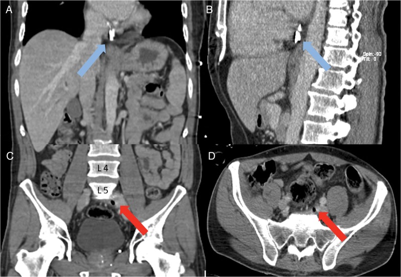
A spiral computed tomography (CT) scan of the abdomen and the thorax was performed to define the exact location of the tip and to evaluate possible complications. The foreign body was indeed located at the junction of inferior vena cava and right atrium (Fig. 1A and B). There was no evidence of retroperitoneal haematoma nor pericardial effusion. The entry site of the corpus alienum into the circulation was suggested by an aberrant air pocket posterior and adjacent to the left common iliac vein (Fig. 1C and D).
Figure 1:
Preoperative CT scan. A and B demonstrating the position of the scalpel (blue arrow) at the junction of the inferior caval vein and right atrium. C and D showing the air pocket (red arrow) in between the spine (L5–S1) and left common iliac vein suggesting the site of perforation.
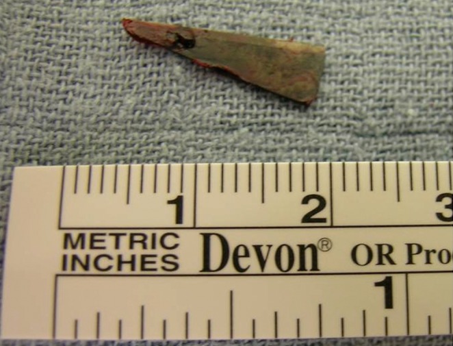
The patient was brought to the operation theatre. A median sternotomy was performed to assess the mediastinum. A purse string was placed at the level of the right atrium through which a Tennet clamp was entered without support of cardiopulmonary bypass (CPB). The bistoury was catched by using fluoroscopic guidance (Video 1). We exteriorized the tip successfully (Fig. 2). Total blood loss was estimated around 250 ml.
Video 1: Complete procedure, starting at the moment of the introduction of the Tennet-clamp till removal of the scalpel.
Figure 2:

The scalpel after removal.
After surgery, the patient was transferred to the intensive care unit without any haemodynamic support. He had an uneventful recovery and could be discharged 7 days after initial operation.
DISCUSSION
This case reports the coincidence of two rare events, a vascular complication during PLIF with migration of a knife into the heart. Indeed, vascular injuries in the PLIF are rare. Papadoulas et al. identified 99 cases of iatrogenic vascular complications counting for an incidence of 1–5 in 10 000 procedures. The most common vascular injury was the right common iliac artery, and the level of operation was associated with the injury of a specific vessel [1]. Secondly, the presence of a corpus alienum in the great vessels and the heart are seldom seen. Most common iatrogenic foreign bodies include stents and catheter fragments, although a diversity of objects are found in the literature [2]. Complications due to broken scalpels are rare, and migration into the blood circulation or heart has, to our knowledge, never been described.
We opted for a median sternotomy to have good access in the case of perforation and pericardial effusion. Since this was ruled out, we continued the procedure without CPB. The position of the scalpel was prone for this strategy. Moreover, the cannula could dislocate the knife with further migration. Besides, snaring of the inferior caval vein would be dangerous to cause further perforation into the caval vein/right atrium. As an alternative, a cannula could be installed into the femoral vein with the use of an occlusive balloon, but this was assessed as not necessary in our case.
Iatrogenic foreign bodies in the heart can be treated either conservatively, endovascularly or surgically. Most data regarding foreign bodies in the heart are case reports. This makes a general recommendation difficult. There are no current guidelines for the treatment of a foreign body in the heart. Therapy should therefore be individualized. First, we need to respond to the question if the object should be retrieved? [2] Conservative treatment is an option in asymptomatic foreign bodies without associated risks, particularly if they are small (<1 cm), smooth and minimal contaminated and embedded in the myocardium or pericardium. Intervention is necessary if symptomatic, irrespective of their location, or asymptomatic patients with associated risk factors and possible complications such as tamponade, endocarditis, arrhythmia and thrombi [3–5]. Life expectancy and morbidity should be also taken into account. Endovascular retrieval needs to be the first choice if technically feasible and safe [2]. Owing to the risk of perforation or embolization to the pulmonary artery, removal was the only option in our case. An endovascular procedure was not appropriate due to the nature of the object with the risk of perforation during blind endovascular retrieval. Therefore, off-pump removal was the least invasive approach in our patient.
Supplementary Material
REFERENCES
- 1.Papadoulas S, Konstantinou D, Kourea HP, Kritikos N, Haftouras N, Tsolakis JA. Vascular injury complicating lumbar disc surgery. A systematic review. Eur J Vasc Endovasc Surg. 2002;24:189–95. doi: 10.1053/ejvs.2002.1682. [DOI] [PubMed] [Google Scholar]
- 2.Schechter MA, O'Brien PJ, Cox MW. Retrieval of iatrogenic intravascular foreign bodies. J Vasc Surg. 2013;57:276–81. doi: 10.1016/j.jvs.2012.09.002. [DOI] [PubMed] [Google Scholar]
- 3.Actis Dato GM, Arslanian A, Di Marzio P, Filosso PL, Ruffini E. Posttraumatic and iatrogenic foreign bodies in the heart: report of fourteen cases and review of the literature. J Thorac Cardiovasc Surg. 2003;126:408–14. doi: 10.1016/s0022-5223(03)00399-4. [DOI] [PubMed] [Google Scholar]
- 4.LeMaire SA, Wall MJ, Jr, Mattox KL. Needle embolus causing cardiac puncture and chronic constrictive pericarditis. Ann Thorac Surg. 1998;65:1786–7. doi: 10.1016/s0003-4975(98)00246-x. [DOI] [PubMed] [Google Scholar]
- 5.Thanavaro KL, Shafi S, Roberts C, Cowley M, Arrowood J, Cassano A, et al. An unusual presentation of chest pain: needle perforation of the right ventricle. Heart Lung. 2013;42:218–20. doi: 10.1016/j.hrtlng.2013.02.002. [DOI] [PubMed] [Google Scholar]
Associated Data
This section collects any data citations, data availability statements, or supplementary materials included in this article.


