Abstract
Helicobacter pylori (H. pylori) has become accepted as a human pathogen for the development of gastritis and gastroduodenal ulcer. To develop a simple rat model of chronic H. pylori infection, male Sprague-Dawley rats were pretreated with streptomycin suspended in tap water (5 mg/mL) for 3 d. The rats were inoculated by gavage at 1 mL/rat with H. pylori suspension (5 × 10⁸-5 × 10¹⁰ CFU/mL) twice daily at an interval of 4 h for three consecutive days. Two weeks after inoculation, rats were sacrificed and the stomachs were removed. Antral biopsies were performed for urease test and the stomachs were taken for histopathology. Successful H. pylori inoculation was defined as a positive urease test and histopathology. We reported a 69.8%-83.0% success rate for H. pylori infection using the urease test, and hematoxylin and eosin staining confirmed the results. Histopathological analysis detected bacteria along the mucous lining of the surface epithelium and crypt lumen and demonstrated mild to moderate gastric inflammation in successfully inoculated rats. We developed a simple rat model of chronic H. pylori infection for research into gastric microcirculatory changes and therapy with plant products.
Keywords: Helicobacter pylori, Rat model, Chronic infection
Core tip: Helicobacter pylori (H. pylori) causes significant gastroduodenal diseases. Experimental animal models play an important role in helping us understand the pathogenesis and discovering new therapeutic strategies. Previous H. pylori-associated gastritis candidate animal models have included gnotobiotic piglets, non-human primates, pigs, dogs, cats, gerbils, and mice. Rat models of H. pylori infection use a difficult technique and take a long time to establish. In this study, we developed a simple model of H. pylori infection in rats for further research.
INTRODUCTION
Helicobacter pylori (H. pylori) is a Gram-negative spiral bacterium that causes infection with many different clinical outcomes. It has been established as a major etiological agent of chronic gastritis and peptic ulcer disease, which includes duodenal and gastric ulcer[1]. The role of H. pylori infection in gastric adenocarcinoma and mucosa-associated lymphoid tissue (MALT) lymphoma has also been recognized[2].
ANIMAL MODELS OF H. PYLORI INFECTION
Increasing evidence reveals that H. pylori is a significant gastroduodenal pathogen. Experiment animal models are needed to help us understand better its pathogenic mechanisms, and to verify the pathogenesis as well as the relationship of this bacterium to gastric injury. Experimental animal models also play an important role in discovering new therapeutic strategies including application of plant products for efficient treatment against H. pylori infection[3]. Previous H. pylori-associated gastritis candidate animal models have included gnotobiotic piglets, non-human primates, pigs, dogs, cats, gerbils, and mice[4-7].
Some studies have been successful for development of models of H. pylori infection in rats, pretreated with oral omeprazole to reduce acidic conditions in the stomach, and intragastric administration of H. pylori to colonize the stomach[7].
These animal models were designed and used to establish gastritis that closely resembled the disease commonly found in humans. Animal models offer many benefits and have proved useful in conducting studies to understand better human gastritis in animal counterparts. Rats are one of the most commonly used laboratory animals in gastrointestinal research, and their gastric physiology has been thoroughly investigated. Even though other Helicobacter-infected animal models have yielded important information, an H. pylori-infected rat model would be useful for studying pathophysiological events in the gastrointestinal tract during chronic H. pylori infection[8].
In the past, H. pylori bacteria or bacterium-free H. pylori filtrates have been used to inoculate rats with normal mucosa and surgically produced gastric ulcers[3]. Recently, rat models to study reactions from rat gastric mucosa during long-term H. pylori infection have been established[9]. Another model of H. pylori infection in rats was also reported by Zeng et al[10], who developed mouse and rat models of H. pylori infection by using the Sydney strain 1 H. pylori (SS1 Hp) to colonize the stomach. They used a difficult technique over a long period of time and found that H. pylori could lead to chronic active gastritis after 8, 12 and 24 wk.
Our model was a simple rat model of chronic H. pylori infection developed to research gastric microcirculatory changes and treatment with plant products. Sprague-Dawley rats (120-150 g) were pretreated with streptomycin suspended in tap water (5 mg/mL) for 3 d before the first H. pylori inoculation. The rats were then inoculated by gavage at 1 mL/rat with H. pylori suspension (5 × 10⁸-5 × 10¹⁰ CFU/mL) twice daily at an interval of 4 h for three consecutive days (Figure 1). We reported a 69.8-83.0% success rate of H. pylori infection[11-13] using the urease test. Hematoxylin and eosin staining confirmed these results. The level of bacterial colonization was evaluated by using a grading system and gastric inflammation levels were scored following the updated Sydney System (Figure 2).
Figure 1.
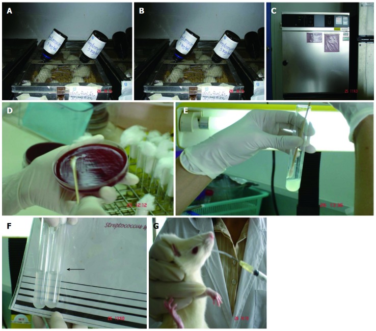
Illustration of Helicobacter pylori rat inoculation. A: Male Sprague-Dawley rats 120-150 g; B: Pretreatment with streptomycin (5 mg/kg) for three consecutive days; C and D: H. pylori in microaerophilic condition: 5% O2, 50% CO2, 37 °C; E and F: H. pylori 108-1010 CFU/mL, suspended in saline; G: Gavage, 1 mL/rat twice daily at an interval of 4 h, for three consecutive days. H. pylori: Helicobacter pylori.
Figure 2.
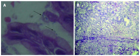
Antral mucosa from Helicobacter pylori-infected rats with hematoxylin and eosin staining. A: Helicobacter pylori organism in the gastric mucosa (arrow) (600 ×); B: Gastric mucosa with erosion and scattered infiltration of inflammatory cells (250 ×).
GASTRIC MICROCIRCULATORY CHANGES IN A RAT MODEL OF H. PYLORI INFECTION
The effects of topical administration of H. pylori on the mesenteric microcirculation were detected by using intravital microscopy[14]. The exposed mesentery that was subjected to H. pylori extracts showed an increase in leukocyte adhesion and emigration in the venules. H. pylori extracts exhibited changes in the rat mesenteric microcirculation. However, H. pylori infection was localized in the stomach, and the leukocyte involvement demonstrated within the mesentery may not be mirrored in the gastric mucosa. Kalia et al[14-16] studied gastric mucosal microcirculation changes caused by H. pylori extracts using intravital fluorescent in vivo microscopy. H. pylori water extracts were applied to rat gastric mucosa and macromolecular leakage, leukocyte adherence, leukocyte rolling, and platelet activity were observed for 90 min. H. pylori induced increases in macromolecular leakage after 5 min, and induced adherent platelet thrombi and circulating platelet emboli after 5 and 15 min, respectively.
In our study, we explored the effects of in vivo chronic H. pylori infection on changes in rat gastric microcirculation using intravital fluorescent microscopy to understand better the pathogenic mechanism of inflammation by monitoring macromolecular leakage and leukocyte-endothelium interaction[12,13]. Twenty-four male Sprague-Dawley rats were divided into two groups (12 control and 12 H. pylori infected). In the H. pylori-infected group, rats were inoculated by gavage with bacterial suspension (5 × 10⁸-5 × 10¹⁰ CFU/mL) twice daily at an interval of 4 h for three consecutive days. Two weeks after inoculation, intravital fluorescence microscopy was performed to examine leukocyte adhesion to post-capillary venules. Macromolecular leakage was examined at 0 and 30 min after fluorescein isothiocyanate-dextran average molecular weight 250K (FITC-dx-250) injection on the posterior surface of the stomach. In the H. pylori-infected group, leukocyte adhesion per 100 μm vessel length was 13.40 ± 1.0 cells, which increased significantly (P < 0.01) compared with the control group (2.47 ± 0.62 cells). The average macromolecular leakage was 15.41% ± 2.83% and 10.69% ± 1.43% in H. pylori-infected and control groups, respectively (P < 0.01) (Figures 3-5).
Figure 3.
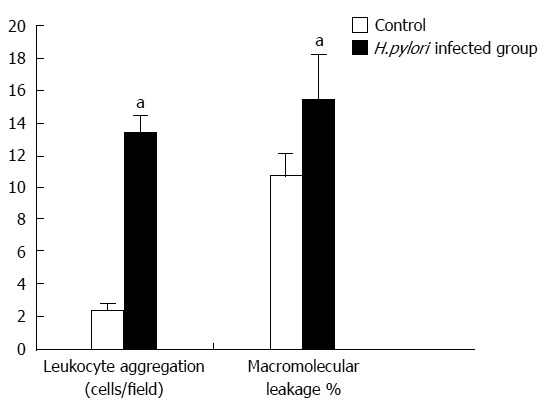
Bar graph of the mean ± SE of adherent leukocytes and macromolecular leakage of control group compared with Helicobacter pylori-infected group. The leukocyte adhesion and macromolecular leakage in the H. pylori-infected group were significantly increased compared with the control group (aP < 0.01 vs control group). H. pylori: Helicobacter pylori.
Figure 5.
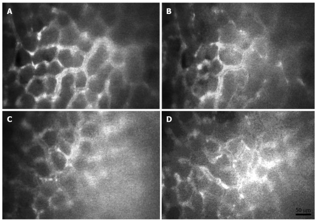
Intravital fluorescent microscopic images (20 ×) demonstrate macromolecular leakage from vessels to the interstitial fluid at 0 and 30 min after injection of control group (A and B) and Helicobacter pylori-infected group (C and D). FITC-dx-250 injection (0 min) (A and C); same area at 30 min after FITC-dx-250 injection (B and D).
Figure 4.
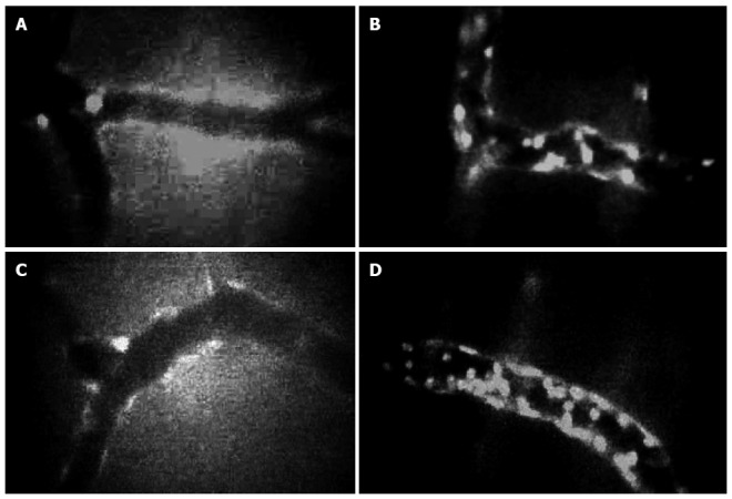
Intravital microscopy demonstrated leukocyte adhesion in control group (A and C) and Helicobacter pylori-infected group (B and D) (40 ×).
The strain of H. pylori plays an important role in the pathogenesis of gastroduodenal diseases. H. pylori obtained from peptic ulcer patients or other pathogenic strains can increase infection rates and develop pathogenesis in the animal stomach. In contrast, intragastric administration of a nontoxigenic strain to normal rat stomach was unsuccessful at inducing chronic inflammation, and only resulted in low-level colonization[3]. Gastric infection with H. pylori expressing cagA- and vacA-encoded cytotoxins delayed healing of ischemia/reperfusion-induced acute gastric lesions due to impairment of gastric microcirculation[17]. To conclude, after 2 wk inoculation, H. pylori successfully colonized Sprague-Dawley rats, with development of mild to moderate gastric inflammation[18].
Footnotes
P- Reviewers: Kim YJ, Luo JC, Pierzchalski P, Suk KT S- Editor: Wen LL L- Editor: Kerr C E- Editor: Wang CH
References
- 1.Tytgat GN, Rauws EA. Campylobacter pylori and its role in peptic ulcer disease. Gastroenterol Clin North Am. 1990;19:183–196. [PubMed] [Google Scholar]
- 2.Suerbaum S, Michetti P. Helicobacter pylori infection. N Engl J Med. 2002;347:1175–1186. doi: 10.1056/NEJMra020542. [DOI] [PubMed] [Google Scholar]
- 3.Ross JS, Bui HX, del Rosario A, Sonbati H, George M, Lee CY. Helicobacter pylori. Its role in the pathogenesis of peptic ulcer disease in a new animal model. Am J Pathol. 1992;141:721–727. [PMC free article] [PubMed] [Google Scholar]
- 4.Fox JG. In vivo models of gastric Helicobacter infections. In: Hunt RH, Tytgat GNJ, editors. Helicobacter pylori. Basic mechanisms to clinical cure. Lancaster, UK: Kluwer Academic Publishers; 1994. pp. 3–27. [Google Scholar]
- 5.Fox JG, Batchelder M, Marini R, Yan L, Handt L, Li X, Shames B, Hayward A, Campbell J, Murphy JC. Helicobacter pylori-induced gastritis in the domestic cat. Infect Immun. 1995;63:2674–2681. doi: 10.1128/iai.63.7.2674-2681.1995. [DOI] [PMC free article] [PubMed] [Google Scholar]
- 6.Hirayama F, Takagi S, Yokoyama Y, Iwao E, Ikeda Y. Establishment of gastric Helicobacter pylori infection in Mongolian gerbils. J Gastroenterol. 1996;31 Suppl 9:24–28. [PubMed] [Google Scholar]
- 7.Li H, Kalies I, Mellgård B, Helander HF. A rat model of chronic Helicobacter pylori infection. Studies of epithelial cell turnover and gastric ulcer healing. Scand J Gastroenterol. 1998;33:370–378. doi: 10.1080/00365529850170991. [DOI] [PubMed] [Google Scholar]
- 8.Lee A, O’Rourke J, De Ungria MC, Robertson B, Daskalopoulos G, Dixon MF. A standardized mouse model of Helicobacter pylori infection: introducing the Sydney strain. Gastroenterology. 1997;112:1386–1397. doi: 10.1016/s0016-5085(97)70155-0. [DOI] [PubMed] [Google Scholar]
- 9.Li H, Andersson EM, Helander HF. Reactions from rat gastric mucosa during one year of Helicobacter pylori infection. Dig Dis Sci. 1999;44:116–124. doi: 10.1023/a:1026662402734. [DOI] [PubMed] [Google Scholar]
- 10.Zeng Z, Hu P, Chen M. Development of mouse and rat model of Helicobacter pylori infection. Zhonghua Yixue Zazhi. 1998;78:494–497. [PubMed] [Google Scholar]
- 11.Thong-Ngam D, Prabjone R, Videsopas N, Chatsuwan T. A simple rat model of chronic Helicobacter pylori infection for research study. Thai J Gastroenterol. 2005;6:3–7. [Google Scholar]
- 12.Prabjone R, Thong-Ngam D, Wisedopas N, Chatsuwan T, Patumraj S. Anti-inflammatory effects of Aloe vera on leukocyte-endothelium interaction in the gastric microcirculation of Helicobacter pylori-infected rats. Clin Hemorheol Microcirc. 2006;35:359–366. [PubMed] [Google Scholar]
- 13.Sintara K, Thong-Ngam D, Patumraj S, Klaikeaw N, Chatsuwan T. Curcumin suppresses gastric NF-kappaB activation and macromolecular leakage in Helicobacter pylori-infected rats. World J Gastroenterol. 2010;16:4039–4046. doi: 10.3748/wjg.v16.i32.4039. [DOI] [PMC free article] [PubMed] [Google Scholar]
- 14.Kalia N, Jacob S, Brown NJ, Reed MW, Morton D, Bardhan KD. Studies on the gastric mucosal microcirculation. 2. Helicobacter pylori water soluble extracts induce platelet aggregation in the gastric mucosal microcirculation in vivo. Gut. 1997;41:748–752. doi: 10.1136/gut.41.6.748. [DOI] [PMC free article] [PubMed] [Google Scholar]
- 15.Kalia N, Bardhan KD, Atherton JC, Brown NJ. Toxigenic Helicobacter pylori induces changes in the gastric mucosal microcirculation in rats. Gut. 2002;51:641–647. doi: 10.1136/gut.51.5.641. [DOI] [PMC free article] [PubMed] [Google Scholar]
- 16.Kalia N, Bardhan KD, Reed MW, Jacob S, Brown NJ. Effects of chronic administration of Helicobacter pylori extracts on rat gastric mucosal microcirculation in vivo. Dig Dis Sci. 2000;45:1343–1351. doi: 10.1023/a:1005504019868. [DOI] [PubMed] [Google Scholar]
- 17.Konturek SJ, Brzozowski T, Konturek PC, Kwiecien S, Karczewska E, Drozdowicz D, Stachura J, Hahn EG. Helicobacter pylori infection delays healing of ischaemia-reperfusion induced gastric ulcerations: new animal model for studying pathogenesis and therapy of H. pylori infection. Eur J Gastroenterol Hepatol. 2000;12:1299–1313. doi: 10.1097/00042737-200012120-00007. [DOI] [PubMed] [Google Scholar]
- 18.Sunanliganon C, Thong-Ngam D, Tumwasorn S, Klaikeaw N. Lactobacillus plantarum B7 inhibits Helicobacter pylori growth and attenuates gastric inflammation. World J Gastroenterol. 2012;18:2472–2480. doi: 10.3748/wjg.v18.i20.2472. [DOI] [PMC free article] [PubMed] [Google Scholar]


