Abstract
Background
The treatment of knee arthritis with coexistent bone or joint sepsis is challenging. Despite the condition causing considerable morbidity, there is no generally agreed-upon approach to its treatment.
Description of Technique
We used aggressive débridement of the knee and implantation of intraoperatively molded articulating antibiotic cement spacers with 4 g vancomycin and 2 g streptomycin per bag of cement for patients with unknown organisms as a first stage. When the infecting organism was known, organism-specific antibiotics were used. For fungal infections, 400 mg amphotericin B was added per bag of cement. This was followed by TKA as a second stage once soft tissues had healed 2 to 29 months later, (mean, 6 months) and return of laboratory parameters to within a normal range. One patient underwent two débridement and spacer procedures for suspected persistent infection.
Methods
To determine whether this approach resulted in adequate control of infection and satisfactory scores for pain and function, we retrospectively reviewed 15 patients who presented with infected arthritic knees between 2001 and 2009; all patients with infected arthritic knees were treated with this same technique during this period. We assessed knee ROM, Knee Society scores, WOMAC scores, and VAS scores preoperatively and during followup. Followup was at a mean of 4 years (range, 2–7 years); No patient was lost to followup before 2 years.
Results
Two of the 15 patients were comfortable with the spacers and declined a more definitive reconstruction, and no patient had a recurrent infection after TKA. Before spacer placement, the mean ROM was 103.° (range, 60°–150°), with the spacers in place it decreased to a mean 87° (range, 60°–135°), and after TKA it improved to a mean of 115° (range, 75°–150°). The mean Knee Society Knee and Function scores progressed from 41 and 43 preoperatively to 85 and 83 at latest followup, respectively. The WOMAC scores improved from 51 initially to 18 after TKA. The mean VAS scores improved from 66 preoperatively to 18 after the TKA.
Conclusions
In this small proof-of-concept series, we found that joint débridement and use of intraoperatively molded articulating antibiotic cement spacers as part of a staged approach to treat the infected arthritic knee before TKA resulted in infection control in all patients at a minimum of 2 years’ followup, reduction of knee pain, and restoration of knee function. We suggest that larger, comparative series be performed to further validate these results.
Introduction
When patients present with advanced knee arthritis and coexistent bone or joint sepsis, the treatment is complex, and these patients are at greater risk of infection after TKA than are patients who undergo otherwise uncomplicated primary TKA [10]. The treatment strategy for arthritis with intractable current knee sepsis is not well established. One approach for patients with a history of septic arthritis of the knee is thorough débridement and administration of a high concentration of systemic and local antibiotics as a first stage before TKA is considered as the definitive second-stage procedure [8, 14]. Although a two-stage approach using antibiotic-loaded cement spacers followed by delayed revision TKA has been studied extensively [1, 7, 11, 12, 15, 16], there have been only a couple reports on attempts of two-stage TKA for patients with advanced knee arthritis with coexistent bone or joint sepsis [8, 14]. In addition, all of those reports used nonarticulating spacers.
By contrast, we have used a two-stage TKA technique using intraoperatively molded articulating cement spacers to treat patients with a history of joint infection and severe knee arthritis, using the same basic principles of management as those accepted for the treatment of an infected TKA. We call it the native intraoperatively molded articulating cement spacer technique to differentiate it from the technique using intraoperatively molded articulating cement spacers for two-stage revision of an infected TKA.
We present our technique to treat advanced knee arthritis with coexistent bone or joint sepsis. We sought to determine whether this approach would effectively control infection and improve pain relief and function in patients with this difficult-to-manage problem.
Surgical Technique
The first stage of surgery consisted of complete débridement of all necrotic soft tissue and/or bone (Fig. 1). Bone cuts were made on the distal femur and proximal tibia just like in preparation for a TKA (Fig. 2). Then antibiotic-impregnated cement spacers were constructed using Palacos® R (Biomet Inc, Warsaw, IN, USA) polymethylmethacrylate cement powder. For the patients whose infectious organism was unknown, 4 g vancomycin and 2 g streptomycin were added to each 40 g bag of cement. For patients in whom the infecting organism(s) was/were known, the antibiotics used were modified to achieve organism-specific coverage. For fungal infections, 400 mg amphotericin B was added to each 40 g bag of cement. Usually, two bags of cement were required to make each spacer for the femur or tibia. The trial components of the TKA instrumentation sets were used to manufacture the intraoperative cement molds, which then were used to create the spacers according to a previously described technique [5]. The only difference was that we used trial components to make the molds rather than using the prostheses removed in the previously described technique. A bolus of bone cement in a late doughy stage was placed on the upper surface of the tibia for a brief time. The intraoperatively manufactured mold was coated with lubricant (sterile mineral oil) and pressed firmly on the cement bolus. Excess cement was removed. The appropriate thickness of the cement spacer was achieved by controlling the pressure on the bolus. Complete bonding of the cement spacer and the underlying bone was prevented by applying the cement bolus on a nondried underlying bone surface and taking the cement construct out of the tibia a few times before complete setting of the bone cement (Fig. 3). A similar procedure was performed on the femoral side.
Fig. 1.
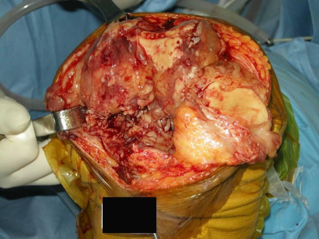
An intraoperative view of a knee before débridement shows grossly infected synovium and necrotic bone.
Fig. 2.
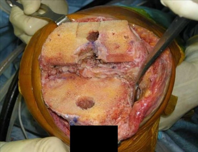
An intraoperative view shows the knee after débridement and femoral and tibial resection.
Fig. 3.
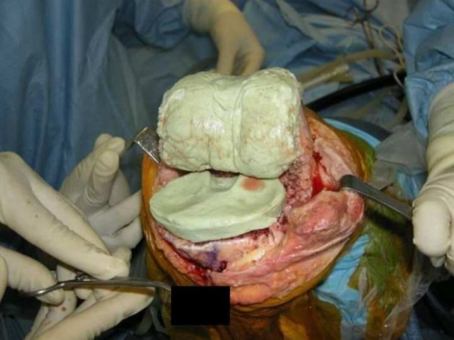
An intraoperative view shows the antibiotic-impregnated articulating cement spacers in situ with the knee in flexion. The flexion gap has been kept loose to facilitate postoperative flexion.
During the postoperative period, continuous passive motion is used for physiotherapy, for approximately 3 hours a day (1 hour at a time, three times per day) until the patient can bend the knee approximately 90o. The patients were allowed to bear weight and bend their knees. They were instructed to continue their daily activities and use a hinged PCL brace to prevent posterior subluxation of the tibia. Parenteral antibiotics were continued according to the results of the preoperative or intraoperative culture and sensitivity results. The choice of antibiotics was based on the results of preoperative cultures if the infecting organism was identified. If not, broad-spectrum antibiotics were started and then modified based on the intraoperative culture results. The patients were discharged 2 weeks postoperatively with appropriate oral antibiotics or transferred to a local hospital for appropriate intravenous antibiotics if susceptible oral antibiotics were not available (Fig. 4).
Fig. 4A–B.
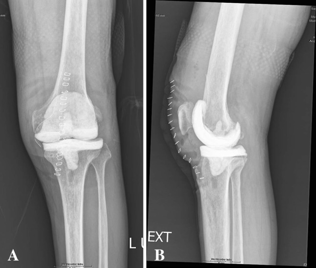
(A) A postoperative AP view shows the complete coverage of the femoral and tibial condyles by the articulating spacers. (B) A postoperative lateral view shows the stability and congruence of the spacers.
The primary TKA was performed when the laboratory markers of infection were normalized or near normalized and no clinical evidence of infection was observed. Preoperative aspiration before conversion to TKA was not performed provided the blood test results had normalized and clinical examination was not suggestive of ongoing infection. The spacer was removed and samples were collected from the joint fluid and deep tissues for bacteriologic culture and sensitivity. Pathologic examination of frozen section tissue specimens was performed. Our protocol was to perform repeat débridement and implantation of new spacers if there was suspicion of residual infection by blood laboratory results (erythrocyte sedimentation rate, C-reactive protein, and white blood cell count) or clinical findings. One patient underwent repeat débridement and implantation of new spacers.
When patients were believed to be free of infection, based on preoperative and intraoperative testing, the cement spacers were removed, thorough débridement was performed again, and then the TKA prostheses were implanted. Adequate ligament balancing and equalization of the flexion and extension gaps were performed. The NexGen® Legacy® Constrained Condylar Knee (LCCK) system (Zimmer Inc, Warsaw, IN, USA) was used with antibiotic-impregnated bone cement. One gram vancomycin was added per each 40-g batch of cement. Patellae were not resurfaced according to our routine in primary TKAs.
Patients and Methods
Between 2001 and 2009, 15 patients presented with advanced knee arthritis and coexistent bone or joint sepsis. All were treated with débridement and articulating cement spacer placement according to the procedure described above. All 15 patients (100%) had at least 2 years of followup and were included in this series. The mean duration of followup was 4 years (range, 2–7 years) (Table 1). Among the 15 patients, two were satisfied with their knee function and pain relief with the articulating antibiotic-loaded cement spacer in situ and declined to undergo a second surgery (TKA). These two patients consequently were excluded from the subsequent assessment in this study. Thus, 13 patients subsequently underwent a primary TKA. There were five male and eight female patients with a mean age of 65 years (range, 39–81 years). There were nine patients with chronic infections whose mean duration of infection was 7 months (range, 3–18 months) before the first-stage procedure and four patients in the acute stage whose mean duration of infection was 11 days (range, 7–14 days).
Table 1.
Demographics and history of patients in the study
| Patient number | Age (years)/sex | Comorbidities | Previous surgeries/any history of sepsis | Preoperative organism isolated from bacterial culture |
|---|---|---|---|---|
| 1 | 58/F | None | Arthroscopic débridement for septic knee | None |
| 2 | 80/F | None | Arthrotomy for knee sepsis; arthroscopic débridement for knee sepsis | Methicillin-sensitive Staphylococcus aureus |
| 3 | 70/F | None | Arthroscopic débridement for septic knee | Methicillin-resistant Staphylococcus aureus |
| 4 | 66/M | Diabetes mellitus | None | Candida spp |
| 5 | 81/F | Bronchial asthma | None | Candida spp |
| 6 | 73/F | Polytrauma, spine infection, and Addison crisis | L2 corpectomy and posterior instrumentation | Methicillin-resistant Staphylococcus aureus |
| 7 | 72/F | Diabetes mellitus | Contralateral (left) TKA | None |
| 8 | 63/M | Diabetes mellitus | None | None |
| 9 | 39/M | None | ACL reconstruction; knee synovectomy and ACL graft removal | Pseudomonas aeruginosa |
| 10 | 71/M | None | Arthrotomy for knee sepsis | None |
| 11 | 48/F | None | ACL reconstruction; knee synovectomy | None |
| 12 | 64/F | None | None | None |
| 13 | 66/M | Diabetes mellitus | Arthrotomy for knee sepsis | None |
* Patients 14 and 15 were not included because they declined definitive reconstruction with TKA.
Nine patients had advanced arthritis with isolated septic arthritis and the remaining four patients had periarticular osteomyelitis as well. Of the nine patients with isolated septic arthritis, two had a positive fungal culture and one of these two patients also had a tuberculous infection. Potential immunity-compromising factors were identified in six patients: four patients had diabetes mellitus, one had bronchial asthma with long-term steroid use, and one had recent polytrauma, spinal surgery, and hypopituitarism on a chronic basis and was using steroids.
Before transferring to our institution, seven patients had previous knee surgery which failed to control the infection. Two of them had ACL reconstruction, which was complicated by infection; one patient was treated with open débridement and graft removal, whereas the other was treated with arthroscopic débridement. The remaining five patients had septic arthritis of the arthritic knee; they had at least one previous surgery for treatment of sepsis (three knee arthrotomies and four arthroscopic débridements). One patient also underwent spinal surgery resulting from infection. All these seven patients had received antibiotics for a mean of 13 weeks (range, 4–20 weeks) before transferring to our institution. Among them, one patient with combined tuberculosis infection had been receiving long-term antituberculosis medication as well.
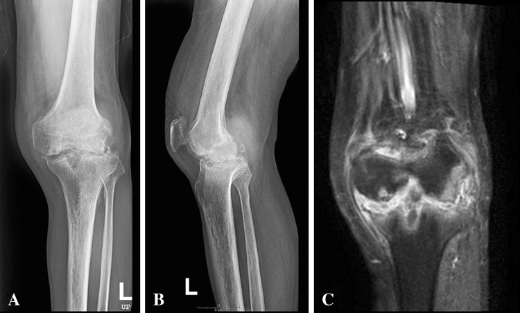
Preoperative analysis included joint ROM, Knee Society scores, 100-mm VAS on pain, WOMAC scores, radiologic examination (weightbearing posteroanterior view, lateral view, and Merchant’s view) (Fig. 5A–B), and laboratory analysis including total and differential white cell count, erythrocyte sedimentation rate, and C-reactive protein. Results of synovial fluid culture were positive in six (46%) of the 13 patients (Table 1). The four patients who had a septic knee combined with osteomyelitis had a draining sinus. In these patients, MRI was done to determine the extent of the spread of infection around the joint. All showed evidence of infection in the knee and adjacent bones on MR images (Fig. 5C).
Fig. 5A–C.
(A) A preoperative AP radiograph shows advanced arthritis, subchondral bone destruction, and soft tissue swelling. The preoperative (B) lateral radiograph shows irregular destruction of bone, and the (C) MR image shows irregular destruction of subchondral bone with increased signal intensity of the surrounding tissues.
Our criteria for the diagnosis of current infection of the knee in this study was one or more of the following: aspiration of infective joint fluid (determined by white blood cell count and percent of polymorphonuclear leukocytes in the differential count), isolation of organism(s) from aspirated joint fluid, presence of a draining sinus, or MRI evidence of a septic knee combined with osteomyelitis. Ten of the 13 patients in this study had their diagnosis made preoperatively according to these criteria and were treated with our native intraoperatively molded articulating cement spacer technique. Based on the results of joint fluid cultures alone, six patients had active infection while for the remaining three patients, the initial plan was to perform TKA. When suspicious findings of infection were observed during surgery or for any patient with a previous history of an infection of the joint that had resolved, five tissue samples were taken from each patient and sent for frozen section examination. Two or more of the five samples were positive for our criteria suggesting infection (more than 10 polymorphonuclear leukocytes per high-power field) in all these patients. Therefore, after resecting the bone ends as is done in a TKA, thorough débridement was performed and intraoperatively molded articulating cement spacers were inserted instead of prostheses. The mean interval between spacer insertion and the TKA was 5.6 months (median, 4 months; range, 2–29 months). Ten patients underwent TKA within 4 months after insertion of the spacers. Two patients chose to undergo the TKA, one after 10 months and the other after 29 months, because of personal reasons. Infection control was not achieved in one patient after the initial spacer implantation. Four months after spacer insertion, the patient still had some knee effusion and pain, with elevated laboratory results. The patient’s intraoperative frozen section examination during the second-stage surgery suggested residual infection. According to our protocol, repeat débridement and insertion of a new spacer were performed. TKA was performed 1 month later when the patient had normal laboratory and frozen section results. The patients with fungal infection had 12 week and 16 weeks of antifungal treatment each after the first-stage procedure to undergo TKA. The patient with combined tuberculosis infection received antituberculosis medication for 12 months after the TKA.
Parenteral antibiotics were used after the TKA for approximately 2 weeks, and then changed to oral antibiotics if available. Antibiotics were maintained based on the clinical course and laboratory results, which varied from 4 weeks to 12 weeks.
The patients were evaluated at 6 weeks, then every 3 months until 1 year postoperatively, and then yearly thereafter. Knee ROM, VAS scores, Knee Society scores, and WOMAC scores were recorded at every visit. The ROM and Knee Society scores were measured in a blinded manner by a research assistant (T-HS) responsible for collecting clinical data before and after joint arthroplasty. All patients were followed up with blood laboratory tests until normalized and careful clinical observation for any symptoms or signs of infection thereafter for a minimum of 2 years (range, 2–7 years) after the TKA.
Results
As noted earlier, two of the 15 patients were comfortable with the spacers and declined a more definitive reconstruction, and one patient had the débridement and spacer procedure performed a second time based on suspected persistence of infection, but after implantation of the definitive revision TKA prosthesis, no patients had recurrent infection.
The mean knee ROM at initial presentation was 103° (range, 60°–155°), which decreased after the spacer implantation (before the TKA) to a mean of 87 ° (range, 60°–135°), and then improved to a mean of 115° (range, 75°–150°) after TKA. Twelve of the 13 patients had knee flexion of 90° or greater and six of these patients had knee flexion of 120° or greater at the last followup. The patient who had a second débridement and spacer placement according to our protocol had ROM from 10° to 90° at last followup.
The mean Knee Society Knee scores improved from 41(range, 26–73) at the initial visit to 85 (range, 46–93) after TKA. The Knee Society Function scores increased from 43 (range, 27– 73) at the initial visit to 83 (range, 47–92) after TKA. The mean WOMAC scores improved from 51 (range, 40–65) at the first visit to 18 (range, 11–31) after the TKA. Similarly, the mean VAS for pain improved from 66 (range, 50–75) at the initial presentation to 18 (range, 0–40) after the TKA. The patient who had a second débridement and spacer placement according to our protocol had WOMAC, Knee Society, and VAS scores of 31, 63, and 30, respectively, at last followup.
Discussion
The treatment of advanced knee arthritis with coexistent bone or joint sepsis is different and more complex than the treatment of either knee arthritis or knee sepsis. There are no fixed guidelines for this challenging situation because of the paucity of treatment strategies that have been reported [6, 10, 12]. A good technique is needed that deals with all aspects of this problem. We therefore share our results using aggressive débridement of the knee and implantation of intraoperatively molded articulating antibiotic cement spacers; using this approach in a small series we found consistent infection eradication at 2-year minimum followup, with knee scores that compare favorably with those of staged revision surgery for other indications [3, 4] and with those of other series of treatment for infected, arthritic knees [6, 12].
Our study has some limitations. First, this is a retrospective study with a limited number of patients. However, a septic knee combined with a knee with already disabling arthritis is not common but is difficult to treat. Our study included only patients meeting these criteria, so although we have a limited number of patients, we believe it provides valuable information. Second, we had no control group. However, as all patients in this series were free of clinical infection at a minimum of 2 years followup, there seems little reason to consider the use of static spacers, which is an alternative reported by others [14], given that articulating spacers have shown improved function when used in revision TKAs [3, 4, 16]. Vancomycin and aminoglycosides carry the risk of systemic toxicity; we do not routinely follow serum levels of antibiotics in these patients, but it may be reasonable to do so in patients with other comorbidities such as renal insufficiency.
There are a few reports regarding the treatment of arthritis combined with intractable infection. Nazarian et al. [14] reported a 100% success rate with a two-stage approach to primary TKA in 14 patients with chronic knee sepsis with the use of a nonarticulating cement spacer. Kirpalani et al. [8] suggested that a staged two-stage procedure is a valuable procedure with good results in patients with diabetes mellitus with arthritic knees and septic arthritis. Matsumoto et al. [12] reported the case of a patient with methicillin-resistant Staphylococcus aureus and knee osteoarthritis treated with a two-stage procedure that included placement of vancomycin-coated hydroxyapatite blocks and immobilization using an external fixator for 3 months followed by TKA. Mirza et al. [13] reported a cohort of three patients with knee sepsis treated with débridement and insertion of gentamicin beads and a methylmethacrylate “hamburger”. In all these previous studies, the knees were immobilized in some form during the period of infection control. Several studies have proven the benefits of using an articulating spacer over a static spacer during the treatment of an infected TKA [2–4, 16]. Because ROM is encouraged with an articulating spacer, the final ROM after the eventual TKA may be better with an articulating spacer than with a static spacer. In addition, knee function during treatment is superior with an articulating spacer than with a static spacer. Two of our patients declined definitive reconstruction with TKA because they were so satisfied with their knee function and pain relief after implantation of the articulating spacer. We do not advocate this because of the high likelihood that the articulating spacer eventually will fracture and can cause bone loss, but ultimately it is the patient’s decision.
The results of our study show that the use of this two-stage approach (native intraoperative molded articulating cement spacer technique) is an effective method to eliminate infection and maintain knee function and mobility during and after infection control in an infected arthritic joint. Infection control is critically important in these patients, because primary TKA in patients with a history of deep infection is associated with a high risk of recurrent infection. In a retrospective study of 65 TKAs in knees with prior infection, Jerry et al. [6] reported a deep infection rate of 4% with prior joint sepsis and 15% with prior osteomyelitis. Lee et al. [9] reported a 5% reinfection risk in 16 primary TKAs with a 5-year followup in patients with a history of prior sepsis of the knee. Moreover, the patients in our study had good functional results after treatment, and they were mobile throughout the treatment. Thus, our results are encouraging because they derive from challenging cases: advanced knee arthritis with intractable coexisting infection and/or periarticular osteomyelitis (nine of the 13 patients had mean duration of 7 months [range, 3–18 months] of infection with failed eradication of the infection, and seven of the nine patients had undergone previous knee surgery for the infection). We believe there are multiple reasons for our encouraging results. First, a large amount of antibiotics can be inserted in the joint with the intraoperatively molded articulating cement spacers, which can provide a high local concentration of antibiotics. Second, the spacer is applied to the bone in a moderately doughy phase and takes the exact shape of the irregular surface of the underlying bone after débridement. This eliminates any dead space between the cement-bone interfaces and may help by reducing any nidi of recurrent infection. Moreover, the interdigitation of cement in these spaces enhances the stability of this construct, which seems to result in less pain and better function and mobility during the period of infection control.
There could be concern regarding the hole made for intramedullary alignment guides. We performed thorough débridement of the intramedullary canal and massive irrigation during the first-stage procedure. However, we have not placed antibiotic cement sticks or beads in the medullary canal, even for treatment of an infected prosthetic joint. Instead, we make the part of the spacer that protrudes into the medullary canal opening area sufficient to fill some portion of the canal adjacent to the spacer. Eradication of infection in the medullary canal is important. However, Freeman et al. [4] reported good results without placing antibiotic cement sticks or beads in the medullary canal, which mirrors our experience, and to our knowledge, we do not have evidence that placing antibiotic cement sticks or beads in the medullary canal is required or that it improves the likelihood of eradication of infection. Regarding the kind of antibiotics impregnated in the cement spacer, gentamicin or tobramycin may be used instead of vancomycin or streptomycin, depending on the clinical situation. The incidence of methicillin-resistant organisms is relatively high in our patient population, thus our choice of antibiotics was vancomycin for patients with unknown organisms. In addition, although infection with tuberculosis is uncommon, it often is missed, and the diagnosis can be difficult to make. Therefore, considering the potential risk of needing an additional stage if appropriate antibiotic coverage is not achieved, we use vancomycin and streptomycin when the infecting organism is unknown, which is similar to our approach for prosthetic joint infection.
This two-stage technique using aggressive joint débridement and placement of an intraoperatively molded antibiotic cement spacer, using the basic principles used in the revision of infected TKAs, appears to be an option for the treatment of patients with advanced knee arthritis coexistent with sepsis. In this small series, this approach achieved infection control at 2-year minimum followup while maintaining knee function throughout treatment. This technique is not a substitute for open or arthroscopic débridement in an acute septic knee. We advocate this technique only in patients with a current septic knee with already disabling arthritis.
Acknowledgments
We thank our research assistant, Tae-Hee Seo BS, Samsung Biomedical Research Institute, Department of Orthopedic Surgery, Samsung Medical Center, Seoul, Korea for her tremendous contribution in collecting the clinical data used for this study.
Footnotes
Each author certifies that he or she, or a member of his or her immediate family, has no funding or commercial associations (eg, consultancies, stock ownership, equity interest, patent/licensing arrangements, etc) that might pose a conflict of interest in connection with the submitted article.
All ICMJE Conflict of Interest Forms for authors and Clinical Orthopaedics and Related Research editors and board members are on file with the publication and can be viewed on request.
Clinical Orthopaedics and Related Research neither advocates nor endorses the use of any treatment, drug, or device. Readers are encouraged to always seek additional information, including FDA-approval status, of any drug or device prior to clinical use.
Each author certifies that his or her institution approved the human protocol for this investigation, that all investigations were conducted in conformity with ethical principles of research.
References
- 1.Chiang ER, Su YP, Chen TH, Chiu FY, Chen WM. Comparison of articulating and static spacers regarding infection with resistant organisms in total knee arthroplasty. Acta Orthop. 2011;82:460–464. doi: 10.3109/17453674.2011.581266. [DOI] [PMC free article] [PubMed] [Google Scholar]
- 2.Emerson RH, Jr, Muncie M, Tarbox TR, Higgins LL. Comparison of a static with a mobile spacer in total knee infection. Clin Orthop Relat Res. 2002;404:132–138. doi: 10.1097/00003086-200211000-00023. [DOI] [PubMed] [Google Scholar]
- 3.Fehring TK, Odum S, Calton TF, Mason JB. Articulating versus static spacers in revision total knee arthroplasty for sepsis: The Ranawat Award. Clin Orthop Relat Res. 2000;380:9–16. doi: 10.1097/00003086-200011000-00003. [DOI] [PubMed] [Google Scholar]
- 4.Freeman MG, Fehring TK, Odum SM, Fehring K, Griffin WL, Mason JB. Functional advantage of articulating versus static spacers in 2-stage revision for total knee arthroplasty infection. J Arthroplasty. 2007;22:1116–1121. doi: 10.1016/j.arth.2007.04.009. [DOI] [PubMed] [Google Scholar]
- 5.Ha CW. A technique for intraoperative construction of antibiotic spacers. Clin Orthop Relat Res. 2006;445:204–209. doi: 10.1097/01.blo.0000201161.52196.c5. [DOI] [PubMed] [Google Scholar]
- 6.Jerry GJ, Jr, Rand JA, Ilstrup D. Old sepsis prior to total knee arthroplasty. Clin Orthop Relat Res. 1988;236:135–140. [PubMed] [Google Scholar]
- 7.Jia YT, Zhang Y, Ding C, Zhang N, Zhang DL, Sun ZH, Tian MQ, Liu J. Antibiotic-loaded articulating cement spacers in two-stage revision for infected total knee arthroplasty: individual antibiotic treatment and early results of 21 cases. Chin J Traumatol. 2012;15:212–221. [PubMed] [Google Scholar]
- 8.Kirpalani PA, In Y, Choi NY, Koh HS, Kim JM, Han CW. Two-stage total knee arthroplasty for non-salvageable septic arthritis in diabetes mellitus patients. Acta Orthop Belg. 2005;71:315–320. [PubMed] [Google Scholar]
- 9.Lee GC, Pagnano MW, Hanssen AD. Total knee arthroplasty after prior bone or joint sepsis about the knee. Clin Orthop Relat Res. 2002;404:226–231. doi: 10.1097/00003086-200211000-00036. [DOI] [PubMed] [Google Scholar]
- 10.Lützner J, Hübel U, Kirschner S, Günther KP, Krummenauer F. [Long-term results in total knee arthroplasty: a meta-analysis of revision rates and functional outcome][in German] Chirurg. 2011;82:618–624. doi: 10.1007/s00104-010-2001-8. [DOI] [PubMed] [Google Scholar]
- 11.Mahmud T, Lyons MC, Naudie DD, Macdonald SJ, McCalden RW. Assessing the gold standard: a review of 253 two-stage revisions for infected TKA. Clin Orthop Relat Res. 2012;470:2730–2736. doi: 10.1007/s11999-012-2358-8. [DOI] [PMC free article] [PubMed] [Google Scholar]
- 12.Matsumoto K, Itokazu M, Uemura S, Takigami I, Naganawa T, Shimizu K. Successful joint arthroplasty after treatment of destructive MRSA arthritis of the knee using antibiotic loaded hydroxyapatite blocks: a case report. Arch Orthop Trauma Surg. 2007;127:47–50. doi: 10.1007/s00402-006-0212-8. [DOI] [PubMed] [Google Scholar]
- 13.Mirza AH, Noble J, Teanby D. Infected knee treated by total knee arthroplasty. Knee. 2000;7:171–174. doi: 10.1016/S0968-0160(00)00045-4. [DOI] [PubMed] [Google Scholar]
- 14.Nazarian DG, de Jesus D, McGuigan F, Booth RE., Jr A two-stage approach to primary knee arthroplasty in the infected arthritic knee. J Arthroplasty. 2003;18(7 suppl 1):16–21. doi: 10.1016/S0883-5403(03)00343-7. [DOI] [PubMed] [Google Scholar]
- 15.Qiu XS, Sun X, Chen DY, Xu ZH, Jiang Q. Application of an articulating spacer in two-stage revision for severe infection after total knee arthroplasty. Orthop Surg. 2010;2:299–304. doi: 10.1111/j.1757-7861.2010.00103.x. [DOI] [PMC free article] [PubMed] [Google Scholar]
- 16.Romanò CL, Gala L, Logoluso N, Romanò D, Drago L. Two-stage revision of septic knee prosthesis with articulating knee spacers yields better infection eradication rate than one-stage or two-stage revision with static spacers. Knee Surg Sports TraumatolArthrosc. 2012;20:2445–2453. doi: 10.1007/s00167-012-1885-x. [DOI] [PubMed] [Google Scholar]


