Abstract
Acute rejection continues to present a major obstacle to successful lung transplantation. While CD4+ T lymphocytes are critical for the rejection of some solid organ grafts the role of CD4+ T cells in the rejection of lung allografts is largely unknown. In this study we demonstrate in a novel model of orthotopic vascularized mouse lung transplantation that acute rejection of lung allografts is independent of CD4+ T cell-mediated allorecognition pathways. CD4+ T cell-independent rejection occurs in the absence of donor-derived graft-resident hematopoietic antigen presenting cells. Furthermore, blockade of the CD28/B7 costimulatory pathways attenuates acute lung allograft rejection in the absence of CD4+ T cells, but does not delay acute rejection when CD4+ T cells are present. Our results provide new mechanistic insight into the acute rejection of lung allografts and highlight the importance of identifying differences in pathways that regulate the rejection of various organs.
While it is well established that T lymphocytes play a critical role in mediating allograft rejection, it is unclear how CD4+ and CD8+ T cells differentially regulate this immune process. The vast majority of studies in mice have suggested that CD4+ T lymphocytes are both necessary and sufficient for the acute rejection of cardiac allografts (1, 2). To this end, CD4+ T cell depletion can lead to long term survival of cardiac allografts indicating that CD8+ T cells alone can not mediate this rejection (1). Moreover, we and others have shown that activation of the CD4+ T cells is dependent on the direct allorecognition pathway through graft-resident hematopoietic cells (3, 4). Analogous to heart allograft rejection, most reports indicate that the rejection of pancreatic islet allografts by unprimed recipients requires CD4+ T cells (5–8). Similarly, earlier studies have indicated that the rejection of corneal allografts is dependent on CD4+ T cells (9). However, this notion was challenged by newer reports that show that the rejection of allogeneic corneas can be mediated in the absence of CD4+ T cells (10). While it is well established that CD4+ T cells can independently reject skin allografts, numerous papers indicate that CD8+ T cells can also reject allogeneic skin grafts in the absence of CD4+ T cell help (11–14). Both CD4+ and CD8+ T cells can independently reject intestinal allografts and allogeneic hepatocytes in mice (15–19). Taken together, these studies suggest that the characteristics of the specific allograft tissue may influence the relative contribution of CD4+ and CD8+ T cells to acute allograft rejection.
Importantly, there are differences between various organs regarding their susceptibility to rejection. Skin and small bowel appear to be most susceptible to rejection in mice, whereas pancreatic islets, hearts, kidneys and livers are progressively more easily accepted (20). There is also evidence that CD4+ and CD8+ T cell-mediated rejection responses against allografts may depend on different costimulatory signals and have different susceptibilities to immunosuppression. It is therefore critical to define the relative contributions of CD4+ and CD8+ T cells in mediating allograft rejection in specific organs (12, 21–23).
Similar to mice, organs differ with regard to their immunogenicity in humans as well. To this end, outcomes after lung transplantation in humans are far worse than those after transplantation of other solid organs (24). The five year survival is less than 50% and the majority of patients succumb to obliterative bronchiolitis. This condition is characterized by progressive fibrous obliteration of the distal airways and is thought to be a manifestation of chronic rejection. The frequency and severity of acute rejection has been shown to be a risk factor for the development of chronic rejection (25). Interestingly, a single episode of acute rejection was found to be a predictor of bronchiolitis obliterans independent of other risk factors (26). Mechanisms that lead to both acute and chronic lung allograft rejection are poorly understood as their study has been hampered by the lack of a physiologically relevant mouse model of lung transplantation. To date, the most frequently used model to study immunological pathways leading to lung allograft rejection has been the heterotopic transplantation of tracheal allografts. While both CD4+ and CD8+ T cells have been shown to contribute to obliteration of the airway, this model only employs the proximal airway and is not vascularized. Therefore it may not be representative of the pathophysiology of lung allografts (27, 28). We have recently developed a novel model of orthotopic vascularized lung transplantation in the mouse and have described marked differences between outcomes of orthotopic vascularized mouse lung transplants and heterotopic tracheal transplants (29, 30).
In this study we examined the role of CD4+ T cells in the acute rejection of vascularized lung allografts. We show that lung allografts are acutely rejected in the absence of CD4+ T cells. Similar to our previous findings in a heart transplant model triggering of CD4+ T cell-independent rejection can occur in the absence of graft-resident hematopoietic cells (29). Moreover, blockade of the CD28/B7 costimulatory pathways delays lung allograft rejection in the absence of CD4+ T cells, but does not alter the tempo of acute rejection when CD4+ T cells are present.
Materials and Methods
Animals
Male inbred C57BL/6 (H-2Kb) (designated as B6), CD4-knockout B6 (B6.129S2-Cd4tm1Mak/J), Balb/c (H-2Kd) and CBA/Ca (H-2Kk) (designated as CBA) mice were purchased from Jackson Laboratory, Ben Harbor, ME. Male MHC class II-deficient B6 mice (B6.129-H2-Ab1tm1GruN12) were purchased from Taconic Farms, Germantown, NY. Ten to twelve week old animals weighing 25–30 grams were used as both donors and recipients.
Bone Marrow Transplantation
Bone marrow chimeras were created as previously described (31). Briefly, for bone marrow transplantation, male wild-type B6 were reconstituted with marrow from CBA mice. Bone marrow was harvested from the femora of donor mice and T lymphocytes were depleted with anti-CD90 (Thy)-labeled magnetic microbeads (Miltenyi Biotec, Auburn, CA). Recipient mice received an inoculum of 1 × 10⁷ T cell-depleted donor bone marrow via lateral tail vein injection 6 hours after lethal irradiation (10 Gy). These bone marrow chimeras were used as lung donors at least 90 days after the bone marrow transplant. The following designation is used to describe chimeric organs: nonhematopoietic cells (hematopoietic antigen presenting cells).
Replacement of lung-resident hematopoietic antigen presenting cells in bone marrow chimeras was assessed by flow cytometry. Lung tissue derived from wildtype B6 and CBA mice as well as B6(CBA) bone marrow chimeras was cut into 2mm slices and digested by placing them into a RPMI 1640 solution containing Type 2 collagenase (0.5 mg/ml) (Worthington Biochemical Corporation, Lakewood, NJ) and 5 U/ml DNAse (Sigma, St. Louis, MO) for 90 minutes. The digested lung tissue was then passed through a 70 μm cell strainer and treated with ACK lysing buffer. Replacement of hematopoietic antigen presenting cells was evaluated by staining with fluorochrome-labeled anti-CD45 (clone 30-F11), anti-I-Ab (clone 25-9-17), anti-I-Ak (clone 11-5.2) and respective isotype control antibodies (BD Biosciences, San Jose, CA).
Lung Transplantation
Left orthotopic vascularized lung transplants were performed utilizing cuff techniques as previously described (29). In some experimental groups CD4+ T lymphocytes were depleted as previously described (GK1.5 100 μg intraperitoneally at days −2 and −1) (BIOExpress, West Lebanon, NH) and depletion was maintained with weekly injections (31). Some animals were treated with CTLA4Ig (200 μg intraperitoneally) on day 2 or days 0, 2, 4 and 6 (BIOExpress, West Lebanon, NH). Control Ig was purchased from Jackson Immuno Research Laboratories (West Grove, PA).
Histology
Portions of the transplanted lungs were fixed in formaldehyde, sectioned and stained with Hematoxylin and Eosin. Grading for acute cellular rejection was performed in a blinded fashion by a pathologist (F.H.K.) using standard criteria developed by the Lung Rejection Study Group (32). The grading scale was as follows: grade A0: no cellular infiltrates; grade A1: minimal acute rejection with perivascular infiltrates comprising only one or two layers of predominantly small lymphocytes; grade A2: mild acute rejection with several layers of small and large lymphoid cells cuffing vessels and endothelialitis; grade A3: moderate acute rejection with spread of infiltrates into the interstitium of alveolar septae and spaces as well as predominance of large lymphoid cells and intra-alveolar accumulation of macrophages and neutrophils; grade A4: severe acute rejection characterized by vascular thrombosis with massive intra-alveolar hemorrhage and infarction resulting in diffuse alveolar damage with necrosis of alveolar epithelium.
Flow Cytometry
Seven days after transplantation, portions of transplanted lungs were prepared for flow cytometric analysis. Cellular infiltration into lungs was assessed by staining with fluorochrome-labeled anti-CD90.2 (clone 30-H12), anti-CD4 (clone RM4-5), anti-CD8 (clone 53-6.7), anti-NK1.1 (clone PK136), anti-γδ T-cell receptor (clone GL3), anti-CD25 (clone PC61), anti-CD62Ligand (clone MEL-14) and anti-CD44 (clone IM7) antibodies as well as their respective isotype controls (BD Biosciences, San Jose, CA and eBioscience, San Diego, CA). Intracellular staining was performed with anti-IFN-γ (clone XMG1.2), anti-IL4 (clone 11B11) (BD Pharmingen, San Jose, CA), anti-Foxp3 (clone FJK-16s) (eBioscience, San Diego, CA) and their respective isotype controls. For intracellular cytokine staining of graft-infiltrating T lymphocytes, cells were cultured after lung digestion with 20 ng/ml PMA (Sigma-Aldrich, St. Louis, MO) and 1 μM ionomycin (Calbiochem, La Jolla, CA) for 4 hours. Two μM monensin (Sigma, St. Louis, MO) was added for the last 3 hours of culture. Cells were subsequently surface stained for 30 minutes in a small volume of cold PBS, followed by a 60-minute period of fixation in 2.5% paraformaldehyde (Sigma, St. Louis, MO) on ice. Cells were then permeabilized with 0.1% saponin (Sigma, St. Louis, MO) in PBS containing 2% FCS for minutes on ice. Intracellular staining was performed for 30 minutes with the cells resuspended in a small volume of 0.1% saponin. After two washes in 0.01% saponin, the cells were resuspended in PBS containing 2% FCS and analyzed by flow cytometry. For absolute live cell counts 50,000 CD45.1+ splenocytes labeled with fluorochrome-labeled anti-CD45.1 mAb (A20: BD Pharmingen, San Jose, CA.) were also added to stained T cell sample FACS tubes just prior to FACS analysis. Live CD4+ or CD8+ T cell counts were calculated by taking the ratio of the number of CD4+ or CD8+ CD45.1− live events collected, respectively to the number CD45.1+ events collected and multiplying by 50,000.
Statistical Analysis
Histological scores, numbers of graft-infiltrating T lymphocytes and intracellular IFN-γ levels are expressed as the mean ± SEM. Statistical analysis to assess significant differences between experiment groups was performed by using the Student’s t test. Results were considered statistically different if p < 0.05.
Results
Graft-Infiltrating CD8+ T cells predominate over CD4+ T cells in Rejected Mouse Lung Allografts
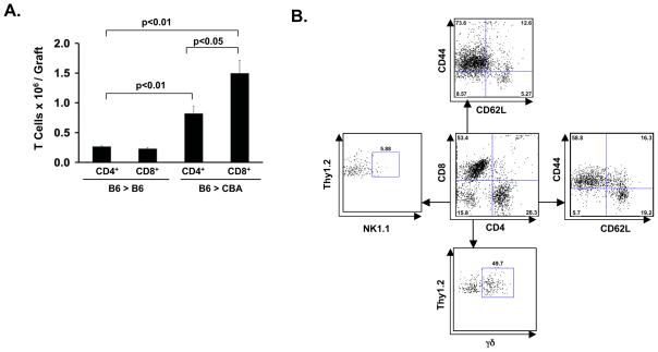
We have previously shown that – similar to the rejection of cardiac allografts in this strain combination – B6 lungs show histological evidence of acute vascular rejection 7 days after transplantation into CBA recipients (29). We first wanted to assess the absolute numbers of graft-infiltrating CD4+ and CD8+ T cells in vascularized lung transplants at the time of acute allograft rejection. For this purpose we performed orthotopic vascularized lung transplants in the syngeneic B6 → B6 and allogeneic B6 → CBA strain combinations. The absolute number of both graft-infiltrating CD4+ and CD8+ T cells was significantly lower in syngeneic grafts than allogeneic grafts (Figure 1A). The ratio of CD8+ to CD4+ T cells was 0.9 ± 0.2 in syngeneic grafts. Interestingly, in allogeneic grafts there were twice as many CD8+ T cells than CD4+ T cells with a ratio of 1.9 ± 0.3. We further evaluated graft-infiltrating CD4+ and CD8+ T cells in allogeneic transplants (Figure 1B). Naive T cells express the CD62Lhigh CD44low phenotype, whereas the effector memory T cells express the CD62LlowCD44high phenotype (33). The majority of both CD4+ and CD8+ T cells within the B6 → CBA lung grafts were CD62LlowCD44high indicating an effector memory phenotype. Of note, a larger proportion of CD8+ T cells were CD62LlowCD44hi when compared to CD4+ T cells. A small portion of graft-infiltrating Thy1.2 cells expressed neither CD4 nor CD8. Further analysis of this subset revealed that only a small percentage of these cells expressed NK1.1. Notably, up to half of these Thy1.2+CD4−CD8− cells expressed the γδ-T cell receptor. Thus, our data indicate that, in contrast to other acutely rejected murine allografts, CD8+ T cells are more numerous than CD4+ T cells in acutely rejected mouse lung allografts (34).
Figure 1.
(A) Absolute numbers of infiltrating CD4+ and CD8+ T cells in syngeneic (B6 → B6) and allogeneic lung grafts (B6 → CBA) 7 days following engraftment. T cell infiltrate is represented as mean absolute numbers ± S.E.M of intragraft CD4+ and CD8+ T cells (n=4 per experimental group). Statistically significant differences between experimental groups are indicated and were calculated using the Student’s t test. (B) Characterization of live intragraft Thy1.2+ cells from B6 → CBA lung grafts 7 days following transplantation. Results are representative of 4 independent experiments.
Mouse Lung Allografts are Acutely Rejected in the Absence of CD4+ Direct Allorecognition
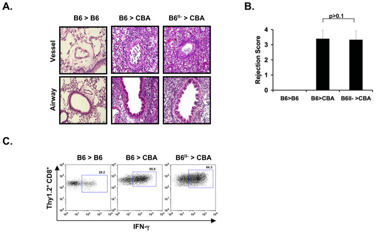
Having shown that CD8+ T lymphocytes are more predominant than CD4+ T lymphocytes in acutely rejected mouse lung allografts we next set out to examine whether the acute rejection of mouse lung allografts was dependent on CD4+ T cells. To test the role of CD4+ T cell direct allorecognition in the acute rejection of vascularized mouse lung transplants we transplanted B6 MHC class II-deficient (designated as B6II-) lungs into CBA hosts. Analogous to wild type B6 lungs that were transplanted into CBA hosts, B6II- lungs demonstrated acute cellular rejection with perivascular cuffing and subepithelial lymphocytic infiltrates in the small airways 7 days after transplantation (Figure 2A). Rejection scores were analogous for B6 → CBA and B6II- → CBA lung grafts (Figure 2B). We also analyzed cytokine expression in graft-infiltrating CD8+ T cells (Figure 2C). The majority of graft-infiltrating CD8+ T cells expressed IFN-γ in both B6 → CBA and B6II- → CBA combinations at comparable levels. We could not detect expression of IL-4 in graft-infiltrating CD8+ T cells indicating a polarization towards the Tc1 phenotype (data not shown).
Figure 2.
Histological and flow cytometric analysis of B6 → B6 (n=3), B6 → CBA (n= 4) and B6 II- → CBA (n=4) lung grafts 7 days after transplantation. (A) H&E slides are represented at 400X magnification. (B) Rejection scores represented as mean ± S.E.M. Statistical analysis was conducted with the Student’s t test. (C) Flow cytometric plots depict the percentage of live intragraft Thy1.2+ CD8+ cells that have the capacity to produce IFN-γ. Mean percentage ± S.E.M of IFN-γ producing Thy1.2+ CD8+ cells were 23.9 ± 5.1 for B6 → B6, 65.3 ± 5.5 for B6 → CBA and 67.6 ± 9.0 for B6 II- → CBA lung transplants. Statistical analysis was conducted with the Student’s t test; B6 → CBA vs. B6 → B6 p<0.05, B6II- → CBA vs. B6 → B6 p<0.05, B6II- → CBA vs. B6 → CBA p>0.1.
CD4+ T cells are Not Necessary for the Acute Rejection of Vascularized Mouse Lung Allografts
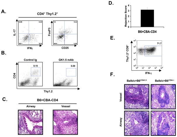
We next set out to characterize the graft-infiltrating CD4+ T cells. Th1 and Th2 cells have been characterized as two classic effector CD4+ T cell subsets that secrete pro-inflammatory cytokines, such as IFN-γ, or anti-inflammatory cytokines, such as IL-4, respectively. Recent studies have demonstrated that CD4+ T cells can also differentiate towards an independent lineage of IL-17-producing Th17 cells, which has been shown to play an important role in inflammatory responses (35). In addition, CD4+Foxp3+ T cells have been well described as a distinct lineage of regulatory CD4+ T cells. At 7 days after engraftment approximately one third of the graft-infiltrating CD4+ T cells in B6 → CBA lung transplants express IFN-γ consistent with a Th1 phenotype and only very few CD4+ T cells express IL-17 (Figure 3A). We were unable to detect any IL-4 expression in graft-infiltrating CD4+ T cells. Notably, approximately 25% of graft-infiltrating CD4+ T cells in acutely rejected B6 → CBA lungs express Foxp3 indicating a regulatory phenotype. Of note, a large portion of the CD4+Foxp3+ T cells lack expression of CD25 consistent with observations previously made for lung-resident regulatory CD4+ T cells (36). Thus, the population of graft-infiltrating CD4+ T cells is heterogeneous with presence of both IFN-γ-expressing Th1 cells and Foxp3-expressing cells.
Figure 3.
Histological and flow cytometric analysis of B6 → CD4+-depleted CBA/Ca lung grafts 7 days after transplantation (n=4). (A) Flow cytometric analysis of live Thy1.2+ CD4+ T cells in B6 → CBA lungs transplants 7 days after engraftment. Plots are representative of 3 independent experiments. (B) Intragraft live Thy1.2+ CD4+ cells in control Ig-treated (left) and GK1.5-treated (right) CBA recipients of B6 lungs 7 days after engraftment. (C) H&E slides are represented at 400X magnification. (D) Rejection score represented as a mean ± S.E.M. recipient. Statistical analysis was conducted with the Student’s t test; B6 → CBA-CD4 vs. B6 → B6 p<0.05, B6 → CBA-CD4 vs. B6 → CBA p>0.1, B6 → CBA-CD4 vs. B6II- → CBA p>0.1. (E) Flow cytometric plots depict the percentage of live intragraft Thy1.2+ CD8+ cells that have the capacity to produce IFN-γ. Mean percentage ± S.E.M of IFN-γ producing Thy1.2+ CD8+ cells was 68.5 ± 4.1. Statistical analysis was conducted with the Student’s t test; B6 → CBA-CD4 vs. B6 → B6 p<0.05, B6 → CBA-CD4 vs. B6 → CBA p>0.1, B6 → CBA-CD4 vs. B6II- → CBA p>0.1. (F) H&E slides of Balb/c → B6CD4+/+ (n=4) and Balb/c → B6CD4−/− (n=2) 7 days after transplantation. Rejection scores were A3 for all recipients in both experimental groups.
Having shown that the CD4+ T cell direct allorecognition pathway is not necessary for the acute rejection of lung allografts we next wanted to determine whether CD4+ T cells are required to trigger acute rejection of lung allografts. For this purpose, we depleted CD4+ T cells in recipient CBA mice through the administration of a short course of preoperative CD4-specific antibodies. We confirmed depletion of CD4+ T cells in the allografts by flow cytometrical analysis of rejected lungs (Figure 3B). At 7 days after transplantation these lung allografts showed acute cellular rejection with rejection scores analogous to B6 → CBA and B6II- → CBA transplants (Figure 3C and D). Moreover, similar to what we observed in lung transplants in the B6 → CBA and B6II- → CBA combinations, graft-infiltrating CD8+ T cells were polarized towards Tc1 with the majority of CD8+ T cells expressing IFN-γ (Figure 3E). There were no significant differences in the percentage of CD8+ T cells expressing IFN-γ between B6→CBA, B6II-→CBA and B6→CD4-depleted CBA recipients. Again, IL-4 was not detected in graft-infiltrating CD8+ T cells (data not shown). Next, we wanted to confirm these observations utilizing an alternative approach. For this purpose, we transplanted Balb/c lungs into CD4 knockout B6 recipients (Figure 3F). We have previously shown that Balb/c lungs are acutely rejected when transplanted into B6 recipients (30). Similarly, Balb/c lungs showed analogous acute rejection after transplantation into CD4 knockout B6 recipients. Taken together, these results show that the rejection of vascularized mouse lungs is independent of CD4+ T cells. Moreover, histological analysis revealed that the rejection scores were equivalent independent of the presence of CD4+ T cells (B6 → CBA vs. B6 → CBA + GK1.5 and Balb/c → B6 vs. Balb/c → B6 CD4 KO) indicating that depletion of regulatory CD4+ T cells does not result in worsening of the acute rejection.
CD4+ T cell-Independent Acute Rejection of Lung Allografts does not require Graft-Resident Donor-Derived Hematopoietic Antigen Presenting Cells
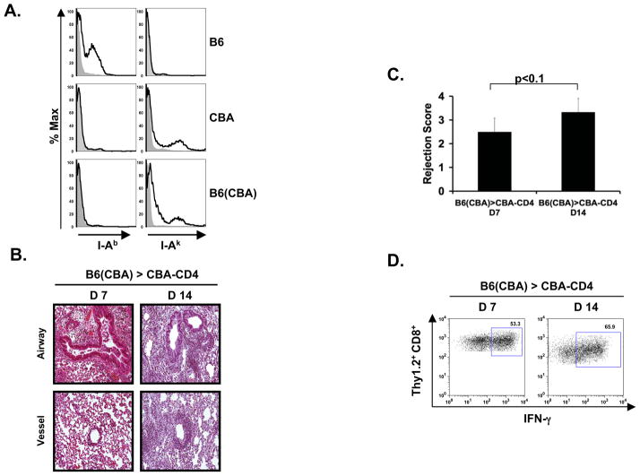
We have previously shown that non-hematopoietic cells can activate alloreactive CD8+ T cells in a B7-dependent fashion in vitro and in vivo (37). Furthermore, our group has demonstrated in a T cell receptor transgenic model that cardiac allografts can be rejected via CD8+ T cell direct allorecognition in the absence of antigen presentation by hematopoietic antigen presenting cells (38). We next set out to determine whether CD4+ T cell-independent acute rejection of lung allografts was dependent on graft-resident donor-derived hematopoietic antigen presenting cells. For this purpose we used B6 bone marrow chimeras that had been reconstituted with CBA bone marrow as lung donors (designated as B6(CBA)). We have previously shown that bone marrow transplantation leads to near complete replacement of hematopoietic antigen presenting cells such as dendritic cells and macrophages (31). We confirmed these findings in the B6(CBA) bone marrow chimeras (Figure 4A). While the lung-resident hematopoietic cells expressed I-Ak we were unable to detect expression of I-Ab. These lungs were transplanted into CBA hosts that had been depleted of CD4+ T cells. At 7 days after engraftment we observed histological signs of acute rejection (Figure 4B). However, the rejection score was less severe when compared to lung transplants performed in the B6 → CD4-depleted CBA strain combination and the percentage of graft-infiltrating CD8+ T cells that expressed IFN-γ in chimeric grafts was significantly lower when compared to B6 grafts (Figure 4C and 4D). We did not detect IL-4 in the graft-infiltrating CD8+ T cells at 7 days after engraftment (data not shown). At 14 days after engraftment into CD4-depleted CBA hosts B6(CBA) grafts had histological evidence of worsening acute rejection with extensive perivascular cuffing and subepithelial lymphocytic infiltrates in the small airways and there was a trend towards a higher percentage of graft-infiltrating CD8+ T cells expressing IFN-γ (Figure 4B, C, D). Moreover, at 14 days after engraftment the rejection scores of B6(CBA) → CD4-depleted CBA lungs were analogous to the ones observed in B6 → CD4-depleted CBA at 7 days after transplantation (Figure 4C). Moreover, there were no statistically significant differences between the percentage of IFN-γ producing CD8+ T cells in B6(CBA) → CD4-depleted CBA lungs at day 14 compared to in B6 → CD4-depleted CBA at 7 days after transplantation (Figure 4D). Thus, the activation of graft-infiltrating CD8+ T cells and the tempo of rejection are delayed in the absence of graft-resident donor-derived hematopoietic antigen presenting cells. However, donor-derived hematopoietic antigen presenting cells are not necessary for the CD4+ T cell-independent activation of CD8+ T cells and acute rejection of lung allografts.
Figure 4.
Histological and flow cytometric analysis of B6(CBA) → CD4+ T cell-depleted CBA 7 (n= 4) and 14 days (n=3) after transplantation. (A) Flow cytometric analysis of I-Ab and I-Ak expression on lung-resident live CD45+ cells in B6, CBA and B6(CBA) mice. Shaded curves represent isotype controls (B) H&E slides are represented at 400X magnification. (C) Rejection scores represented as mean ± S.E.M. Statistical analysis was conducted with the Student’s t test; B6(CBA) → CBA-CD4 (day7) vs. B6 → CBA-CD4 (day7) p<0.1, B6(CBA) → CBA-CD4 (day7) vs. B6(CBA) → CBA-CD4 (day14) p<0.1, B6(CBA) → CBA-CD4 (day14) vs. B6 → CBA-CD4 (day7) p>0.1 (D) Flow cytometric plots depict the percentage of live intragraft Thy1.2+ CD8+ cells that have the capacity to produce IFN-γ. Mean percentage ± S.E.M of IFN-γ producing Thy1.2+ CD8+ cells were 55.9 ± 9.3 and 62.9 ± 8.9 for days 7 and 14, respectively. Statistical analysis was conducted with the Student’s t test; B6(CBA) → CBA-CD4 (day7) vs. B6 → CBA-CD4 (day7) p<0.05, B6(CBA) → CBA-CD4 (day7) vs. B6(CBA) → CBA-CD4 (day14) p>0.1, B6(CBA) → CBA-CD4 (day14) vs. B6 → CBA-CD4 (day7) p>0.1.
In the Absence of CD4+ T Cells Acute Lung Allograft Rejection can be Attenuated by CD28/B7 Costimulatory Blockade
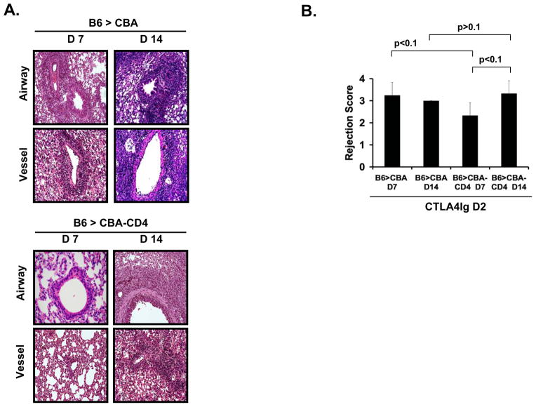
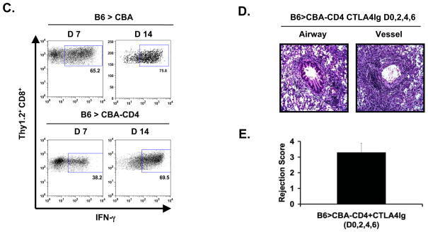
Blockade of the B7:CD28 costimulatory pathway through treatment with a single dose of CTLA4Ig has been shown to lead to prolongation of survival of mouse cardiac allografts (39, 40). We next set out to investigate whether analogous treatment with a single dose of CTLA4Ig had any impact on the rejection response of lung allografts. To this end, CBA mice were treated with a single dose of CTLA4Ig on day 2 after transplantation of B6 lung allografts (Figure 5A). Seven days after transplantation these allografts were acutely rejected with perivascular cuffing and subepithelial lymphocytic infiltrates in the small airways. The rejection scores were analogous to those observed in untreated CBA recipients of B6 lung allografts (Figure 5B). Analogous to our findings in B6 allografts that were transplanted into untreated CBA recipients, the majority of graft-infiltrating CD8+ T cells in B6 lung allografts of CTLA4Ig-treated CBA recipients expressed IFN-γ (Figure 5C). We also evaluated such grafts at 14 days after transplantation. Again, we observed severe acute rejection and the majority of graft-infiltrating CD8+ T cells expressed IFN-γ (Figures 5A, B and C). Thus, unlike the case for heart transplantation, a single dose of CTLA4Ig does not impact the tempo of lung allograft rejection.
Figure 5.
Histological and flow cytometric analysis of CTLA4Ig-treated B6 → CBA 7 days (n=3) and 14 days (n=3) after transplantation as well as CTLA4Ig-treated B6 → CD4+ T cell-depleted CBA 7 days (n=5) and 14 days (n=5) after transplantation. (A) H&E slides are represented at 400X magnification. (B) Rejection scores represented as a mean ± S.E.M. Statistical analysis was conducted with the Student’s t test; B6 → CBA (day 7) vs. B6 → CBA +CTLA4Ig (day 7) p>0.1. B6 → CBA-CD4 (day 7) vs. B6 → CBA-CD4+CTLA4Ig (day 7) p=0.05. (C) Flow cytometric plots depict the percentage of live intragraft Thy1.2+ CD8+ cells that have the capacity to produce IFN-γ. In the case of CTLA4Ig-treated B6 → CBA transplants the mean percentage ± S.E.M of IFN-γ producing Thy1.2+ CD8+ cells were 66.1 ± 9.2 seven days after engraftment and 73.8 ± 1.9 fourteen days after engraftment. In the case of CD4-depleted CTLA4Ig-treated B6 → CBA transplants the mean percentage ± S.E.M of IFN-γ producing Thy1.2+ CD8+ cells were 38.2 ± 5.1 seven days after engraftment and 59.1 ± 5.7 fourteen days after engraftment. Statistical analysis was conducted with the Student’s t test; B6 → CBA-CD4+CTLA4Ig (day 7) vs. B6 → CBA +CTLA4Ig (day 7) p<0.005, B6 → CBA-CD4+CTLA4Ig (day 7) vs. B6 → CBA-CD4+CTLA4Ig (day 14) p<0.005, B6 → CBA+CTLA4Ig (day 7) vs. B6 → CBA+CTLA4Ig (day 14) p>0.1, B6 → CBA-CD4+CTLA4Ig (day 7) vs. B6 → CBA-CD4 (day 7) p<0.001, B6 → CBA vs. B6 → CBA+CTLA4Ig (day 7) p>0.1. (D) Histological analysis of B6→ CD4-depleted CBA lung transplants that received CTLA4Ig (200 μg) on days 0, 2, 4 and 6. H&E slides are represented at 400X magnification. (E) Rejection scores represented as a mean ± S.E.M. Statistical analysis was conducted with the Student’s t test; B6 → CBA-CD4 + CTLA4Ig (D 0, 2, 4 and 6) (day 14) vs. B6 → CBA-CD4 + CTLA4Ig (D 2) (day 14), p>0.1.
Having shown that blockade of the B7:CD28 costimulatory pathway through administration of a single dose of CTLA4Ig was not effective at preventing the activation of CD8+ T cells or lung allograft rejection in the presence of CD4+ T cells, we next wanted to examine whether this costimulatory blockade had any impact on the activation of CD8+ T cells or lung allograft rejection in the absence of CD4+ T cells. To this end, we treated CD4+ T cell-depleted CBA recipients of B6 lung allografts with a single dose of CTLA4Ig 2 days after transplantation. Compared to our observations with CTLA4Ig treatment in the presence of CD4+ T cells, we observed milder rejection in lung allografts 7 days after transplantation when CD4+ T cells were absent (Figures 5A and 5B). The percentage of graft-infiltrating CD8+ T cells that expressed IFN-γ 7 days after transplantation was also significantly lower when compared to CTLA4Ig-treated wild-type recipients that had not received CD4-depleting antibodies or CD4+ T cell-depleted animals that had not received costimulatory blockade (Figure 5C). Previous reports have indicated that inhibition of allograft rejection by CTLA4Ig can be associated with a shift towards production of Th2 cytokines (41). We have not observed a shift towards Tc 2 in the graft-infiltrating CD8+ T cells (data not shown). At 14 days after transplantation, however, lung allografts in CD4+ T cell-depleted CTLA4Ig-treated recipients had worsening rejection with severe perivascular lymphocytic cuffing and subepithelial mononuclear infiltrates in the airways (Figure 5A and B). In addition, a significantly larger percentage of graft-infiltrating CD8+ T cells expressed IFN-γ at 14 days when compared to 7 days after transplantation (Figure 5C). To evaluate whether earlier initiation and prolonged administration of CTLA4Ig enhances the protective effect and further attenuates the acute rejection we treated CD4-depleted CBA recipients of B6 lungs with CTLA4Ig on days 0, 2, 4 and 6 (Figures 5D and E). The histological changes and rejection scores of these grafts at 14 days after transplantation were equivalent to those seen in animals that received a single dose of CTLA4-Ig on day 2. Thus, unlike our observations in the presence of CD4+ T cells, treatment with CTLA4Ig leads to an attenuation of the rejection response when CD4+ T cells are absent but cannot prevent the ultimate rejection of the lung allografts.
Discussion
Acute rejection continues to contribute to the morbidity of lung transplant recipients. In addition, acute rejection has also been demonstrated to be an important risk factor for the development of chronic lung allograft rejection (26). Elucidating mechanisms that lead to the rejection of lung allografts is of critical importance in order to improve outcomes after lung transplantation. However, immune pathways leading to the rejection of lung allografts are poorly understood, in part because such studies have been hampered by the lack of a physiological mouse model of lung transplantation. This study utilizes a novel mouse model of orthotopic vascularized lung transplantation and offers new mechanistic insight into in vivo mechanisms of lung allograft rejection. To our knowledge, this study shows for the first time that the acute rejection of lung allografts is not dependent on CD4+ T lymphocytes. This report illustrates several important issues that have not been previously addressed.
The relative composition of graft-infiltrating T lymphocytes varies considerably between solid organ grafts. Clinical studies have shown that CD4+ T cells are more numerous than CD8+ T cells in heart allografts undergoing acute rejection (42). Studies analyzing graft-infiltrating lymphocytes in acutely rejected mouse cardiac allografts as well pancreatic islet allografts have shown equivalent percentages of CD4+ T cell and CD8+ T cells (34, 43, 44). While CD8+ T cells predominate over CD4+ T cells at early time points in heterotopically transplanted allogeneic murine tracheas, at later time points CD4+ T cells outnumber CD8+ T cells (45, 46). The composition of graft-infiltrating T lymphocytes in vascularized lung transplants had not been previously explored. We show that graft-infiltrating CD8+ T cells predominate over CD4+ T cells in acutely rejected mouse lung allografts. Interestingly, the analysis of bronchoalveolar lavage fluid from lung transplant recipients, who were diagnosed with acute rejection, has similarly demonstrated that CD8+ T cells were more abundant than CD4+ T cells (47, 48).
This predominance of CD8+ T cells over CD4+ T cells in acutely rejected mouse lung allografts led us to examine whether CD4+ T cells were necessary for the acute rejection of murine lungs. Previous studies have shown that transplantation of MHC class II-deficient cardiac allografts leads to long-term survival demonstrating that CD4+ T cell-mediated direct allorecognition is critically important for the acute rejection of cardiac allografts (2, 49). In contrast to these findings we observed that lung allografts were acutely rejected when the CD4+ direct allorecognition pathway was eliminated. Furthermore, while the majority of studies indicate that depletion of CD4+ T cells leads to prolonged or indefinite survival of allogeneic murine hearts and pancreatic islets, vascularized lung allografts are rejected in the absence of CD4+ T cells (1, 6, 8). CD4+ T cell-independent acute rejection has also been observed in the case of murine skin and intestinal allografts (11, 15). Our group has previously shown that graft-resident donor-derived hematopoietic antigen presenting cells are not necessary to trigger CD4+ T cell-independent rejection of murine cardiac allografts (38). We have now extended these observations and show that lung allografts that carry recipient-type hematopoietic antigen presenting cells are still able of undergoing acute rejection when transplanted into a CD4+ T cell-depleted host - albeit at a delayed tempo when compared to lung allografts with graft-resident donor-derived hematopoietic antigen presenting cells.
While a single dose of CTLA4Ig has been shown to lead to prolonged survival of mouse cardiac allografts, this regimen does not alter the rejection tempo of mouse lung transplants when CD4+ T cells are present (39, 40). Of note, similar to our observation in lung transplants, blockade of the B7:CD28 costimulatory pathway does not lead to prolongation of survival of skin allografts or small bowel transplants in wild-type murine recipients (15, 50). In the absence of CD4+ T cells, however, blockade of the B7:CD28 costimulatory pathway delays the tempo of rejection suggesting that CD4+ T cells contribute to acute rejection in CTLA4Ig-treated animals. These observations are supported by previous reports describing the dependence of CD8+ T cell activation on B7/28 costimulation in the absence of CD4+ T cells (51, 52). Furthermore, we have previously shown that non-hematopoietic cells such as vascular endothelial cells can activate alloreactive CD8+ T cells in B7-dependent fashion both in vitro and in vivo (37). Various tissue and solid organ grafts differ with regard to their quantity and composition of non-hematopoietic cells. Compared to other grafts such as skin and hepatocytes lungs are rich in non-hematopoietic cells such as vascular endothelial cells and airway epithelial cells. Interestingly, bronchial epithelial cells, a non-hematopoietic cell population unique to lung grafts, have been shown to express B7 after lung transplantation in humans (53). Thus, attenuated rejection after CD28/B7 blockade in CD4-depleted lung transplant recipients may at least in part be due to inhibition of CD8+ T cell activation by non-hematopoietic allograft cells.
This is the first study that examines the T cell-dependent mechanisms of acute rejection in vascularized mouse lung allografts. We demonstrate that the acute rejection of lung allografts is independent of CD4+ T cells, which is in contrast to observations made in other cellular and solid organ allografts. Our observations extend previous findings that the mechanisms that control rejection vary between transplanted organs (20). Therefore, lessons learned from the rejection of one specific organ can not be necessarily applied to other organs. In conclusion, this study provides new mechanistic insight into the acute rejection of lung allografts, which should help support investigative efforts to design strategies to improve short and long term outcomes following lung transplantation.
References
- 1.Krieger NR, Yin DP, Fathman CG. CD4+ but not CD8+ cells are essential for allorejection. The Journal of Experimental Medicine. 1996;184:2013–2018. doi: 10.1084/jem.184.5.2013. [DOI] [PMC free article] [PubMed] [Google Scholar]
- 2.Pietra BA, Wiseman A, Bolwerk A, Rizeq M, Gill RG. CD4 T cell-mediated cardiac allograft rejection requires donor but not host MHC class II. The Journal of Clinical Investigation. 2000;106:1003–1010. doi: 10.1172/JCI10467. [DOI] [PMC free article] [PubMed] [Google Scholar]
- 3.Grazia TJ, Pietra BA, Johnson ZA, Kelly BP, Plenter RJ, Gill RG. A two-step model of acute CD4 T-cell mediated cardiac allograft rejection. J Immunol. 2004;172:7451–7458. doi: 10.4049/jimmunol.172.12.7451. [DOI] [PMC free article] [PubMed] [Google Scholar]
- 4.Kreisel D, Krasinskas AM, Krupnick AS, Gelman AE, Balsara KR, Popma SH, Riha M, Rosengard AM, Turka LA, Rosengard BR. Vascular endothelium does not activate CD4+ direct allorecognition in graft rejection. J Immunol. 2004;173:3027–3034. doi: 10.4049/jimmunol.173.5.3027. [DOI] [PubMed] [Google Scholar]
- 5.Lunsford KE, Gao D, Eiring AM, Wang Y, Frankel WL, Bumgardner GL. Evidence for tissue-directed immune responses: analysis of CD4- and CD8-dependent alloimmunity. Transplantation. 2004;78:1125–1133. doi: 10.1097/01.tp.0000138098.19429.99. [DOI] [PubMed] [Google Scholar]
- 6.Makhlouf L, Grey ST, Dong V, Csizmadia E, Arvelo MB, Auchincloss H, Jr, Ferran C, Sayegh MH. Depleting anti-CD4 monoclonal antibody cures new-onset diabetes, prevents recurrent autoimmune diabetes, and delays allograft rejection in nonobese diabetic mice. Transplantation. 2004;77:990–997. doi: 10.1097/01.tp.0000118410.61419.59. [DOI] [PubMed] [Google Scholar]
- 7.Shizuru JA, Gregory AK, Chao CT, Fathman CG. Islet allograft survival after a single course of treatment of recipient with antibody to L3T4. Science. 1987;237:278–280. doi: 10.1126/science.2955518. [DOI] [PubMed] [Google Scholar]
- 8.Gill RG, Rosenberg AS, Lafferty KJ, Singer A. Characterization of primary T cell subsets mediating rejection of pancreatic islet grafts. J Immunol. 1989;143:2176–2178. [PubMed] [Google Scholar]
- 9.Boisgerault F, Liu Y, Anosova N, Ehrlich E, Dana MR, Benichou G. Role of CD4+ and CD8+ T cells in allorecognition: lessons from corneal transplantation. J Immunol. 2001;167:1891–1899. doi: 10.4049/jimmunol.167.4.1891. [DOI] [PubMed] [Google Scholar]
- 10.Niederkorn JY, Stevens C, Mellon J, Mayhew E. CD4+ T-cell-independent rejection of corneal allografts. Transplantation. 2006;81:1171–1178. doi: 10.1097/01.tp.0000203140.70742.cb. [DOI] [PubMed] [Google Scholar]
- 11.Jones ND, Carvalho-Gaspar M, Luo S, Brook MO, Martin L, Wood KJ. Effector and memory CD8+ T cells can be generated in response to alloantigen independently of CD4+ T cell help. J Immunol. 2006;176:2316–2323. doi: 10.4049/jimmunol.176.4.2316. [DOI] [PubMed] [Google Scholar]
- 12.Vu MD, Amanullah F, Li Y, Demirci G, Sayegh MH, Li XC. Different costimulatory and growth factor requirements for CD4+ and CD8+ T cell-mediated rejection. J Immunol. 2004;173:214–221. doi: 10.4049/jimmunol.173.1.214. [DOI] [PubMed] [Google Scholar]
- 13.Haskova Z, Usiu N, Pepose JS, Ferguson TA, Stuart PM. CD4+ T cells are critical for corneal, but not skin, allograft rejection. Transplantation. 2000;69:483–487. doi: 10.1097/00007890-200002270-00004. [DOI] [PubMed] [Google Scholar]
- 14.Habiro K, Shimmura H, Kobayashi S, Kotani M, Ishida Y, Tanabe K, Toma H, Abe R. Effect of inflammation on costimulation blockade-resistant allograft rejection. Am J Transplant. 2005;5:702–711. doi: 10.1111/j.1600-6143.2005.00768.x. [DOI] [PubMed] [Google Scholar]
- 15.Newell KA, He G, Guo Z, Kim O, Szot GL, Rulifson I, Zhou P, Hart J, Thistlethwaite JR, Bluestone JA. Cutting edge: blockade of the CD28/B7 costimulatory pathway inhibits intestinal allograft rejection mediated by CD4+ but not CD8+ T cells. J Immunol. 1999;163:2358–2362. [PubMed] [Google Scholar]
- 16.Guo Z, Wang J, Meng L, Wu Q, Kim O, Hart J, He G, Zhou P, Thistlethwaite JR, Jr, Alegre ML, Fu YX, Newell KA. Cutting edge: membrane lymphotoxin regulates CD8+ T cell-mediated intestinal allograft rejection. J Immunol. 2001;167:4796–4800. doi: 10.4049/jimmunol.167.9.4796. [DOI] [PubMed] [Google Scholar]
- 17.Wang J, Guo Z, Dong Y, Kim O, Hart J, Adams A, Larsen CP, Mittler RS, Newell KA. Role of 4-1BB in allograft rejection mediated by CD8+ T cells. Am J Transplant. 2003;3:543–551. doi: 10.1034/j.1600-6143.2003.00088.x. [DOI] [PubMed] [Google Scholar]
- 18.Bumgardner GL, Gao D, Li J, Baskin JH, Heininger M, Orosz CG. Rejection responses to allogeneic hepatocytes by reconstituted SCID mice, CD4, KO, and CD8 KO mice. Transplantation. 2000;70:1771–1780. doi: 10.1097/00007890-200012270-00017. [DOI] [PubMed] [Google Scholar]
- 19.Lunsford KE, Horne PH, Koester MA, Eiring AM, Walker JP, Dziema HL, Bumgardner GL. Activation and maturation of alloreactive CD4-independent, CD8 cytolytic T cells. Am J Transplant. 2006;6:2268–2281. doi: 10.1111/j.1600-6143.2006.01479.x. [DOI] [PubMed] [Google Scholar]
- 20.Jones ND, Turvey SE, Van Maurik A, Hara M, Kingsley CI, Smith CH, Mellor AL, Morris PJ, Wood KJ. Differential susceptibility of heart, skin, and islet allografts to T cell-mediated rejection. J Immunol. 2001;166:2824–2830. doi: 10.4049/jimmunol.166.4.2824. [DOI] [PubMed] [Google Scholar]
- 21.Ito T, Ueno T, Clarkson MR, Yuan X, Jurewicz MM, Yagita H, Azuma M, Sharpe AH, Auchincloss H, Jr, Sayegh MH, Najafian N. Analysis of the role of negative T cell costimulatory pathways in CD4 and CD8 T cell-mediated alloimmune responses in vivo. J Immunol. 2005;174:6648–6656. doi: 10.4049/jimmunol.174.11.6648. [DOI] [PubMed] [Google Scholar]
- 22.Gao D, Li J, Orosz CG, Bumgardner GL. Different costimulation signals used by CD4+ and CD8+ cells that independently initiate rejection of allogenic hepatocytes in mice. Hepatology. 2000;32:1018–1028. doi: 10.1053/jhep.2000.19325. [DOI] [PubMed] [Google Scholar]
- 23.Trambley J, Bingaman AW, Lin A, Elwood ET, Waitze SY, Ha J, Durham MM, Corbascio M, Cowan SR, Pearson TC, Larsen CP. Asialo GM1+ CD8+ T cells play a critical role in costimulation blockade-resistant allograft rejection. The Journal of Clinical Investigation. 1999;104:1715–1722. doi: 10.1172/JCI8082. [DOI] [PMC free article] [PubMed] [Google Scholar]
- 24.Trulock EP, Edwards LB, Taylor DO, Boucek MM, Keck BM, Hertz MI. Registry of the International Society for Heart and Lung Transplantation: twenty-third official adult lung and heart-lung transplantation report-- 2006. J Heart Lung Transplant. 2006;25:x880–892. doi: 10.1016/j.healun.2006.06.001. [DOI] [PubMed] [Google Scholar]
- 25.Khalifah AP, Hachem RR, Chakinala MM, Yusen RD, Aloush A, Patterson GA, Mohanakumar T, Trulock EP, Walter MJ. Minimal acute rejection after lung transplantation: a risk for bronchiolitis obliterans syndrome. Am J Transplant. 2005;5:2022–2030. doi: 10.1111/j.1600-6143.2005.00953.x. [DOI] [PubMed] [Google Scholar]
- 26.Hachem RR, Khalifah AP, Chakinala MM, Yusen RD, Aloush AA, Mohanakumar T, Patterson GA, Trulock EP, Walter MJ. The significance of a single episode of minimal acute rejection after lung transplantation. Transplantation. 2005;80:1406–1413. doi: 10.1097/01.tp.0000181161.60638.fa. [DOI] [PubMed] [Google Scholar]
- 27.Higuchi T, Jaramillo A, Kaleem Z, Patterson GA, Mohanakumar T. Different kinetics of obliterative airway disease development in heterotopic murine tracheal allografts induced by CD4+ and CD8+ T cells. Transplantation. 2002;74:646–651. doi: 10.1097/00007890-200209150-00010. [DOI] [PubMed] [Google Scholar]
- 28.Higuchi T, Maruyama T, Jaramillo A, Mohanakumar T. Induction of obliterative airway disease in murine tracheal allografts by CD8+ CTLs recognizing a single minor histocompatibility antigen. J Immunol. 2005;174:1871–1878. doi: 10.4049/jimmunol.174.4.1871. [DOI] [PubMed] [Google Scholar]
- 29.Okazaki MKA, Kornfeld CG, Lai JM, Ritter JH, Richardson SB, Huang HJ, Das NA, Patterson GA, Gelman AE, Kreisel D. A Mouse Model of Orthotopic Vascularized Aerated Lung Transplantation. American Journal of Transplantation. 2007;7:1672–1679. doi: 10.1111/j.1600-6143.2007.01819.x. [DOI] [PubMed] [Google Scholar]
- 30.Okazaki M, Gelman AE, Tietjens JR, Ibricevic A, Kornfeld CG, Huang HJ, Richardson SB, Lai J, Garbow JR, Patterson GA, Krupnick AS, Brody SL, Kreisel D. Maintenance of airway epithelium in acutely rejected orthotopic vascularized mouse lung transplants. American Journal of Respiratory Cell and Molecular Biology. 2007;37:625–630. doi: 10.1165/rcmb.2007-0257RC. [DOI] [PMC free article] [PubMed] [Google Scholar]
- 31.Krasinskas AM, Eiref SD, McLean AD, Kreisel D, Gelman AE, Popma SH, Moore JS, Rosengard BR. Replacement of graft-resident donor-type antigen presenting cells alters the tempo and pathogenesis of murine cardiac allograft rejection. Transplantation. 2000;70:514–521. doi: 10.1097/00007890-200008150-00020. [DOI] [PubMed] [Google Scholar]
- 32.Yousem SA, Berry GJ, Cagle PT, Chamberlain D, Husain AN, Hruban RH, Marchevsky A, Ohori NP, Ritter J, Stewart S, Tazelaar HD. Revision of the 1990 working formulation for the classification of pulmonary allograft rejection: Lung Rejection Study Group. J Heart Lung Transplant. 1996;15:1–15. [PubMed] [Google Scholar]
- 33.Dutton RW, Bradley LM, Swain SL. T cell memory. Annual Review of Immunology. 1998;16:201–223. doi: 10.1146/annurev.immunol.16.1.201. [DOI] [PubMed] [Google Scholar]
- 34.Spriewald BM, Hara M, Bushell A, Jenkins S, Morris PJ, Wood KJ. Differential role for competitive reverse transcriptase-polymerase chain reaction and intracellular cytokine staining as diagnostic tools for the assessment of intragraft cytokine profiles in rejecting and nonrejecting heart allografts. The American Journal of Pathology. 2000;157:1453–1458. doi: 10.1016/S0002-9440(10)64783-9. [DOI] [PMC free article] [PubMed] [Google Scholar]
- 35.Weaver CT, Harrington LE, Mangan PR, Gavrieli M, Murphy KM. Th17: an effector CD4 T cell lineage with regulatory T cell ties. Immunity. 2006;24:677–688. doi: 10.1016/j.immuni.2006.06.002. [DOI] [PubMed] [Google Scholar]
- 36.Fontenot JD, Rasmussen JP, Williams LM, Dooley JL, Farr AG, Rudensky AY. Regulatory T cell lineage specification by the forkhead transcription factor foxp3. Immunity. 2005;22:329–341. doi: 10.1016/j.immuni.2005.01.016. [DOI] [PubMed] [Google Scholar]
- 37.Kreisel D, Krupnick AS, Balsara KR, Riha M, Gelman AE, Popma SH, Szeto WY, Turka LA, Rosengard BR. Mouse vascular endothelium activates CD8+ T lymphocytes in a B7-dependent fashion. J Immunol. 2002;169:6154–6161. doi: 10.4049/jimmunol.169.11.6154. [DOI] [PubMed] [Google Scholar]
- 38.Kreisel D, Krupnick AS, Gelman AE, Engels FH, Popma SH, Krasinskas AM, Balsara KR, Szeto WY, Turka LA, Rosengard BR. Non-hematopoietic allograft cells directly activate CD8+ T cells and trigger acute rejection: an alternative mechanism of allorecognition. Nature Medicine. 2002;8:233–239. doi: 10.1038/nm0302-233. [DOI] [PubMed] [Google Scholar]
- 39.Sayegh MH, Zheng XG, Magee C, Hancock WW, Turka LA. Donor antigen is necessary for the prevention of chronic rejection in CTLA4Ig-treated murine cardiac allograft recipients. Transplantation. 1997;64:1646–1650. doi: 10.1097/00007890-199712270-00003. [DOI] [PubMed] [Google Scholar]
- 40.Kishimoto K, V, Dong M, Issazadeh S, Fedoseyeva EV, Waaga AM, Yamada A, Sho M, Benichou G, Auchincloss H, Jr, Grusby MJ, Khoury SJ, Sayegh MH. The role of CD154-CD40 versus CD28-B7 costimulatory pathways in regulating allogeneic Th1 and Th2 responses in vivo. The Journal of Clinical Investigation. 2000;106:63–72. doi: 10.1172/JCI9586. [DOI] [PMC free article] [PubMed] [Google Scholar]
- 41.Sayegh MH, Akalin E, Hancock WW, Russell ME, Carpenter CB, Linsley PS, Turka LA. CD28-B7 blockade after alloantigenic challenge in vivo inhibits Th1 cytokines but spares Th2. The Journal of Experimental Medicine. 1995;181:1869–1874. doi: 10.1084/jem.181.5.1869. [DOI] [PMC free article] [PubMed] [Google Scholar]
- 42.van Besouw NM, Balk AH, Mochtar B, Vaessen LM, Weimar W. Phenotypic analysis of lymphocytes infiltrating human cardiac allografts during acute rejection and the development of graft vascular disease. Transpl Int. 1996;9(Suppl 1):S234–236. doi: 10.1007/978-3-662-00818-8_60. [DOI] [PubMed] [Google Scholar]
- 43.Huser N, Tertilt C, Gerauer K, Maier S, Traeger T, Assfalg V, Reiter R, Heidecke CD, Pfeffer K. CCR4-deficient mice show prolonged graft survival in a chronic cardiac transplant rejection model. European Journal of Immunology. 2005;35:128–138. doi: 10.1002/eji.200324745. [DOI] [PubMed] [Google Scholar]
- 44.Kaufman DB, Platt JL, Rabe FL, Dunn DL, Bach FH, Sutherland DE. Differential roles of Mac-1+ cells, and CD4+ and CD8+ T lymphocytes in primary nonfunction and classic rejection of islet allografts. The Journal of Experimental Medicine. 1990;172:291–302. doi: 10.1084/jem.172.1.291. [DOI] [PMC free article] [PubMed] [Google Scholar]
- 45.Neuringer IP, Mannon RB, Coffman TM, Parsons M, Burns K, Yankaskas JR, Aris RM. Immune cells in a mouse airway model of obliterative bronchiolitis. American Journal of Respiratory Cell and Molecular Biology. 1998;19:379–386. doi: 10.1165/ajrcmb.19.3.3023m. [DOI] [PubMed] [Google Scholar]
- 46.Richards DM, Dalheimer SL, Hertz MI, Mueller DL. Trachea allograft class I molecules directly activate and retain CD8+ T cells that cause obliterative airways disease. J Immunol. 2003;171:6919–6928. doi: 10.4049/jimmunol.171.12.6919. [DOI] [PubMed] [Google Scholar]
- 47.Donnenberg VS, Burckart GJ, Zeevi A, Griffith BP, Iacono A, McCurry KR, Wilson JW, Donnenberg AD. P-glycoprotein activity is decreased in CD4+ but not CD8+ lung allograft-infiltrating T cells during acute cellular rejection. Transplantation. 2004;77:1699–1706. doi: 10.1097/01.tp.0000131163.43015.85. [DOI] [PubMed] [Google Scholar]
- 48.Slebos DJ, Postma DS, Koeter GH, Van Der Bij W, Boezen M, Kauffman HF. Bronchoalveolar lavage fluid characteristics in acute and chronic lung transplant rejection. J Heart Lung Transplant. 2004;23:532–540. doi: 10.1016/j.healun.2003.07.004. [DOI] [PubMed] [Google Scholar]
- 49.Campos L, Naji A, Deli BC, Kern JH, Kim JI, Barker CF, Markmann JF. Survival of MHC-deficient mouse heterotopic cardiac allografts. Transplantation. 1995;59:187–191. [PubMed] [Google Scholar]
- 50.Larsen CP, Elwood ET, Alexander DZ, Ritchie SC, Hendrix R, Tucker-Burden C, Cho HR, Aruffo A, Hollenbaugh D, Linsley PS, Winn KJ, Pearson TC. Long-term acceptance of skin and cardiac allografts after blocking CD40 and CD28 pathways. Nature. 1996;381:434–438. doi: 10.1038/381434a0. [DOI] [PubMed] [Google Scholar]
- 51.Zhan Y, Corbett AJ, Brady JL, Sutherland RM, Lew AM. CD4 help-independent induction of cytotoxic CD8 cells to allogeneic P815 tumor cells is absolutely dependent on costimulation. J Immunol. 2000;165:3612–3619. doi: 10.4049/jimmunol.165.7.3612. [DOI] [PubMed] [Google Scholar]
- 52.Zhan Y, Brady JL, Sutherland RM, Lew AM. Without CD4 help, CD8 rejection of pig xenografts requires CD28 costimulation but not perforin killing. J Immunol. 2001;167:6279–6285. doi: 10.4049/jimmunol.167.11.6279. [DOI] [PubMed] [Google Scholar]
- 53.Elssner A, Jaumann F, Wolf WP, Schwaiblmair M, Behr J, Furst H, Reichenspurner H, Briegel J, Niedermeyer J, Vogelmeier C. Bronchial epithelial cell B7-1 and B7-2 mRNA expression after lung transplantation: a role in allograft rejection? Eur Respir J. 2002;20:165–169. doi: 10.1183/09031936.02.00268102. [DOI] [PubMed] [Google Scholar]


