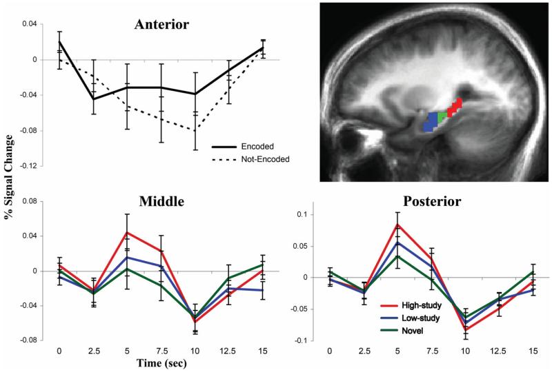
FIGURE 3.
Hippocampal BOLD responses. Anterior (blue), middle (green) and posterior (red) hippocampal ROIs are overlaid on a sagittal cross-section of the mean anatomical image of all subjects (top right). Impulse-response plots display the time-course of the percent signal change (± standard error) in anatomically-defined bilateral anterior, middle and posterior hippocampus. Anterior hippocampus was more deactivated during word recognition trials when pictures were not-encoded than encoded (P < 0.05, top left). Middle and posterior hippocampus, which were not influenced by incidental encoding during retrieval (Ps > 0.10), were more activated during high-study than novel word recognition trials (P < 0.01, bottom). [Color figure can be viewed in the online issue, which is available at wileyonlinelibrary.com.]

