Summary
How different cell types with the same genotype are formed and heritability maintained is a fundamental question in biology. We utilized white-opaque switching in Candida albicans as a system to study mechanisms of cell type formation and maintenance. Each cell type has tractable characters, which are maintained over many cell divisions. Cell type specification is under the control of interlocking transcriptional feedback loops, with Wor1 being the master regulator of the opaque cell type. Here we show that deletion of RTT109, encoding the acetyltransferase for histone H3K56, impairs stochastic and environmentally stimulated white-opaque switching. Ectopic expression of WOR1 mostly bypasses the requirement for RTT109, but opaque cells lacking RTT109 cannot be maintained. We have also discovered that nicotinamide induces opaque cell formation, and this activity of nicotinamide requires RTT109. Reducing the copy number of HST3, which encodes the H3K56 deacetylase, also leads to increased opaque formation. We further show that the Hst3 level is down regulated in the presence of genotoxins and ectopic expression of HST3 blocks genotoxin induced switching. This finding links genotoxin induced switching to Hst3 regulation. Together these findings suggest RTT109 and HST3 genes play an important role in the regulation of white-opaque switching in C. albicans.
Introduction
Epigenetic regulation is fundamental for adaptation to environmental changes and cell specialization/differentiation. Epigenetic states are heritably maintained, allowing cells to “remember” past changes of the external environment or developmental cues. Understanding molecular mechanisms underlining epigenetic states is important for regenerative medicine and cancer treatment. Despite this importance, studies of cell fate specialization in higher eukaryotes are often hindered by a high degree of heterogeneity and sensitivity to a multitude of extrinsic signaling. An alternative, but unicellular eukaryotic system of epigenetic inheritance of specialized cell types is seen in Candida albicans, the most significant human fungal pathogen. C. albicans is able to reversibly switch between two visibly different cell types: white and opaque (Slutsky et al., 1987). White and opaque cells differ in cell shape, the genes they express (Lan et al., 2002, Tsong et al., 2003, Tuch et al., 2010), their mating behaviors (Miller & Johnson, 2002), and virulence properties (Lohse & Johnson, 2008, Kvaal et al., 1999, Morschhauser, 2010). Switching is stochastic and infrequent, with a switching frequency about 10-4 per cell division (Rikkerink et al., 1988). Certain conditions have been reported to increase the switching frequency. Genotoxic stress is long known for promoting the white to opaque switch (Alby & Bennett, 2009, Morrow et al., 1989). Recently, incubation of white cells under anaerobic conditions, high CO2 level or N-acetylglucosamine was shown to induce switching to the opaque cell type (Huang et al., 2009, Ramírez-Zavala et al., 2008, Huang et al., 2010). The opaque phase is sensitive to temperature. Upon shifting the growth temperature from room temperature to 37°C, opaque cells switch en masse to white cells (Bergen et al., 1990). The ease of cell culturing and efficient homologous recombination for gene manipulation in C. albicans make the white-opaque switching system one of the most tractable eukaryotic systems for studying epigenetic regulation in a biomedical relevant context.
Different cell types are determined by specific gene expression programs, which are controlled by transcription factors and chromatin regulators. White and opaque cell type specification in C. albicans is under the control of interlocking transcriptional feedback loops with Wor1 being a master regulator of the white-opaque switch (Huang et al., 2006, Srikantha et al., 2006, Zordan et al., 2006, Zordan et al., 2007). Deletion of WOR1 blocks opaque cell type formation, whereas ectopic expression of WOR1 switches white cells to opaque. WOR1 is preferentially expressed in opaque cells and the positive feedback regulation of WOR1 expression from Wor1 and another transcription factor, Wor2, is believed to provide the bistable expression of WOR1 that is essential for the existence of the stable opaque cell type. Despite the large evolutionary distance between mammals and fungi, transcriptome analysis combined with Wor1 ChIP-chip suggests that the Wor1 circuit shares several characteristics with the transcriptional circuits for pluripotency of mammalian embryonic stem cells (Tuch et al., 2010, Zordan et al., 2007). In addition to the transcriptional loops, histone-modifying enzymes constitute another layer of regulation for the white and opaque cell types. Lack of the histone deacetylases HDACs HDA1 and RPD3 modify frequencies of switching (Srikantha et al., 2001). More recently, a comprehensive deletion analysis focusing on catalytic subunits of histone-modifying enzymes identified several enzymes that can modulate frequencies of switching (Hnisz et al., 2009). Epistasis analysis mapped them into at least two independent pathways in relation to the known transcriptional regulators, and they all seem to act upstream of WOR1 (Hnisz et al., 2009).
Acetylation within the globular core domain of histone H3 on lysine 56 (H3K56) plays a critical role in chromatin disassembly during transcriptional activation (Williams et al., 2008, Xu et al., 2005) and packing DNA into chromatin following DNA replication and repair in yeast (Chen et al., 2008a, Li et al., 2008, Masumoto et al., 2005). H3K56 acetylation also plays a role in anti-silencing, and is required for transcription at heterochromatin loci (Varv et al., 2010, Xu et al., 2007). H3K56 acetylation in yeast is catalyzed by the fungal-specific histone acetyltransferase (HAT) Rtt109 (Driscoll et al., 2007, Han et al., 2007). RTT109 expression and H3K56Ac peaks during S phase when new histones are synthesized (Masumoto et al., 2005). H3K56Ac facilitates histone deposition onto replicating DNA and nucleosome assembly (Chen et al., 2008a, Li et al., 2008). Replication-independent dynamic histone exchange has been mapped to promoters and the newly deposited histones are marked by acetylation at H3K56 before deposition (Rufiange et al., 2007, Kaplan et al., 2008, Dion et al., 2007). In yeast H3K56 is deacetylated by sirtuin class HDACs, Hst3 and Hst4, with Sir2 contributing some activity (Celic et al., 2006, Maas et al., 2006) (Xu et al., 2007). H3 lysine 56 acetylation marks correlate with developmentally important loci upon human embryonic stem cell differentiation, and co-localizes with key pluripotency regulators (Xie et al., 2009). Recently, fungal Rtt109 was found to be an unexpected structural homolog of metazoan CBP/p300 (Tang et al., 2008), and H3K56 acetylation in multicellular organisms is shown to be catalyzed by CBP/p300 (Das et al., 2009) but competing reports conclude that GCN5/KAT2A is the main acetylase responsible for H3K56Ac in human cells (Tjeertes et al., 2009). p300 was found to map with the core pluripotency regulators in embryonic stem cells by ChIP-seq (Chen et al., 2008b), and in vivo mapping of p300 binding sites by ChIP-seq in mouse embryonic tissues has accurately predicted tissue-specific activity of enhancers (Visel et al., 2009, Blow et al., 2010), consistent with the critical roles of p300 in mouse embryonic development (Yao et al., 1998). Significance of H3K56 acetylation in fungal development and in white-opaque cell type specialization in C. albicans is not known.
Recent studies have shown that H3K56 acetylation in C. albicans is regulated by Rtt109 and Hst3, and both genes are important to maintain genome integrity as in S. cerevisiae (Lopes da Rosa et al., 2010, Wurtele et al., 2010). These studies were performed in a/α strains, thus the function of RTT109 and HST3 in white and opaque cell type formation was not determined. In identifying conditions that promoted white-to-opaque switching, we discovered that nicotinamide (NAM) is a potent stimulator of white-opaque switching. HST3/hst3 haploinsufficiency in the presence of NAM indicates that nicotinamide targets Hst3. We find that reducing HST3 copy number also led to an increased frequency of opaque cell formation. We also discovered that genotoxic stress induces white-to-opaque switching by reducing Hst3 level. Unlike all other characterized histone-modifying enzymes (Hnisz et al., 2009), we find that cells lacking RTT109 have inefficient opaque cell type formation and fail to properly maintain the opaque cell type. Together, our data suggest that RTT109 and HST3 genes are important for opaque cell type formation and maintenance. This offers new insight into the basis of cell type formation and maintenance in a unicellular eukaryote.
Results
rtt109 mutants do not efficiently undergo white-to-opaque switching
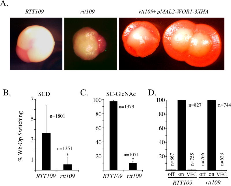
To investigate the importance of RTT109 in cell type formation from white-to-opaque, we converted the rtt109 mutant PKA13 (Lopes da Rosa et al., 2010) from a/α mating type to α/α mating type by sorbose selection as described in (Kabir et al., 2005). Quantitative switching assays were performed to determine the frequency of opaque formation from white cells for the rtt109 mutant. Deletion of RTT109 resulted in a reduction in spontaneous opaque cell type formation compared to the wild-type α/α strain (Figure 1B). The rare rtt109 opaque sectors observed do stain with phloxine B indicating genuine opaque cells (Figure 1A). When cells from this opaque sector were plated, only a few opaque-like colonies were observed among mostly white colonies, suggesting that many cells in those opaque sectors were white cells or have reverted to white cells at a high frequency after plating. The lack of opaque colonies and the very low frequency of opaque sectors formed from the white cells of the rtt109 mutant suggest that the H3K56 acetylation or chromatin constructed with H3K56Ac is important for spontaneous white-to-opaque switching.
Figure 1. rtt109 mutants are impaired in white-to-opaque switching.
(A) Colony morphology of wild type (HLY3555) and rtt109 (HLY3997) white colonies with spontaneous opaque sectors on SCD media, and rtt109 (HLY4057) ectopically expressing WOR1. 5 μg/ml phloxine B is used in the plates. (B) Quantitative switching assay on SCD media of wild type (HLY3555) and rtt109 (HLY3997). (C) Quantitative switching assay on SC+2%GlcNAc media of wild type (HLY3555) and rtt109 (HLY3997) to determine GlcNAc stimulated white-opaque switching. (D) Quantitative switching assay when ectopically expressing WOR1. “off” corresponds to wild type (HLY4055) and rtt109 (HLY4057) strains grown on YEP+2% dextrose while “on” corresponds to growth on YEP 2% maltose. “VEC” refers to wild type (HLY4025) and rtt109 (HLY4027) containing the control vector grown on YEP+2% maltose. Data is from at least two independent quantitative switching assays started on different days with different starter cultures. Abbreviations: Wh, white; Op, opaque; SCD, synthetic complete 2% dextrose; GlcNAc, N-acetylglucosamine; n are the total number of colonies scored; error bars are differences in phenotypic scores between replicate tests; * is p-value < 0.01 compared to wild type.
Spontaneous opaque cell type formation is low in frequency even for wild-type strains under normal growth conditions. To further characterize the importance of RTT109 in white-to-opaque switching, we performed quantitative switching assays under conditions known to promote white-to-opaque switching. N-acetylglucosamine (GlcNAc) has been shown to be a potent inducer of opaque cell type formation on Lee's media (Huang et al., 2010). Therefore, we performed a quantitative switching assay of wild type and rtt109 on synthetic complete media, a defined media similar to Lee's media, containing either 2% dextrose (SCD) or 2% GlcNAc (SC-GlcNAc). After 7-days of growth on SC-GlcNAc, 98% of wild-type colonies have efficiently switched, whereas only 10% of rtt109 colonies switched. (Figure 1C). It should be noted that the fold-increase in GlcNAc stimulated switching for rtt109 is about 50% of wild type. Longer incubation did not improve the switching frequency for the rtt109 mutant (data not shown). Growth, as measured by colony size, was not significantly different between SCD and SC-GlcNAc plates for both the wild-type strain and rtt109 mutants but rtt109 does grow slower than wild type. This result demonstrates that RTT109 is important for opaque formation under the GlcNAc induction. RTT109 is also important for opaque cell type formation under 5% CO2 (data not shown), a condition reported to promote white-to-opaque switching (Huang et al., 2009). Genotoxins and hydrogen peroxide also induce opaque cell-type formation in C. albicans (Alby & Bennett, 2009). The rtt109 mutant is sensitive to genotoxic and oxidative stress (Lopes da Rosa et al., 2010, Wurtele et al., 2010), so switching could not be reliably assessed under those conditions. However, persistent DNA damage signals exist in the rtt109 mutant (Lopes da Rosa et al., 2010), yet a high frequency of white-to-opaque switching was not observed in the mutant suggesting that upstream DNA damage signals function through RTT109 or that opaque cells are not well maintained in the rtt109 mutant. Together, the quantitative switching assays conducted under various media or environmental conditions identify RTT109 as a key contributor in opaque cell formation.
Ectopic WOR1 expression can bypass the requirement for RTT109 in opaque cell type formation
WOR1 is the master regulator of white-to-opaque switching. Wor1 is necessary to establish and maintain opaque cell types, and ectopic expression of WOR1 is able to switch white cells opaque on solid media (Huang et al., 2006, Srikantha et al., 2006, Zordan et al., 2006). To determine the genetic relationship between WOR1 and RTT109, a C-terminal 3xHA tagged Wor1 was placed under the glucose-repressible, maltose-inducible MAL2 promoter (pMAL2-WOR1-3XHA). We found that both wild-type cells and rtt109 cells ectopically expressing WOR1 switched efficiently after one week (Figure 1D). Wild-type cells with the control vector did not appreciably switch on YEP maltose plates nor did wild-type strains carrying the MAL2-WOR1-3XHA transgene switch while grown on YEPD. Our data suggest that Wor1 overexpression could bypass the requirement of RTT109 in opaque cell formation. However, the extent of opaque cell formation in the rtt109 mutant may depend on the level of ectopic Wor1 expression, as we had seen reduced switching proficiency in the rtt109 mutant depending on the induction system and growth media used.
The rtt109 mutant is unable to maintain the opaque cell type
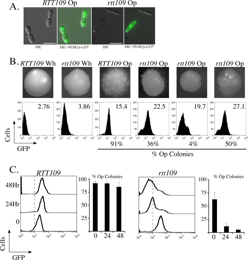
To differentiate white and opaque cells at single cell resolution, a reporter gene with GFP under the WOR1 promoter (Huang et al., 2006) was used to differentiate between white and opaque cells as WOR1 is highly expressed in opaque cells. Using the reporter gene, we observed rtt109 opaque cells were slightly larger than wild-type opaque cells (Figure 2A). They also exhibited the typical elongated phenotype of wild type opaque cells but we found that some elongated cells did not express pWOR1-GFP, indicating that cell shape alone was not sufficient to distinguish between true opaque form and other morphologies. The reporter gene would also allow us to determine if the level of WOR1 expression is reduced in the rtt109 mutant in the opaque cell type, which would be a contributing factor to the opaque cell type establishment and maintenance.
Figure 2. Maintenance of the opaque cell type is lost in the rtt109 mutant.
(A) Wild type (HLY3555) and rtt109 (HLY3998) opaque cells expressing the pWOR1-GFP reporter gene. Scale bar is 10 micrometers. (B) Colony phenotypes and whole colony FACS plots of pWOR1-GFP fluorescence from wild type (HLY3555) and rtt109 (HLY3998) opaque strains. Mean GFP signals are shown in the FACS plots. The % Op colonies that originated from the same colonies used in FACS analysis are indicated. Approximately 500 colonies were scored from each opaque colony for wild type and rtt109. (C) rtt109 opaque cells are unstable during liquid growth. At indicated times, aliquots of cells were removed for FACS analysis and plating assays. Adjoining graphs are colony phenotypes scored 3-5 days after plating. For (C) Wild type: 0-Hr n=1242, 24-Hr n=1463, 48-Hr n=1838. rtt109: 0-Hr n=577, 24-Hr n=1101, 48-Hr n=1346. error bars are differences in phenotypic scores between at least two independent tests. Dashed lines in FACS plots for (C) roughly demarcate white and opaque cell types based on fluorescent signal.
To obtain opaque rtt109 colonies, cells from the rare opaque sectors were replated onto SCD plates. They grew up as obvious white colonies or opaque colonies that had an atypical phenotype and no white sectors (Figure 2B). The lack of white sectors in those opaque-like colonies and the high percentage of white colonies when replated suggested that each opaque colony might consist of a heterogeneous cell population of both white and opaque cell types. Fluorescent activated cell sorting (FACS) was used to assay pWOR1-GFP expression levels in cells collected from several independent colonies (Figure 2B). As determined by FACS, cells from a wild-type opaque colony gave predominantly a single peak that represented similar WOR1 expression levels within the colony population of cells. In contrast, cells collected from the rtt109 opaque-like colonies display a wider range of WOR1 expression levels, and a significant proportion of the cell population had low levels of WOR1 expression similar to that from white cells (Figure 2B). This was confirmation that cells in individual opaque-like colonies of rtt109 mutants were heterogeneous in cell types but with no obvious white sectors. We also took rtt109 opaque cells formed by ectopic expression of WOR1 and determined that the opaque stability was also not maintained (data not shown). In addition, the percentages of white colonies generated from each of the three independent opaque rtt109 colonies were consistently higher than the corresponding percentages of cells with low levels of WOR1 expression by FACS. This suggested that a significant portion of the WOR1 expressing cells had switched to white cells when plated on SCD during the quantitative switching assay. To explore this observation further, we assayed the stability of the rtt109 mutant opaque cell type in liquid culture (Figure 2C). Wild-type opaque cells retained a sharp peak of WOR1 expression characteristic of opaque cells after 24- and 48-hours. The maintenance of the opaque cell type was demonstrated because subsequent plating of liquid grown wild-type cells grew up as opaque colonies. In contrast the pWOR1-GFP expression in the rtt109 opaque population shifted mostly to the level of white cells after 24- and 48-hours. Subsequent plating of the rtt109 cells after 24- and 48-hours confirmed that there was a rapid loss of the opaque cell type when grown in liquid culture.
Nicotinamide induces white-to-opaque switching in an RTT109 dependent manner
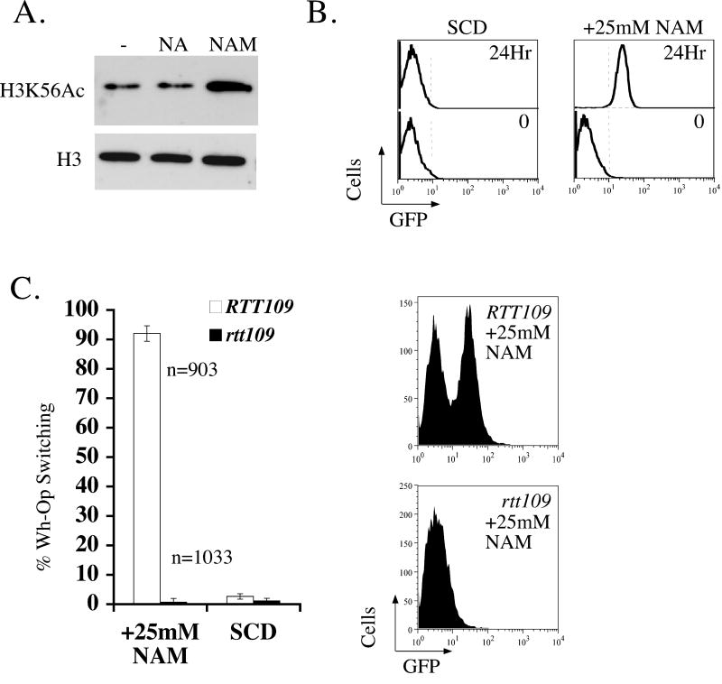
Based on the impaired switching phenotype of the rtt109 mutant, we reasoned that increased levels of H3K56Ac might lead to increased spontaneous white-to-opaque switching. Nicotinamide is a potent non-competitive inhibitor of the Sirtuin family of HDACs, and treatment of S. cerevisiae cells with nicotinamide increases H3K56Ac in vivo (Celic et al., 2006, Landry et al., 2000). Treatment of C. albicans with nicotinamide, but not nicotinic acid, increased H3K56 acetylation levels (Figure 3A), a finding also made by (Wurtele et al., 2010). Encouraged that nicotinamide increases H3K56Ac in vivo, we next examined whether nicotinamide treatment activates WOR1 expression. Wild-type white cells were grown in liquid SCD in the presence of 25mM nicotinamide at room temperature. After 24-hours they expressed the pWOR1-GFP reporter at a level similar to opaque cells, whereas untreated cells did not (Figure 3B). The culture treated with 25mM nicotinamide had a significantly reduced density of cells compared to the untreated culture. When cells from the nicotinamide treated culture were plated, only about 50% of the expected cells plated formed colonies. Within the population of colonies that did grow up, only about 10% were actually opaque. This percentage of opaque colonies was reproducibly higher than cells without nicotinamide treatment, but much lower than expected based on the pWOR1-GFP profile. The lower than expected switching frequency could be caused by the inhibition of growth and cell division by nicotinamide or the use of liquid culture to induce switching. Therefore we examined the nicotinamide effect on solid media. Wild-type white cells were plated on SCD agar plates with and without 25mM nicotinamide and incubated at room temperature. 25mM nicotinamide slowed growth significantly, and colonies after one week on nicotinamide were too small to score cell types faithfully. To prevent the agar plates form drying out, test plates were poured thick. After 2-weeks of incubation at room temperature, we noticed that >90% of colonies that grew up formed opaque sectors (Fig. 3C, left panel), but there was about a 10% reduction in cell density compared to untreated cells (data not shown). FACS analysis of cells from the colonies with opaque sectors indicated the presence of opaque cells (Fig. 3C, right panel). This switch was heritable because cells from several independent sectors were plated to SCD plates lacking nicotinamide and a majority of colonies that grew up remained opaque (data not shown). We also titrated nicotinamide concentrations and found that 0.05mM nicotinamide can still induce switching at an appreciable level (data not shown). In accordance with the postulation that chromatin constructed with higher amounts of H3K56Ac is a driver of nicotinamide-induced switching, rtt109 mutants failed to switch in the presence of nicotinamide (Figure 3C). The growth inhibition by nicotinamide is also completely abolished by the deletion of RTT109 (data not shown and (Wurtele et al., 2010)).
Figure 3. RTT109 is required for nicotinamide induced switching.
(A) Western blot of wild-type white cells (HLY3879) untreated, nicotinic acid treated (25mM), and nicotinamide (25mM) treated. H3K56Ac and H3 protein levels were probed after approximately six hours of growth at 30°C in YEPD media. (B) FACS plots of wild-type white cells grown in liquid SCD culture in the presence or absence of 25mM nicotinamide. (C) Quantitative switching assay of wild type (HLY3555) and rtt109 (HLY3998) in the presence of 25mM nicotinamide. Adjoining FACS plots are of representative nicotinamide treated colonies of either wild type (HLY3555) or rtt109 (HLY3998). For figure (C), the SCD without NAM switching frequency data are from Figure 1B with wild type n=1801 and rtt109 n=1351. Switching data are from at least two independent tests. n are the total number of colonies counted; error bars are differences in phenotypic scores between tests. Dashed lines are added for clarity.
HST3/hst3 mutants have increased white-to-opaque switching
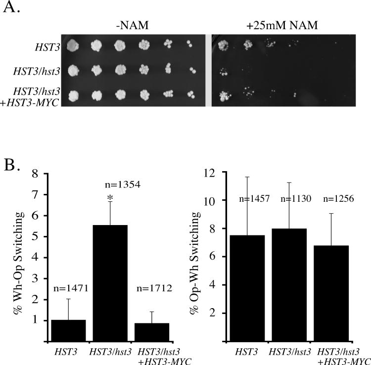
Nicotinamide most likely can inhibit any sirtuin (Landry et al., 2000). The C. albicans genome contains four sirtuin genes: SIR2, HST1, HST2, and HST3 (Skrzypek et al., 2010). Deletions of SIR2 or HST1 did not dramatically increase white-to-opaque switching frequency while deletion of HST2 reduced the frequency of white-to-opaque switching (Hnisz et al., 2009). HST3 has not been characterized in white-opaque switching before (Hnisz et al., 2009). In the course of our study Wurtele et al. also identified HST3 as the H3K56 deacetylase in C. albicans (Wurtele et al., 2010). In that study, mass spectrometry measured double the amount of H3K56Ac levels in the two independently generated HST3/hst3 mutants. HST3 is not an essential gene in S. cerevisiae, but HST3 is an essential gene in C. albicans, yet both copies of HST3 can be deleted in the rtt109 mutant. We constructed two independent heterozygous HST3/hst3∷FRT deletion strains designated HLY3993 and HLY3994 in the WO-1 background using the Flp/FRT sequence specific recombination system (Reuss et al., 2004) . Subsequently, strain HLY3993 was used to generate HLY4041 (HST3/hst3∷HST3-MYC), where HST3 was reintroduced into the disrupted hst3∷FRT locus. Characterization of Wild-type (TS3.3), HST3/hst3∷FRT (HLY3993), and HST3/hst3∷HST3-MYC (HLY4041) strains were carried out. First we tested if nicotinamide targets Hst3 in vivo (Figure 4A) by haploinsufficiency for growth in HST3/hst3 strain (HLY3993), a method used in genome-wide identification of drug targets in S. cerevisiae (Giaever et al., 1999). We found the HST3/hst3 strain was more sensitive to nicotinamide compared to wild-type cells. Restoration of HST3-MYC into the HST3/hst3 strain eliminated the haploinsufficiency. Next we carried out quantitative switching assays in the white-to-opaque and opaque-to-white directions. The heterozygous strain had a 5-fold increase in white-to-opaque switching over the wild-type strain but reintroducing HST3-MYC restored switching frequencies of the HST3/hst3 strain to wild type levels (Figure 4B). This suggests that reducing the copy number of HST3 from two copies to one copy is sufficient to increase white-to-opaque switching frequency. This result, together with the finding that rtt109 mutants are insensitive to nicotinamide-induced switching, suggests that increased levels of H3K56Ac increase spontaneous opaque cell-type formation. We did not detect a difference in the opaque-to-white switch between the wild-type and HST3/hst3 mutants (Figure 4B), indicating additional layers of regulation in opaque cells. Our study identifies a function of HST3 in regulating switching, most likely via H3K56Ac status.
Figure 4. HST3/hst3 mutants have increased white-opaque switching.
(A) Overnight cultures of HST3/HST3 (TS3.3), HST3/hst3 (HLY3993), HST3/hst3∷HST3-MYC (HLY4041) were adjusted to a final OD600 of 0.1, after which five 5-fold serial dilutions were spotted onto SCD with or without 25mM nicotinamide and incubated at 30°C for 4 days. (B) Quantitative switching assays of indicated strains were carried out on SCD to determine the switching phenotypes in the white-to-opaque and opaque-to-white directions. Data are from at least two independent tests; n is the total number of colonies counted; error bars are differences in phenotypic scores between tests. * is p-value < 0.01 compared to wild type.
Genotoxic stress induces white-to-opaque switching by reducing Hst3 level
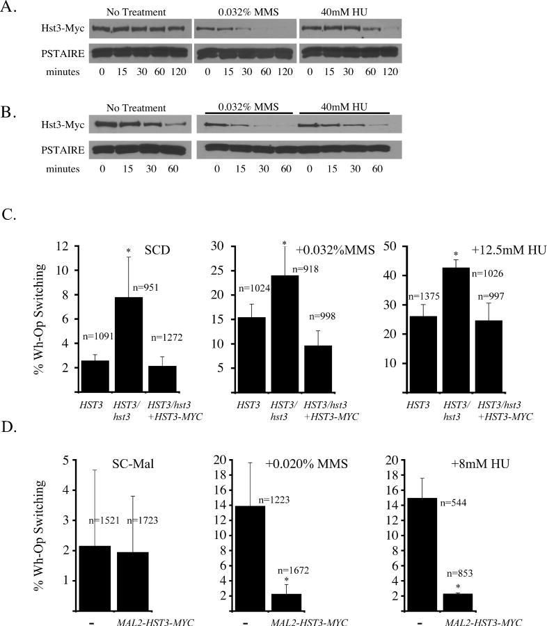
Methyl methanesulfonate (MMS) and hydroxyurea (HU) are known to induce white-to-opaque switching but the underlying molecular mechanism is unknown. In S. cerevisiae, Hst3 is down regulated in response to genotoxic stress (Thaminy et al., 2007). Because tight regulation of H3K56Ac is critical in the DNA damage response in both yeasts, such regulation of Hst3 in response to genotoxic stress may be conserved in C. albicans (Celic et al., 2006, Driscoll et al., 2007, Lopes da Rosa et al., 2010, Masumoto et al., 2005, Wurtele et al., 2010). To test this possibility, we tagged Hst3 C-terminus with 13xMyc under the endogenous HST3 promoter and measured Hst3-Myc levels in cells grown in the presence of MMS or HU. The presence of genotoxins resulted in diminished Hst3-Myc levels within 30-min of treatment with MMS and within 1-hour of treatment with hydroxyurea (Figure 5A). Genotoxin induced loss of Hst3-Myc could be caused by HST3 transcriptional repression, Hst3 protein stability, or a combination of both. To test if Hst3 protein stability is regulated in response to genotoxins, HST3-MYC was placed under the control of the glucose-repressible, maltose-inducible MAL2 promoter. Hst3-Myc protein stability was determined after MAL2 promoter shutdown by adding glucose in the presence or absence of genotoxins. MMS treatment resulted in a loss of Hst3-Myc after promoter shutdown, indicating that Hst3 protein was unstable under this condition. Because the dynamics of Hst3-Myc disappearance under its endogenous promoter and the MAL2 promoter were similar, HST3 transcription is probably repressed during MMS treatment as well. HU treatment resulted in a gradual loss of Hst3-Myc levels after shutdown, somewhat similar to the no treatment samples (Figure 5B).
Figure 5. Genotoxins, Hst3, and switching are linked.
(A) Western blot of Hst3-Myc from wild type (HLY3995) cultures treated with or without genotoxins for the indicated times. (B) Western blot of Hst3-Myc after MAL2 promoter shutdown in the presence or absence of genotoxins, strain (HLY3996). PSTAIRE serves as a loading control. (C) Quantitative white-opaque switching assays of wild type (TS3.3), HST3/hst3 (HLY3993), and HST3/hst3∷HST3-MYC (HLY4041) in the presence of genotoxins. (D) Quantitative white-opaque switching assays on SC maltose agar plates of wild type (TS3.3) and cells ectopically expressing HST3 from the MAL2 promoter strain (HLY3996). SCD is synthetic complete media 2% dextrose. SC-Mal is synthetic complete media 2% maltose. Data are from at least two separate tests; n is the total number of colonies counted; error bars are differences in phenotypic scores between tests. * is p-value < 0.01 compared to wild type.
If genotoxic stress induces the white-to-opaque switching through down-regulating HST3 levels, ectopically expressing HST3 should render cells insensitive to genotoxic stress in cell type switching, and conversely, the heterozygous HST3/hst3 mutant is likely to show increased white-to-opaque switching in the presence of genotoxins. To examine the importance of HST3 regulation by genotoxins in white-opaque switching, quantitative switching assays were performed by growing white cells of the wild type, HST3/hst3 mutants, and the complemented strain on SCD agar plates containing genotoxins MMS or HU (Figure 5C). Under both conditions tested, HST3/hst3 mutants had a higher percentage of opaque cell formation than the wild-type control and the complemented strain. The switching difference between HST3/hst3 baseline vs. genotoxic induced switching is not as dramatic compared to wild-type baseline vs. genotoxin induced switching. This is most likely due to the sensitization of HST3/hst3 and acquired tolerance to constitutively higher H3K56Ac levels. Ectopic expression of HST3 under the MAL2 promoter strongly reduced both MMS and HU induced opaque cell formation (Figure 5D). We observed no significant difference in white-to-opaque switching frequency between wild-type cells in the absence of genotoxins and the pMAL2-driven HST3 strains in the presence of genotoxins. The observation that ectopic expression of HST3 can suppress genotoxin induced switching strongly suggests that the down regulation of HST3 transcription and/or Hst3 protein levels is a key regulatory step in response to genotoxin induced switching. These findings demonstrate that genotoxins, Hst3, and switching work in a similar pathway and provide a molecular mechanism for genotoxin induced switching.
H3K56Ac correlates with transcription
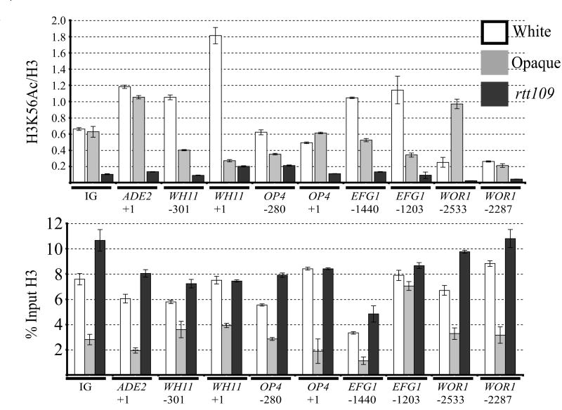
We have identified RTT109 and HST3 genes as contributing regulators of opaque cell type formation and maintenance. Histone H3K56 is a shared target for acetylation/deacetylation reactions by the encoded enzymes. To investigate whether H3K56Ac exhibits any cell-type specific profile or locus specific enrichment, chromatin cross-linking and immunoprecipitation (ChIP) with anti-H3 and anti-H3K56Ac antibodies was performed in wild-type white and opaque cell types, as well as rtt109 white cells (Figure 6). Several non-cell type specific and cell-type specific regions were probed for H3K56Ac enrichment. First, an intergenic region (IG) was probed between ORFs 19.3983 and 19.3984, where Wor1 does not bind and the proximal genes are not differentially expressed between wild-type white and opaque cells, nor is expression different between wild type and rtt109. This IG region would allow an assessment of H3 and H3K56Ac/H3 levels where Wor1 is absent and no transcription is occurring. Secondly, the ADE2 +1 position was probed because it is constitutively expressed, does not have differential expression between white and opaque cells, and is not expressed differently between wild type and rtt109 strains. WH11 and OP4 are genes preferentially expressed in stable white and opaque cell types. Thus regions proximal to the +1 position and at the +1 position of each gene were included to assess if H3K56Ac correlated with cell specific expression. Lastly, EFG1 and WOR1 are key genes controlling switching and were included to determine if H3K56Ac also correlated with their transcriptional activity. In general, the H3K56Ac/H3 ratio correlated with the transcriptional state of the corresponding gene. We could detect enrichment of H3K56Ac/H3 signal at the IG region, indicating that enrichment occurs in the absence of proximal genes and transcription. The ADE2 gene is not cell-type regulated. Accordingly, we did not detect a difference in H3K56Ac/H3 levels between white and opaque cells. Comparing the H3K56Ac/H3 ratio at WH11 and OP4 regions, we could detect a difference that depended on cell type. This difference was more pronounced at WH11 than OP4. The EFG1 and WOR1 regions followed a similar profile as was seen in WH11 and OP4, although a difference between white and opaque cells in the H3K56Ac/H3 ratio was not detected at the transcriptional start site of WOR1. The ChIP experiments also identified that opaque cells have less chromatin bound H3 across several of the regions tested compared to white cells (Figure 6 lower panel). Consistent with the ChIP H3 data, total cell extract from opaque cells showed a lower level of H3 than white cells (data not shown). Numbers of histone transcripts were found to be fewer in opaque cells in a previous RNA (Tuch et al. 2010).
Figure 6. H3K56Ac correlates with cell-type specific transcription.
ChIP of H3 and H3K56Ac in wild type (HLY3555) white, opaque, and rtt109 (HLY3998) white cells. The lower panel is the % input of H3 presented on Log2 scale. The upper panel is the ratio of the % input for individual H3 and H3K56Ac IP. Data presented is the average of two separate ChIP experiments. Error bars shown are propagated from separate ChIP experiments and triplicate reactions in QPCR reactions. The rtt109 strain also serves as a negative control for H3K56Ac enrichment. Numbers below gene names are relative to the +1 position, and are in basepair (bp) units. Amplicon lengths extend 5′-3′ direction approximately 100-250bp from the indicated positions, except for the +1 amplicons, where the +1 position is located in the middle portion of the amplicon. Note, EFG1 and WOR1 contain large 5′-UTRs, and transcription starts at -1173 and -2000 relative to their +1 position, respectively.
Discussion
How do unique and heritable phenotypes emerge from the same genotype is a fundamental question in biology. Here we utilized white-opaque switching in C. albicans as a simple model to study cell type formation and maintenance. We have shown that deletion of the histone acetyltransferase RTT109 for H3K56 reduces both spontaneous opaque cell formation and decreases the efficiency of switching in the presence of environmental conditions known to stimulate the white-to-opaque switch. Ectopic expression of WOR1 can mostly bypass the requirement for the RTT109 gene. Once opaque, cells lacking RTT109 cannot maintain the opaque cell type on solid or in liquid culturing conditions. Conversely, reducing the copy number of HST3 or inhibiting the deacetylase activity by nicotinamide treatment leads to an increase in white-to-opaque switching. Together, these data suggest that modulation of H3K56 acetylation and possibly the subsequent chromatin composition plays a significant role in the opaque cell type formation and maintenance in C. albicans. The importance of this finding is further highlighted by the fact that a comprehensive deletion analysis of most known histone modifying enzymes found only a few deletion mutants that showed changes in switching frequency, but none showed an inability to maintain the opaque cell type (Hnisz et al., 2009). Therefore, acetylation of other sites on histone or non-histone proteins by HATs and HDACS, other than Rtt109 and Hst3, do not seem to play such a dramatic role in opaque cell formation and maintenance, although we could not exclude the possibility that Rtt109 and Hst3 regulate the acetylation of a site other than H3K56 that is important for opaque cell formation.
Acetylation of histone H3 lysine 56 is a mark associated with both replication-dependent and replication-independent histone replacement (Rando & Chang, 2010). In S. cerevisiae, the replication-independent histone replacement is mapped to promoters of both actively transcribed and repressed genes (Rufiange et al., 2007, Kaplan et al., 2008). Cells lacking H3K56Ac have globally reduced histone turnover and Rtt109 preferentially enhances the histone turnover at rapidly replaced nucleosomes (Kaplan et al., 2008). Dynamic histone turnover in promoter regions may transiently expose DNA to DNA-binding factors and regulate transcription. In addition, rapid histone replacement is found at chromatin boundary elements, suggesting a potential function in preventing the lateral spreading of chromatin states and thereby insulating chromatin domains (Rando & Chang, 2010, Dion et al., 2007, Mito et al., 2007, Dhillon et al., 2009). H3K56 acetylation is also suggested to provide a positive-feedback loop because replacement of one nucleosome enhances subsequent nucleosome replacement at the same location (Kaplan et al., 2008). It is possible that the WOR1 promoter and other Wor1-bound promoters are associated with rapid histone turnover and/or eviction and these events are important for the induction or sustained expression of these genes. Consistent with this notion, we find a general correlation between H3K56Ac enrichment and the transcriptional state of the cell-type specific gene. Interestingly, induction from the MAL2 promoter is slower in the rtt109 mutant compared to wild type cells (data not shown). This observation in C. albicans is consistent with the reported role of Rtt109 in chromatin disassembly during transcriptional activation (Williams et al., 2008, Xu et al., 2005). It is conceivable that Rtt109 function is a critical part of the Wor1-mediated positive feedback. We expect these events to work in concert with previously recognized transcriptional loops. The expression of other Wor1-regulated white and opaque genes is likely affected in a similar manner.
Histone H3K56 deacetylation is regulated through the sirtuin HDACs Hst3/Hst4 in S. cerevisiae. HST3 and HST4 are expressed during the G2 and M phases (Celic et al., 2006, Maas et al., 2006), and their protein levels are down regulated in response to genotoxic stress (Thaminy et al., 2007). Treatment of white cells with nicotinamide increased H3K56Ac levels in vivo and activated WOR1 gene expression in liquid culture. Nicotinamide treatment also stimulated opaque cell formation on agar plates but cells lacking RTT109 do not switch. This effect of nicotinamide is likely mediated mostly through the inhibition of Hst3 because deletion of one copy of HST3 is sufficient to increase spontaneous opaque cell formation by 5-fold. In contrast, deletion of other sirtuin genes does not lead to an increase in the frequency of spontaneous opaque cell formation (Hnisz et al., 2009). We further show that genotoxic stress induces white-to-opaque switching by reducing levels of Hst3, and overexpression of HST3 can suppress genotoxin induced cell type switching. Therefore, DNA damage signaling and Hst3 mediated genome integrity might converge on H3K56Ac status, subsequently leading to switching. On the other hand, rtt109 has constitutive DNA damage signaling, yet do not switch. This suggests that H3K56Ac is a key effecter subsequent to the upstream DNA damage-signaling pathway responsible for white-to-opaque switching. The signaling pathways responsible for the genotoxin induced Hst3 down-regulation in C. albicans have not been defined yet. This could be mediated through the genome integrity checkpoint protein Mec1 kinase as in S. cerevisiae (Thaminy et al., 2007), or indirectly through an arrest in cell cycle. In addition to genotoxins, HST3 transcript level is repressed upon high-level of peroxide stress (Enjalbert et al., 2006), and peroxide treatment of white cells enhances opaque cell type formation (Alby & Bennett, 2009), suggesting that peroxide-induced opaque cell formation may also signal through Hst3. Our results provide molecular connections for the metabolite nicotinamide, genotoxic stress and the sirtuin Hst3 in the epigenetic regulation of white-opaque switching.
Experimental procedures
Yeast Strains and Culturing Conditions
C. albicans strains used in this study are listed in Table 1 as following: C. albicans JYC5, an a/a mating type derivative from SC5314 (wild-type clinical isolate (Huang et al., 2006)); C. albicans rtt109 mutant PKA13 (generous gift from Paul Kaufman (Lopes da Rosa et al., 2010)) converted to α/α mating type; C. albicans TS3.3 derived from WO-1 (generous gift from David Soll (Slutsky et al., 1985, Srikantha et al., 1998)). To propagate strains used in experiments, yeast were routinely grown on SCD or YEPD. The pWOR1-GFP reporter gene used was initially constructed in (Huang et al., 2006) but the SAT1 selectable marker (Reuss et al., 2004) replaced the URA3 marker to allow for selection in the rtt109 mutant background. The pMAL2-WOR1-3XHA transgene was constructed in two cloning steps. First, a 3xHA tag from pMG1874 (Gerami-Nejad et al. 2009, obtained from the FGSC) was PCR amplified with (F) 5′-cttctgggtcgtattacACCGGtactggtggtggtcggatccccgggttaattaa-3′ (AgeI site is capitalized) and (R) 5′-tctagaaggaccacctttgattg-3′, sequentially digested first with AgeI, then with BglII, and ligated into an AgeI site found at the very 3′ end of WOR1 and the unique BglII site of the pTET-WOR1 cassette generated before (Ramírez-Zavala et al., 2008). Then genomic DNA from strain HLY3573 (was used to generate a MAL2-WOR1 amplicon, containing 550bp of the MAL2 promoter sequence directly upstream of the entire WOR1 ORF (Huang et al., 2006) with primers (F) 5′-GTCGACaatgattggtttgatatttttgtctagtac-3′ and (R) 5′-gtatgggtatccagtaccggtgtaatacgaccc-3′. An introduced SalI site is capitalized in the forward primer. This amplicon was sequentially digested with AgeI first, then SalI, and ligated into the SalI/AgeI site of pTET-WOR1-3xHA replacing the original copy of WOR1 with the MAL2-WOR1 amplicon thereby forming a MAL2-WOR1-3XHA transgene. Construction of the HST3/hst3 mutants (HLY3993 and HLY3994) was carried out with the strategy used in (Reuss et al., 2004). Primers used to amplify flanking ORF19.1934 (HST3) regions to replace the endogenous gene by homologous recombination are: (F) 5′-aataGGGCCCaaggtgtgcaaagtggaaaa-3′ and (R) 5′-ccatCTCGAGggcatactagtgcctgaa-3′ for the left HR region. Capitalized letters denote designed ApaI and XhoI restriction sites. (F) 5′-ttttCCGCGGgaacctgtggctaaagtgc-3′ and (R) 5′-ctttggttgtaGAGCTCgcggagagacaggaaagaa-3′ for the right HR region. Capitalized letters denote designed SacII and SacI restriction sites. To generate the pMAL2 driven Hst3-Myc, a similar cloning strategy was used as describe before in (Wang et al., 2007). Primers used to amplify ORF19.1934 (HST3) are: (F) 5′ gcTCTAGAatgatactgattgacttacttgacaattcagg-3′ and (R) 5′-cgACGCGTatgctgatccaaagtaattccttttctagc-3′. Capitalized letters denote XbaI and MluI restriction sites. A unique SpeI restriction site located at the 5′ end of the HST3 gene allowed for integration of the Hst3-Myc to HST3 locus, and is under control of the HST3 promoter. The HST3/hst3∷HST3-MYC strain was generated by SpeI digestion of pMAL2-HST3-MYC followed by transformation into HST3/hst3∷FRT (strain HLY3993). Transformants were screened by PCR for proper integration into the disrupted hst3:FRT position. For promoter shutdown and overexpression assays, the pMAL2-HST3-MYC cassette was integrated at an ADE2 gene. All transformations were carried out as described in (Wang et al., 2007).
Table1. Strains used in this study.
| Strain | Parental Strain | Genotype | Source or reference |
|---|---|---|---|
| TS3.3 | Red3/6 |
MTLα/α ade2/ade2 ura3∷ADE2/ura3∷ADE2 |
(Srikantha et al., 2000) |
| HLY3879 | TS3.3 |
MTLα/α ade2/ade2 ura3∷ADE2/ura3∷ADE2 WOR1/WOR1/PWOR1-GFP-URA3-WOR1 |
This study |
| HLY3993 | TS3.3 |
MTLα/α ade2/ade2 ura3∷ADE2/ura3∷ADE2 HST3/hst3∷FRT |
This study |
| HLY3994 | TS3.3 |
MTLα/α ade2/ade2 ura3∷ADE2/ura3∷ADE2 HST3/hst3∷FRT |
This study |
| HLY3995 | HLY3993 |
MTLα/α ade2/ade2 ura3∷ADE2/ura3∷ADE2 HST3-MYC-URA3-MAL2-HST3/hst1∷FRT |
This study |
| HLY3996 | TS3.3 |
MTLα/α ade2/ade2 ura3∷ADE2/ura3∷ADE2 PMAL2-HST3-MYC-URA3 |
This study |
| HLY4041 | HLY3993 |
MTLα/α ade2/ade2 ura3∷ADE2/ura3∷ADE2 HST3/hst3∷HST3-MYC |
This study |
| HLY3555 | JYC5 |
MTL
a/a
ura3∷imm434/ura3∷imm434 WOR1/PWOR1-GFP-URA3-WOR1 |
(Huang et al., 2006) |
| HLY4025 | HLY3555 |
MTL
a/a
ura3∷imm434/ura3∷imm434 WOR1/PWOR1-GFP-URA3-WOR1 ADH1/adh1∷PTET-GFP-SAT1 |
This study |
| HLY4055 | HLY3555 |
MTL
a/a
ura3∷imm434/ura3∷imm434 WOR1/PWOR1-GFP-URA3-WOR1 ADH1/adh1∷PMAL2-WOR1-3XHA-SAT1 |
This study |
| PKA13 | SC5314 | MTL a/α rtt109∷FRT/rtt109∷FRT | (Lopes da Rosa et al., 2010) |
| HLY3997 | PKA13 | MTLα/α rtt109∷FRT/rtt109∷FRT | This study |
| HLY3998 | HLY3997 |
MTLα/α rtt109∷FRT/rtt109∷FRT WOR1/PWOR1-GFP-SAT1-WOR1 |
This study |
| HLY4027 | HLY3997 |
MTLα/α rtt109∷FRT/rtt109∷FRT ADH1/adh1∷PTET-GFP-SAT1 |
This study |
| HLY4057 | HLY3997 |
MTLα/α rtt109∷FRT/rtt109∷FRT ADH1/adh1∷PMAL2-WOR1-3XHA-SAT1 |
This study |
White–Opaque Switching Assays
C. albicans white–opaque and opaque-white switching assays were performed as reported in (Huang et al., 2006) but synthetic complete or yeast extract peptone (YEP) based media was used instead of Lee's medium. For assays where destination plates were synthetic complete, strains were pre-grown on synthetic complete. For assays where destination plates were YEP based, strains were pre-grown on YEP based media. Strains were routinely grown on appropriate solid media for 4-5 days at 25°C before plating to destination plates. Destination plates were: SC 2% dextrose (w/v), SC 2% GlcNAc (w/v), where GlcNAc replaced dextrose, YEP 2% dextrose (w/v) or YEP 2% maltose (w/v), or SC 2% maltose (w/v). Destination plates supplemented with nicotinamide (Sigma-Aldrich), hydroxyurea (Sigma-Aldrich), or methyl methanesulfonate (Sigma-Aldrich) as indicated in the text. Liquid switching assays started off the same as agar based assays but colonies were inoculated into SCD or YEPD media supplemented with nicotinamide (Sigma-Aldrich), or nicotinic acid (Sigma-Aldrich). Subsequent plating was done on SCD agar plates. Routine colony scores were made after one-week incubation at 25°C, or when colonies were large enough to clearly score phenotypes, up to 10 days or two weeks.
FACS analysis
The distribution of fluorescence from the pWOR1-GFP reporter gene across the entire cell population was investigated by using a dual laser fluorescence-activated cell sorter (BD FACS Calibur System, Becton Dickinson, San Jose, CA). A minimum of 10,000 cells was routinely measured. The results were analyzed and mean fluorescence was determined with FlowJo Flow Cytometry Analysis Software (version 8.7 for Mac).
Immunoblots
To detect H3K56 acetylation levels wild type white cells were diluted and grown at 30°C in YEPD to an of OD600 0.2 in which 25mM nicotinamide (Sigma-Aldrich) or nicotinic acid (Sigma-Aldrich) was added. After 6 hours protein was extracted as described in (Kushnirov, 2000) and protein levels were probed. H3 protein was detected by rabbit polyclonal antibody, ab1791, as described by the manufacturer (Abcam). H3K56Ac protein was detected by a rabbit monoclonal anti-acetyl-histone H3 clone EPR996Y as described by the manufacture (Millipore). Hst3-Myc was detected by using a horseradish peroxidase-conjugated anti-myc antibody (Roche). Promoter shutdown assays were performed similar to (Wang et al., 2007) except that methyl methanesulfonate and hydroxyurea were added to a final concentration of 0.032% and 40mM respectively. Aliquots were collected after the times indicated, protein was extracted as described in (Kushnirov, 2000) and Hst3-Myc was detected by using a horseradish peroxidase-conjugated anti-myc antibody (Roche). PSTAIRE incubation (Santa Cruz) followed by Goat anti-rabbit conjugated to HRP (Bio rad) was used as a protein loading control.
Chromatin Crosslinking and Immunoprecipitation
ChIP was carried out essentially as described (Hernday et al., 2010). For H3, 2.5ug of rabbit polyclonal antibody, ab1791H3 (Abcam) was incubated with approximately 900ug of crosslinked protein overnight at 4°C. For H3K56Ac, 2ug of rabbit monoclonal anti-acetyl-Histone H3 (Lys56) clone EPR996Y (Millipore) was incubated with approximately 900ug of crosslinked protein overnight at 4°C. Protein A (GE Healthcare) beads were used to for immunoprecipitation. After decrosslinking at 65C° overnight, material was treated with proteinase K (Roche) for 2 hours at 37°C. Protein and nucleic acid material was separated by phenol:chloroform:isoamyl alcohol (25:24:1) (Sigma-Aldrich) extraction followed by overnight ethanol precipitation at -20°C. DNA was quantified using an iQ SYBR green Supermix (Bio-Rad) on an iCycler iQ detection system (Bio-Rad). Two independent ChIP experiments were conducted and for each set, the % input of H3 and H3K56Ac/H3 ratio were determined based on Ct values. The ChIP data is the average of these two experiments. The H3 % input is on a log base 2 scale (Log2). The H3K56Ac/H3 ratio was determined by dividing the % input of H3K56Ac signal by % input of H3 signal. Error bars are standard deviations propagated from the independent ChIP experiments and triplicate QPCR reactions.
Acknowledgments
We thank Dr. Paul Kaufman for generously providing C. albicans rtt109 strains. We thank Dr. Joachim Morschhäuser for generously providing the TETp-WOR1 plasmid. We thank Shelley Lane for swapping URA3 for SAT1 in the pWOR-GFP reporter plasmid and all other members of the Liu lab for helpful discussions and technical assistance. This work was supported by the National Institute of Health grant R01GM/AI55155 to HL. HL was also supported in part by NIH National Institute of General Medical Sciences Center grant GM76516.
References
- Alby K, Bennett RJ. Stress-Induced Phenotypic Switching in Candida albicans. Molecular Biology of the Cell. 2009;20:3178–3191. doi: 10.1091/mbc.E09-01-0040. [DOI] [PMC free article] [PubMed] [Google Scholar]
- Bergen MS, Voss E, Soll DR. Switching at the cellular level in the white-opaque transition of Candida albicans. J Gen Microbiol. 1990;136:1925–1936. doi: 10.1099/00221287-136-10-1925. [DOI] [PubMed] [Google Scholar]
- Blow MJ, McCulley DJ, Li Z, Zhang T, Akiyama JA, Holt A, Plajzer-Frick I, Shoukry M, Wright C, Chen F, Afzal V, Bristow J, Ren B, Black BL, Rubin EM, Visel A, Pennacchio LA. ChIP-Seq identification of weakly conserved heart enhancers. Nat Genet. 2010;42:806–810. doi: 10.1038/ng.650. [DOI] [PMC free article] [PubMed] [Google Scholar]
- Celic I, Masumoto H, Griffith WP, Meluh P, Cotter RJ, Boeke JD, Verreault A. The Sirtuins Hst3 and Hst4p Preserve Genome Integrity by Controlling Histone H3 Lysine 56 Deacetylation. Current Biology. 2006;16:1280–1289. doi: 10.1016/j.cub.2006.06.023. [DOI] [PubMed] [Google Scholar]
- Chen CC, Carson JJ, Feser J, Tamburini B, Zabaronick S, Linger J, Tyler JK. Acetylated Lysine 56 on Histone H3 Drives Chromatin Assembly after Repair and Signals for the Completion of Repair. Cell. 2008a;134:231–243. doi: 10.1016/j.cell.2008.06.035. [DOI] [PMC free article] [PubMed] [Google Scholar]
- Chen X, Xu H, Yuan P, Fang F, Huss M, Vega VB, Wong E, Orlov YL, Zhang W, Jiang J, Loh YH, Yeo HC, Yeo ZX, Narang V, Govindarajan KR, Leong B, Shahab A, Ruan Y, Bourque G, Sung WK, Clarke ND, Wei CL, Ng HH. Integration of external signaling pathways with the core transcriptional network in embryonic stem cells. Cell. 2008b;133:1106–1117. doi: 10.1016/j.cell.2008.04.043. [DOI] [PubMed] [Google Scholar]
- Das C, Lucia MS, Hansen KC, Tyler JK. CBP/p300-mediated acetylation of histone H3 on lysine 56. Nature. 2009;459:113–117. doi: 10.1038/nature07861. [DOI] [PMC free article] [PubMed] [Google Scholar]
- Dhillon N, Raab J, Guzzo J, Szyjka SJ, Gangadharan S, Aparicio OM, Andrews B, Kamakaka RT. DNA polymerase epsilon, acetylases and remodellers cooperate to form a specialized chromatin structure at a tRNA insulator. EMBO J. 2009;28:2583–2600. doi: 10.1038/emboj.2009.198. [DOI] [PMC free article] [PubMed] [Google Scholar]
- Dion MF, Kaplan T, Kim M, Buratowski S, Friedman N, Rando OJ. Dynamics of Replication-Independent Histone Turnover in Budding Yeast. Science. 2007;315:1405–1408. doi: 10.1126/science.1134053. [DOI] [PubMed] [Google Scholar]
- Driscoll R, Hudson A, Jackson SP. Yeast Rtt109 Promotes Genome Stability by Acetylating Histone H3 on Lysine 56. Science. 2007;315:649–652. doi: 10.1126/science.1135862. [DOI] [PMC free article] [PubMed] [Google Scholar]
- Enjalbert B, Smith DA, Cornell MJ, Alam I, Nicholls S, Brown AJ, Quinn J. Role of the Hog1 stress-activated protein kinase in the global transcriptional response to stress in the fungal pathogen Candida albicans. Mol Biol Cell. 2006;17:1018–1032. doi: 10.1091/mbc.E05-06-0501. [DOI] [PMC free article] [PubMed] [Google Scholar]
- Giaever G, Shoemaker DD, Jones TW, Liang H, Winzeler EA, Astromoff A, Davis RW. Genomic profiling of drug sensitivities via induced haploinsufficiency. Nat Genet. 1999;21:278–283. doi: 10.1038/6791. [DOI] [PubMed] [Google Scholar]
- Han J, Zhou H, Horazdovsky B, Zhang K, Xu RM, Zhang Z. Rtt109 Acetylates Histone H3 Lysine 56 and Functions in DNA Replication. Science. 2007;315:653–655. doi: 10.1126/science.1133234. [DOI] [PubMed] [Google Scholar]
- Hernday AD, Noble SM, Mitrovich QM, Johnson AD. Genetics and molecular biology in Candida albicans. Methods Enzymol. 2010;470:737–758. doi: 10.1016/S0076-6879(10)70031-8. [DOI] [PubMed] [Google Scholar]
- Hnisz D, Schwarzmüller T, Kuchler K. Transcriptional loops meet chromatin: a dual-layer network controls white–opaque switching in Candida albicans. Molecular Microbiology. 2009;74:1–15. doi: 10.1111/j.1365-2958.2009.06772.x. [DOI] [PMC free article] [PubMed] [Google Scholar]
- Huang G, Srikantha T, Sahni N, Yi S, Soll DR. CO2 Regulates White-to-Opaque Switching in Candida albicans. Current Biology. 2009;19:330–334. doi: 10.1016/j.cub.2009.01.018. [DOI] [PMC free article] [PubMed] [Google Scholar]
- Huang G, Wang H, Chou S, Nie X, Chen J, Liu H. Bistable expression of WOR1, a master regulator of white–opaque switching in Candida albicans. Proceedings of the National Academy of Sciences. 2006;103:12813–12818. doi: 10.1073/pnas.0605270103. [DOI] [PMC free article] [PubMed] [Google Scholar]
- Huang G, Yi S, Sahni N, Daniels KJ, Srikantha T, Soll DR. N-Acetylglucosamine Induces White to Opaque Switching, a Mating Prerequisite in Candida albicans. PLoS Pathog. 2010;6:e1000806–e1000806. doi: 10.1371/journal.ppat.1000806. [DOI] [PMC free article] [PubMed] [Google Scholar]
- Kabir MA, Ahmad A, Greenberg JR, Wang YK, Rustchenko E. Loss and gain of chromosome 5 controls growth of Candida albicans on sorbose due to dispersed redundant negative regulators. Proceedings of the National Academy of Sciences of the United States of America. 2005;102:12147–12152. doi: 10.1073/pnas.0505625102. [DOI] [PMC free article] [PubMed] [Google Scholar]
- Kaplan T, Liu CL, Erkmann JA, Holik J, Grunstein M, Kaufman PD, Friedman N, Rando OJ. Cell Cycle- and Chaperone-Mediated Regulation of H3K56ac Incorporation in Yeast. PLoS Genet. 2008;4:e1000270–e1000270. doi: 10.1371/journal.pgen.1000270. [DOI] [PMC free article] [PubMed] [Google Scholar]
- Kushnirov VV. Rapid and reliable protein extraction from yeast. Yeast. 2000;16:857–860. doi: 10.1002/1097-0061(20000630)16:9<857::AID-YEA561>3.0.CO;2-B. [DOI] [PubMed] [Google Scholar]
- Kvaal C, Lachke SA, Srikantha T, Daniels K, McCoy J, Soll DR. Misexpression of the opaque-phase-specific gene PEP1 (SAP1) in the white phase of Candida albicans confers increased virulence in a mouse model of cutaneous infection. Infect Immun. 1999;67:6652–6662. doi: 10.1128/iai.67.12.6652-6662.1999. [DOI] [PMC free article] [PubMed] [Google Scholar]
- Lan CY, Newport G, Murillo LA, Jones T, Scherer S, Davis RW, Agabian N. Metabolic specialization associated with phenotypic switching in Candidaalbicans. Proceedings of the National Academy of Sciences of the United States of America. 2002;99:14907–14912. doi: 10.1073/pnas.232566499. [DOI] [PMC free article] [PubMed] [Google Scholar]
- Landry J, Slama JT, Sternglanz R. Role of NAD+ in the Deacetylase Activity of the SIR2-like Proteins. Biochemical and Biophysical Research Communications. 2000;278:685–690. doi: 10.1006/bbrc.2000.3854. [DOI] [PubMed] [Google Scholar]
- Li Q, Zhou H, Wurtele H, Davies B, Horazdovsky B, Verreault A, Zhang Z. Acetylation of Histone H3 Lysine 56 Regulates Replication-Coupled Nucleosome Assembly. Cell. 2008;134:244–255. doi: 10.1016/j.cell.2008.06.018. [DOI] [PMC free article] [PubMed] [Google Scholar]
- Lohse MB, Johnson AD. Differential Phagocytosis of White versus Opaque Candida albicans by Drosophila and Mouse Phagocytes. PLoS ONE. 2008;3:e1473–e1473. doi: 10.1371/journal.pone.0001473. [DOI] [PMC free article] [PubMed] [Google Scholar]
- Lopes da Rosa J, Boyartchuk VL, Zhu LJ, Kaufman PD. Histone acetyltransferase Rtt109 is required for Candida albicans pathogenesis. Proceedings of the National Academy of Sciences. 2010;107:1594–1599. doi: 10.1073/pnas.0912427107. [DOI] [PMC free article] [PubMed] [Google Scholar]
- Maas NL, Miller KM, DeFazio LG, Toczyski DP. Cell Cycle and Checkpoint Regulation of Histone H3 K56 Acetylation by Hst3 and Hst4. Molecular Cell. 2006;23:109–119. doi: 10.1016/j.molcel.2006.06.006. [DOI] [PubMed] [Google Scholar]
- Masumoto H, Hawke D, Kobayashi R, Verreault A. A role for cell-cycle-regulated histone H3 lysine 56 acetylation in the DNA damage response. Nature. 2005;436:294–298. doi: 10.1038/nature03714. [DOI] [PubMed] [Google Scholar]
- Miller MG, Johnson AD. White-Opaque Switching in Candida albicans Is Controlled by Mating-Type Locus Homeodomain Proteins and Allows Efficient Mating. Cell. 2002;110:293–302. doi: 10.1016/s0092-8674(02)00837-1. [DOI] [PubMed] [Google Scholar]
- Mito Y, Henikoff JG, Henikoff S. Histone replacement marks the boundaries of cis-regulatory domains. Science. 2007;315:1408–1411. doi: 10.1126/science.1134004. [DOI] [PubMed] [Google Scholar]
- Morrow B, Anderson J, Wilson J, Soll DR. Bidirectional stimulation of the white-opaque transition of Candida albicans by ultraviolet irradiation. J Gen Microbiol. 1989;135:1201–1208. doi: 10.1099/00221287-135-5-1201. [DOI] [PubMed] [Google Scholar]
- Morschhauser J. Regulation of white-opaque switching in Candida albicans. Med Microbiol Immunol. 2010;199:165–172. doi: 10.1007/s00430-010-0147-0. [DOI] [PubMed] [Google Scholar]
- Ramírez-Zavala B, Reuβ O, Park YN, Ohlsen K, Morschhäuser J. Environmental Induction of White–Opaque Switching in Candida albicans. PLoS Pathog. 2008;4:e1000089–e1000089. doi: 10.1371/journal.ppat.1000089. [DOI] [PMC free article] [PubMed] [Google Scholar]
- Rando OJ, Chang HY. Genome-Wide Views of Chromatin Structure. Annu Rev Biochem. 2010;78:245–271. doi: 10.1146/annurev.biochem.78.071107.134639. [DOI] [PMC free article] [PubMed] [Google Scholar]
- Reuss O, Vik A, Kolter R, Morschhäuser J. The SAT1 flipper, an optimized tool for gene disruption in Candida albicans. Gene. 2004;341:119–127. doi: 10.1016/j.gene.2004.06.021. [DOI] [PubMed] [Google Scholar]
- Rikkerink EH, Magee BB, Magee PT. Opaque-white phenotype transition: a programmed morphological transition in Candida albicans. J Bacteriol. 1988;170:895–899. doi: 10.1128/jb.170.2.895-899.1988. [DOI] [PMC free article] [PubMed] [Google Scholar]
- Rufiange A, Jacques Pt, Bhat W, Robert Fo, Nourani A. Genome-Wide Replication-Independent Histone H3 Exchange Occurs Predominantly at Promoters and Implicates H3 K56 Acetylation and Asf1. Molecular Cell. 2007;27:393–405. doi: 10.1016/j.molcel.2007.07.011. [DOI] [PubMed] [Google Scholar]
- Skrzypek MS, Arnaud MB, Costanzo MC, Inglis DO, Shah P, Binkley G, Miyasato SR, Sherlock G. New tools at the Candida Genome Database: biochemical pathways and full-text literature search. Nucleic Acids Research. 2010;38:D428–D432. doi: 10.1093/nar/gkp836. [DOI] [PMC free article] [PubMed] [Google Scholar]
- Slutsky B, Buffo J, Soll DR. High-frequency switching of colony morphology in Candida albicans. Science. 1985;230:666–669. doi: 10.1126/science.3901258. [DOI] [PubMed] [Google Scholar]
- Slutsky B, Staebell M, Anderson J, Risen L, Pfaller M, Soll DR. “White-opaque transition”: a second high-frequency switching system in Candida albicans. The Journal of Bacteriology. 1987;169:189–197. doi: 10.1128/jb.169.1.189-197.1987. [DOI] [PMC free article] [PubMed] [Google Scholar]
- Srikantha T, Borneman AR, Daniels KJ, Pujol C, Wu W, Seringhaus MR, Gerstein M, Yi S, Snyder M, Soll DR. TOS9 Regulates White-Opaque Switching in Candida albicans. Eukaryotic Cell. 2006;5:1674–1687. doi: 10.1128/EC.00252-06. [DOI] [PMC free article] [PubMed] [Google Scholar]
- Srikantha T, Tsai L, Daniels K, Enger L, Highley K, Soll DR. The two-component hybrid kinase regulator CaNIKl of Candida albicans. Microbiology. 1998;144:2715–2729. doi: 10.1099/00221287-144-10-2715. [DOI] [PubMed] [Google Scholar]
- Srikantha T, Tsai L, Daniels K, Klar AJS, Soll DR. The Histone Deacetylase Genes HDA1 and RPD3 Play Distinct Roles in Regulation of High-Frequency Phenotypic Switching in Candida albicans. The Journal of Bacteriology. 2001;183:4614–4625. doi: 10.1128/JB.183.15.4614-4625.2001. [DOI] [PMC free article] [PubMed] [Google Scholar]
- Srikantha T, Tsai LK, Daniels K, Soll DR. EFG1 Null Mutants of Candida albicans Switch but Cannot Express the Complete Phenotype of White-Phase Budding Cells. The Journal of Bacteriology. 2000;182:1580–1591. doi: 10.1128/jb.182.6.1580-1591.2000. [DOI] [PMC free article] [PubMed] [Google Scholar]
- Tang Y, Holbert MA, Wurtele H, Meeth K, Rocha W, Gharib M, Jiang E, Thibault P, Verreault A, Cole PA, Marmorstein R. Fungal Rtt109 histone acetyltransferase is an unexpected structural homolog of metazoan p300/CBP. Nat Struct Mol Biol. 2008;15:998–998. doi: 10.1038/nsmb.1448. [DOI] [PMC free article] [PubMed] [Google Scholar]
- Thaminy S, Newcomb B, Kim J, Gatbonton T, Foss E, Simon J, Bedalov A. Hst3 Is Regulated by Mec1-dependent Proteolysis and Controls the S Phase Checkpoint and Sister Chromatid Cohesion by Deacetylating Histone H3 at Lysine 56. Journal of Biological Chemistry. 2007;282:37805–37814. doi: 10.1074/jbc.M706384200. [DOI] [PubMed] [Google Scholar]
- Tjeertes JV, Miller KM, Jackson SP. Screen for DNA-damage-responsive histone modifications identifies H3K9Ac and H3K56Ac in human cells. EMBO J. 2009;28:1878–1889. doi: 10.1038/emboj.2009.119. [DOI] [PMC free article] [PubMed] [Google Scholar]
- Tsong AE, Miller MG, Raisner RM, Johnson AD. Evolution of a combinatorial transcriptional circuit: a case study in yeasts. Cell. 2003;115:389–399. doi: 10.1016/s0092-8674(03)00885-7. [DOI] [PubMed] [Google Scholar]
- Tuch BB, Mitrovich QM, Homann OR, Hernday AD, Monighetti CK, De La Vega FM, Johnson AD. The Transcriptomes of Two Heritable Cell Types Illuminate the Circuit Governing Their Differentiation. PLoS Genet. 2010;6:e1001070–e1001070. doi: 10.1371/journal.pgen.1001070. [DOI] [PMC free article] [PubMed] [Google Scholar]
- Varv S, Kristjuhan K, Peil K, Looke M, Mahlakoiv T, Paapsi K, Kristjuhan A. Acetylation of H3 K56 is required for RNA polymerase II transcript elongation through heterochromatin in yeast. Mol Cell Biol. 2010;30:1467–1477. doi: 10.1128/MCB.01151-09. [DOI] [PMC free article] [PubMed] [Google Scholar]
- Visel A, Blow MJ, Li Z, Zhang T, Akiyama JA, Holt A, Plajzer-Frick I, Shoukry M, Wright C, Chen F, Afzal V, Ren B, Rubin EM, Pennacchio LA. ChIP-seq accurately predicts tissue-specific activity of enhancers. Nature. 2009;457:854–858. doi: 10.1038/nature07730. [DOI] [PMC free article] [PubMed] [Google Scholar]
- Wang A, Lane S, Tian Z, Sharon A, Hazan I, Liu H. Temporal and Spatial Control of HGC1 Expression Results in Hgc1 Localization to the Apical Cells of Hyphae in Candida albicans. Eukaryotic Cell. 2007;6:253–261. doi: 10.1128/EC.00380-06. [DOI] [PMC free article] [PubMed] [Google Scholar]
- Williams SK, Truong D, Tyler JK. Acetylation in the globular core of histone H3 on lysine-56 promotes chromatin disassembly during transcriptional activation. Proceedings of the National Academy of Sciences. 2008;105:9000–9005. doi: 10.1073/pnas.0800057105. [DOI] [PMC free article] [PubMed] [Google Scholar]
- Wurtele H, Tsao S, Lepine G, Mullick A, Tremblay J, Drogaris P, Lee EH, Thibault P, Verreault A, Raymond M. Modulation of histone H3 lysine 56 acetylation as an antifungal therapeutic strategy. Nat Med. 2010;16:774–780. doi: 10.1038/nm.2175. [DOI] [PMC free article] [PubMed] [Google Scholar]
- Xie W, Song C, Young NL, Sperling AS, Xu F, Sridharan R, Conway AE, Garcia BA, Plath K, Clark AT, Grunstein M. Histone H3 Lysine 56 Acetylation Is Linked to the Core Transcriptional Network in Human Embryonic Stem Cells. Molecular Cell. 2009;33:417–427. doi: 10.1016/j.molcel.2009.02.004. [DOI] [PMC free article] [PubMed] [Google Scholar]
- Xu F, Zhang K, Grunstein M. Acetylation in Histone H3 Globular Domain Regulates Gene Expression in Yeast. Cell. 2005;121:375–385. doi: 10.1016/j.cell.2005.03.011. [DOI] [PubMed] [Google Scholar]
- Xu F, Zhang Q, Zhang K, Xie W, Grunstein M. Sir2 Deacetylates Histone H3 Lysine 56 to Regulate Telomeric Heterochromatin Structure in Yeast. Molecular Cell. 2007;27:890–900. doi: 10.1016/j.molcel.2007.07.021. [DOI] [PMC free article] [PubMed] [Google Scholar]
- Yao TP, Oh SP, Fuchs M, Zhou ND, Ch'ng LE, Newsome D, Bronson RT, Li E, Livingston DM, Eckner R. Gene dosage-dependent embryonic development and proliferation defects in mice lacking the transcriptional integrator p300. Cell. 1998;93:361–372. doi: 10.1016/s0092-8674(00)81165-4. [DOI] [PubMed] [Google Scholar]
- Zordan RE, Galgoczy DJ, Johnson AD. Epigenetic properties of white–opaque switching in Candida albicans are based on a self-sustaining transcriptional feedback loop. Proceedings of the National Academy of Sciences. 2006;103:12807–12812. doi: 10.1073/pnas.0605138103. [DOI] [PMC free article] [PubMed] [Google Scholar]
- Zordan RE, Miller MG, Galgoczy DJ, Tuch BB, Johnson AD. Interlocking Transcriptional Feedback Loops Control White-Opaque Switching in Candida albicans. PLoS Biol. 2007;5:e256–e256. doi: 10.1371/journal.pbio.0050256. [DOI] [PMC free article] [PubMed] [Google Scholar]


