Abstract
Objective
This study was undertaken to evaluate the association between components defining insulin resistance and breast cancer in women.
Study Design
We conducted a systematic review of four databases (PubMed-Medline, EMBASE, Web of Science, and Scopus) for observational studies evaluating components defining insulin resistance in women with and without breast cancer. A meta-analysis of the association between insulin resistance components and breast cancer was performed using random effects models.
Results
Twenty-two studies (n = 33,405) were selected. Fasting insulin levels were not different between women with and without breast cancer (standardized mean difference, SMD −0.03, 95%CI −0.32 to 0.27; p = 0.9). Similarly, non-fasting/fasting C-peptide levels were not different between the two groups (mean difference, MD 0.07, −0.21 to 0.34; p = 0.6). Using individual odds ratios (ORs) adjusted at least for age, there was no higher risk of breast cancer when upper quartiles were compared with the lowest quartile (Q1) of fasting insulin levels (OR Q2 vs. Q1 0.96, 0.71 to 1.28; OR Q3 vs. Q1 1.22, 0.91 to 1.64; OR Q4 vs. Q1 0.98, 0.70 to 1.38). Likewise, there were no differences for quartiles of non-fasting/fasting C-peptide levels (OR Q2 vs. Q1 1.12, 0.91 to 1.37; OR Q3 vs. Q1 1.20, 0.91 to 1.59; OR Q4 vs. Q1 1.40, 1.03 to 1.92). Homeostatic model assessment (HOMA-IR) levels in breast cancer patients were significantly higher than in people without breast cancer (MD 0.22, 0.13 to 0.31, p<0.00001).
Conclusions
Higher levels of fasting insulin or non-fasting/fasting C-peptide are not associated with breast cancer in women. HOMA-IR levels are slightly higher in women with breast cancer.
Introduction
Breast cancer is the most common malignancy and the second leading cause of cancer death among women in the US. According to estimates for the year 2014, 235,030 breast cancer cases are expected to be newly diagnosed and 40,000 women will die from the disease in the US [1]. Breast cancer is a global health concern with worldwide estimates of more than one million women diagnosed with breast cancer every year, and more than 410,000 deaths from the disease, representing 14% of all female cancer deaths [2].
Insulin, a peptide hormone secreted by beta cells of the pancreas, promotes glucose absorption by cells and plays a central role in carbohydrate and fat metabolism. High insulin levels are a hallmark of insulin resistance. Insulin resistance is defined clinically as the inability of insulin to increase cellular glucose uptake and utilization, thereby leading to compensatory and chronic hyperinsulinemia [3].
Several epidemiologic studies have shown association between obesity and breast cancer in postmenopausal women [4], [5], [6]. Increased physical activity has been shown to decrease breast cancer risk in both pre and postmenopausal women [7]. Obesity and sedentary lifestyle are two significant predictors of development of insulin resistance and Type 2 diabetes mellitus (T2DM) [8]. The molecular mechanisms for these associations are unknown, but chronic sustained hyperinsulinemia in these insulin-resistant patients appears to play a role in the carcinogenesis. Several possible mechanisms have been proposed. Hyperinsulinemia amplifies bioavailablity of insulin like growth factor-1 (IGF-1), which together with insulin are known to promote human breast cancer [9]. Several studies have also shown an increase in breast cancer risk among women who have increased testosterone levels, reduced levels of sex hormone-binding globulin (SHBG), and hence elevated levels of bioavailable androgens and estrogens not bound to SHBG [10]. Collectively, these observations lead to the hypothesis that breast cancer risk may be increased in women with elevated plasma insulin levels.
Reliability of insulin and/or C-peptide levels as biomarkers of breast cancer has been a subject of controversy. Few studies report an association between these insulin resistance components and risk of breast cancer [11], [12] while other studies demonstrate a lack of an association [13], [14]. A recent meta-analysis of 6 prospective studies, found no evidence of an association between serum insulin or C-peptide concentrations and breast cancer risk [15]. Against this background, further investigation on this topic is warranted. Here we present a systematic review and meta-analysis of the association between components of insulin resistance and breast cancer.
Materials and Methods
Data sources and Searches
A comprehensive literature search using PubMed-Medline from 1960 through December 15, 2012, EMBASE from 1980 through December 15, 2012, The Web of Science from 1980 through December 15, 2012, and Scopus from 1960 through December 15, 2012 was conducted by three authors (AVH, VP and AD). The following keywords were used: hyperinsulinemia, breast cancer and breast carcinoma.
Pubmed search strategy
(“hyperinsulinaemia”[All Fields] OR “hyperinsulinism”[MeSH Terms] OR “hyperinsulinism”[All Fields] OR “hyperinsulinemia”[All Fields]) AND ((“breast neoplasms”[MeSH Terms] OR (“breast”[All Fields] AND “neoplasms”[All Fields]) OR “breast neoplasms”[All Fields] OR (“breast”[All Fields] AND “cancer”[All Fields]) OR “breast cancer”[All Fields]) OR ((“breast”[MeSH Terms] OR “breast”[All Fields]) AND (“carcinoma”[MeSH Terms] OR “carcinoma”[All Fields])))
The following predetermined inclusion criteria was used: (i) observational studies evaluating the risk of components associated with insulin resistance and breast cancer, (ii) study population of patients ≥18 years; (iii) study in any language. Our exclusion criteria were: (i) no control group; (ii) fasting insulin, non-fasting/fasting c-peptide or HOMA-IR (glucose x insulin/a normalizing constant) data were not available or could not extracted for the study groups. Controls are defined as patients without breast cancer.
Study selection and Data extraction
A list of retrieved articles was reviewed independently by 3 investigators (AVH, VP and AD) in order to choose potentially relevant articles, and disagreements about particular studies were discussed and resolved by consensus.
Two reviewers (VP and AD) independently extracted data from studies. The following information was extracted: age, gender, body mass index (BMI), menopausal status, insulin and/or c-peptide levels, method of diagnosis of breast cancer, fasting status when blood samples were collected, and assays for quantifying insulin and c-peptide. Information regarding homeostatic model assessment (HOMA-IR) scores was also collected, whenever available. One other author (AVH) reviewed the extractions for inconsistencies, and the three authors (AVH, VP and AD) reached consensus.
Evaluation of Study Quality
The quality of the selected studies was assessed independently by two authors (V.P. and A.V.H.) using the Newcastle–Ottawa scale (NOS). The NOS uses two different tools for case–control and cohort studies and consists of three parameters of quality: selection, comparability and exposure/outcome assessment. The NOS assigns a maximum of four points for selection, two points for comparability and three points for exposure or outcome. NOS scores of ≥7 were considered as high quality studies and NOS scores of 5–6 were considered moderate quality. Any discrepancies were addressed by a joint re-evaluation of the original article.
Data synthesis and analysis
Our systematic review and meta-analysis follow the recommendations of the Preferred Reporting Items for Systematic Reviews and Meta-Analyses (PRISMA) collaboration. DerSimonian and Laird random effects models were used for all meta-analyses [16]. We used the log odds ratios and their standard errors to combine ORs provided for specific tertiles or quartiles of continuous variables. We used random effects models to combine these log odds ratios, and then back transformed the association measures to provide ORs. When studies provided means of continuous outcomes (C-peptide and HOMA-IR), we used the mean difference to calculate summary statistics. However, when studies assessed the same outcome (fasting insulin) but measured in a variety of ways, we used standardized mean difference to calculate summary statistics. Standardized mean difference = difference in mean outcome between groups/standard deviation of outcome among participants. We evaluated statistical heterogeneity using the tau-squared (Tau2), Cochran Chi-square (χ2) and the I2 statistic [17], [18]. I2 values of 30–60% represented a moderate level of heterogeneity. A P value of <0.1 for χ2 was defined as indicating the presence of heterogeneity. Tau2 provides an estimate of between-study variance in random-effects meta-analysis. If Tau2 is >1, it suggests presence of substantial statistical heterogeneity. Publication bias was explored with the funnel plot and tested with the Egger's test of funnel plot asymmetry [19]. When the median and IQR were provided, the mean was estimated by the formula x = (a+2m+b)/4 using the values of the median (m), P25 and P75 (a and b, respectively).We used Review Manager (RevMan, version 5.0 for Windows, Oxford, UK; The Cochrane Collaboration, 2008).
Results
Eligible studies
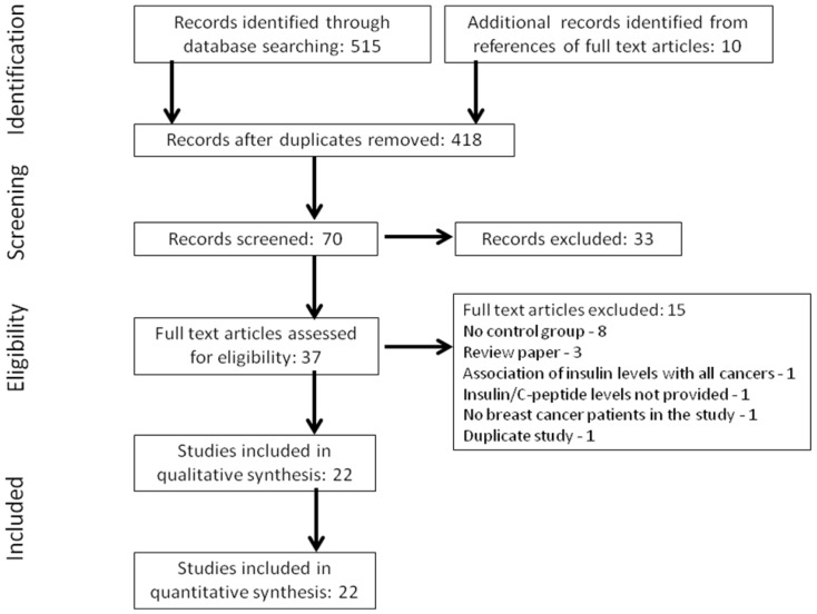
Our search identified 525 publications (Figure 1). After removing duplicates, 418 articles were screened by title for relevance to study topic. Next, 70 articles were screened by abstract following which 37 articles were selected for full-text review based on relevance to the study topic and inclusion/exclusion criteria (Figure 1). Twenty-two studies that reported levels of components that define insulin resistance and their association with breast cancer in women were included in the meta-analysis. The reasons for exclusion of the remaining 15 articles are listed in Figure 1.
Figure 1. Flowchart of selected studies.
Flow diagram showing the number of citations identified, excluded (with reasons for exclusion), and finally included in the meta-analysis.
Study characteristics
Table 1 summarizes the main characteristics of the included studies. Of the 22 studies included, 20 were case-control [11], [12], [13], [14], [20], [21], [22], [23], [24], [25], [26], [27], [28], [29], [30], [31], [32], [33], [34], [35] and 2 cross-sectional studies [36], [37]. Breast cancer cases were identified by pathology/medical records in 12 studies and cancer/tumor registries in 7 studies; no information was provided in 3 studies. Study population included premenopausal women only in 2 studies, postmenopausal women only in 7 studies and both pre- and post-menopausal women in 12 studies; no information was provided in one study. Only one study [23] provided information stratified for menopausal status. In studies providing data as tertiles/quartiles (fasting insulin, n = 7; non-fasting/fasting C-peptide, n = 8), the serologic variables were expressed as tertiles or quartiles based on their distributions in either controls or total study population. Eight studies provided the time lag data between measurement of insulin resistance and the diagnosis of breast cancer. Five studies provided median time interval (range: 2.2 yrs-6 yrs) whereas 3 studies provided mean time interval (range: 2.7 yrs-19 yrs). Of the 22 studies included in the meta-analysis the sample size ranged from 25 to 7,894. Breast cancer cases in the studies ranged from 2.4% to 53.8%.
Table 1. Patient characteristics in studies included in the meta-analysis.
| Study reference, year | Year of publication | Country(ies) | Study design | Study Population | Sample size | Breast cancer cases (%) | Age, Mean (SD) | Blood samples | Biochemical assay | Breast cancer diagnosis | Time between measurement of IR and the diagnosis of cancer | Insulin resistance definition |
| Bruning PF et al 20 | 1992 | Netherlands | CC | Pre and postmenopausal | 664 | 33.6 | NA | NonfastingC-peptide | Radioimmunoassay | NA | NA | NA |
| Yam D et al 11 | 1996 | Israel | CC | NA | 25 | 40.0 | NA | Fasting insulin | Radioimmunoassay | Pathology | NA | NA |
| Del Giudice ME et al 21 | 1998 | Canada | CC | Premenopausal | 198 | 50.0 | 42.5 (5.1) | Fasting insulin | Radioimmunoassay | Pathology | NA | NA |
| Jernstrom H et al 22 | 1999 | USA | CC | Postmenopausal | 438 | 10.3 | 74.3 (9.5) | Fasting insulin | Radioimmunoassay | Pathology | NA | NA |
| Toniolo P et al 23 | 2000 | USA | CC | Pre and postmenopausal | 658 | 26.1 | 49.8 (8.4) | NonfastingC-peptide | Radioimmunoassay | Pathology records | Median: 57 months Range: 7–120 months | NA |
| Kaaks R et al 24 | 2002 | Sweden | CC | Pre and postmenopausal | 700 | 35.1 | 54.4 | Fasting insulin | Immunoradiometric assay | Pathology | Median: 2.2 yrsRange: 1 month-10.6 yrs | NA |
| Mink PJ et al 25 | 2002 | USA | CC | Pre and postmenopausal | 7894 | 2.4 | NA | Fasting insulin | Radioimmunoassay | Medical records and cancer registries | NA | NA |
| Keinan-Boker L et al 26 | 2003 | Netherlands | CC | Postmenopausal | 482 | 30.9 | 57.1 (5.5) | NonfastingC-peptide | Radioimmunoassay | Cancer registry | Median: 28 months Range: 14–73 months | NA |
| Lawlor DA et al 36 | 2004 | UK | CS | Pre and postmenopausal | 3837 | 3.8 | 68.9 (5.5) | Fasting insulin | ELISA assay | Medical records/database | Median: 6 yrs Range: 0.5–36 yrs | product of fasting glucose (mmol/l) and insulin (lU/ml) divided by the constant 22.5 |
| Schairer C et al 27 | 2004 | USA | CC | Postmenopausal | 344 | 53.8 | 64.2 (9.6) | NonfastingC-peptide | Radioimmunoassay | Pathology | Mean: 19 yrs | NA |
| Gonullu G et al 28 | 2005 | Turkey | CC | Postmenopausal | 40 | 50.0 | 47.7 (4.0) | Fasting insulin | Radioimmunoassay | Pathology | NA | [(fasting glucose (mmol/L) x fasting insulin(mU/mL))/22.5]. Value greater than 2.7 was considered as insulin resistance; IR% = 37.5 |
| Falk RT et al 29 | 2006 | USA | CC | Premenopausal | 357 | 47.8 | 38.6 (20-44)* | NonfastingC-peptide | Radioimmunoassay | Pathology | NA | NA |
| Verheus M et al 30 | 2006 | 8 european countries | CC | Pre and postmenopausal | 3345 | 34.1 | 54.5 | Fasting and nonfasting C-peptide | Radioimmunoassay | Cancer and pathology registries | Mean: 2.8 yrs Range: 0.1–6.3 yrs | NA |
| Eliassen AH et al 14 | 2007 | USA | CC | Pre and postmenopausal | 617 (insulin) | 33.7 | 45.0 | Fasting insulin | Radioimmunoassay | Medical records | Mean: 31 months Range: 1–87 months | NA |
| 945 (c-peptide) | 33.4 | Fasting and nonfasting C-peptide | ELISA assay | |||||||||
| Fair AM et al 12 | 2007 | China | CC | Pre and postmenopausal | 794 | 50.0 | 47.7 (7.9) | C-peptide (fasting status NA) | ELISA assay | Cancer registry | NA | NA |
| Garmendia ML et al 13 | 2007 | Chile | CC | Pre and postmenopausal | 340 | 50.0 | 55.8 (11.4) | Fasting insulin | ELISA assay | Pathology | NA | Insulin resistance was measured by HOMA = insulin/(22.5e-lnglucose) and defined as 2.5 or more |
| IR% = 55.3 | ||||||||||||
| Cust AE et al 31 | 2009 | Sweden | CC | Pre and postmenopausal | 1122 | 50.0 | NA | NonfastingC-peptide | Radioimmunoassay | Cancer registry | Median: 4.4 yrs | NA |
| Gunter MJ et al 32 | 2009 | USA | CC | Postmenopausal | 1651 | 50.9 | 63.5 | Fasting insulin | NA | Medical records and tumor registries | NA | HOMA-IR index = fasting insulin [µ IU/mL] × fasting glucose [mg/dL]/22.5 |
| Kabat GC et al 33 | 2009 | USA | CC | Postmenopausal | 5450 | 3.5 | 62.6 (6.6) | Fasting insulin | ELISA assay | Medical records and tumor registries | NA | ([fasting insulin(microIU/mL) x fasting glucose (mg/dL)]/22.5) |
| Abbasi M et al 37 | 2010 | Iran | CS | Pre and postmenopausal | 920 | 8.9 | 38.1 (12.0) | Fasting insulin | Radioimmunoassay | Pathology | NA | fasting insulin (mU/L) x fasting glucose (mg/dL)/405. HOMA-IR values of greater than 1.8 as indicative for insulin resistance |
| Capasso I et al 34 | 2011 | Italy | CC | Postmenopausal | 777 | 37.7 | 57.55 (45–75)* | Insulin (fasting status NA) | NA | NA | NA | NA |
| Sieri S et al 35 | 2012 | Italy | CC | Pre and postmenopausal | 1807 | 20.6 | NA | Fasting insulin | Chemiluminescent immunoassay | NA | NA | Not defined |
NA = not available; CS = cross-sectional; CC = case-control; * = Mean (range); HOMA-IR = Homeostatic model of assessment-insulin resistance; IR = Insulin resistance.
Quality Assessment
Using the NOS scale, all but 2 studies [11], [34] were identified as high quality (reported in Table S1 in File S1). All studies clearly identified the study population and defined the outcome and outcome assessment (reported in Table S2 in File S1). All but 2 studies [11], [34] identified important confounders or prognostic factors and were used for adjustment of the association between insulin/c-peptide levels and breast cancer. There was considerable variation in the selection of confounding variables for adjustment (reported in Table S2 in File S1). It is possible that a few confounding variables were not fully identified and recorded. The most common confounder adjusted was age.
Publication bias
The funnel plot did not suggest the presence of publication bias, and the formal test of asymmetry of this plot was not significant (Egger's p value = 0.8).
Meta-analyses
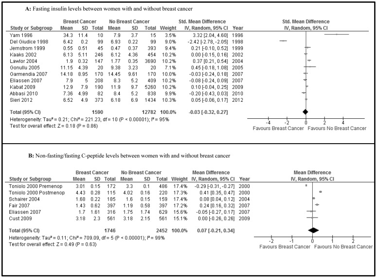
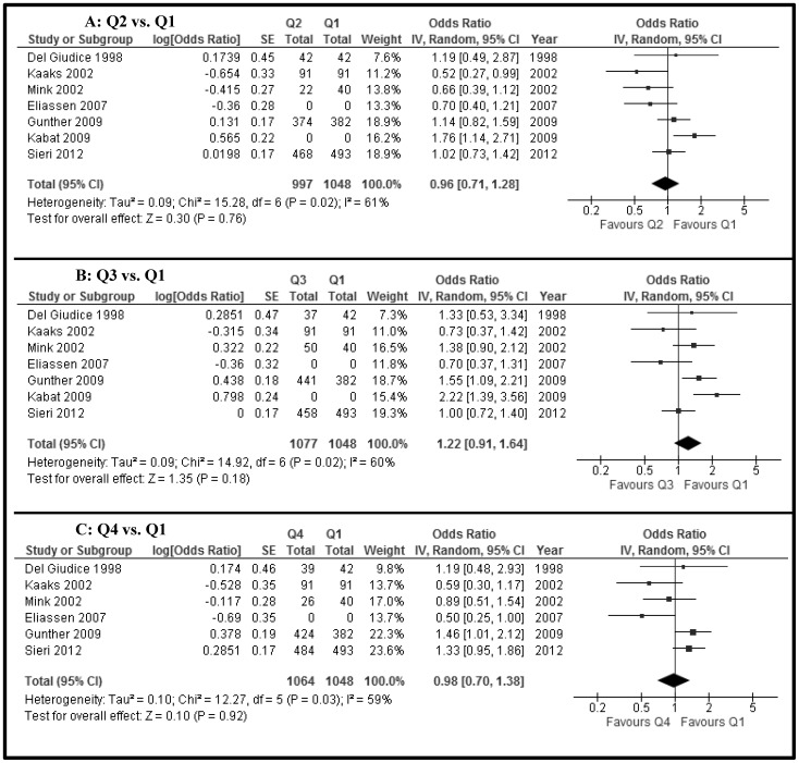
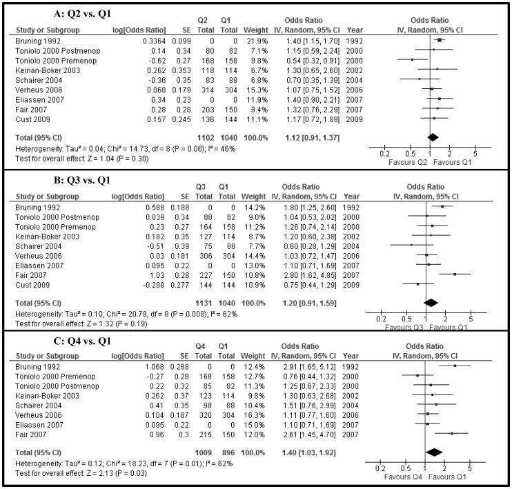
The fasting insulin levels (11 studies, n = 14,372) were not different between women with and without breast cancer (SMD −0.03, 95% CI −0.32 to 0.27, P = 0.9) (Figure 2A). Furthermore, non-fasting/fasting C-peptide levels (5 studies, n = 4,198) were not different between the two groups (MD 0.07, −0.21 to 0.34, p = 0.6) (Figure 2B). There was high heterogeneity (I2 = 95% and 99%, respectively) for these two associations. Using individual ORs adjusted for age at least, there was no increased risk of breast cancer when the higher quartiles were compared with the lowest quartile (Q1) of fasting insulin (Q2 vs Q1, 7 studies, n = 2,045; Q3 vs Q1, 7 studies, n = 2,125; Q4 vs Q1, 6 studies, n = 2,112) (OR Q2 vs Q1 0.96, 0.71 to 1.28; OR Q3 vs Q1 1.22, 0.91 to 1.64; OR Q4 vs. Q1 0.98, 0.70 to 1.38) (Figures 3A, 3B and 3C, respectively). Also, there were no differences for quartiles of non-fasting/fasting C-peptide levels (Q2 vs Q1, 8 studies, n = 2,142; Q3 vs Q1, 8 studies, n = 2,171; Q4 vs Q1, 7 studies, n = 1,905) (OR Q2 vs Q1 1.12, 0.91 to 1.37; OR Q3 vs Q1 1.20, 0.91 to 1.59; OR Q4 vs. Q1 1.40, 1.03 to 1.92) (Figures 4A, 4B and 4C, respectively). The level of heterogeneity on pooling ORs adjusted for age at least was moderate. HOMA-IR levels (5 studies, n = 6,944) in women with breast cancer were significantly and slightly higher than in women without breast cancer (MD 0.22, 0.13 to 0.31, p<0.00001; I2 = 0) (Figure 5).
Figure 2. Forest plot of observational studies comparing women with and without breast cancer for (A) Fasting insulin levels and (B) Non-fasting/fasting C-peptide levels.
(IV, Random = Inverse variance, Random effects model.)
Figure 3. Forest plot of observational studies with adjusted ORs for breast cancer between quartiles of fasting insulin levels (A) Q2 versus Q1; (B) Q3 versus Q1; (C) Q4 versus Q1 ORs.
(IV, Random = Inverse variance, Random effects model.)
Figure 4. Forest plot of observational studies with adjusted ORs for breast cancer between quartiles of non-fasting/fasting C-peptide levels (A) Q2 versus Q1; (B) Q3 versus Q1; (C) Q4 versus Q1.
(IV, Random = Inverse variance, Random effects model.)
Figure 5. Forest plot of observational studies comparing women with and without breast cancer for HOMA-IR levels.

(IV, Random = Inverse variance, Random effects model.)
Discussion
In this systematic review we did not find differences between fasting insulin or non-fasting/fasting C-peptide levels and women with and without breast cancer. There was no higher adjusted risk of breast cancer between higher quartiles of fasting insulin or non-fasting/fasting C-peptide levels and their lowest quartile levels. Finally, fasting HOMA-IR levels were slightly and significantly higher in women with breast cancer in comparison with women without breast cancer.
Insulin and IGF-1 are peptide hormones that stimulate proliferation of tissues and levels higher than normal (i.e. hyperinsulinemia) have been linked with carcinogenic properties in animal models [38]. Hyperinsulinemia occurs in presence of insulin resistance, a key pathophysiological mechanism linked to type 2 diabetes mellitus and obesity. There is substantial epidemiological evidence linking insulin resistance and cancer of the liver, colon and pancreas [39]. However, the association between hyperinsulinemia/insulin resistance and breast cancer has been controversial in humans. Several other mechanisms apart from hyperinsulinemia have been described for breast cancer such as increased angiogenesis, hypoadiponectinemia, and increased bioactivity of estrogens and testosterone [40].
A meta-analysis has linked diabetes mellitus and breast cancer risk [41]. These investigators studied 20 observational studies of diabetic patients with 39,719 cases of breast cancer. There was a modest association between diabetes and breast cancer (OR 1.15, 95% CI 1.12–1.19), which was higher for postmenopausal women than for premenopausal women (OR 1.19, 95%CI 1.15–1.23 and OR 0.94, 95% CI 0.80–1.10, respectively). A high level of heterogeneity was observed among association effects. Obesity and hyperinsulinemia were described as the main potential mechanisms of this positive association.
Another recent meta-analysis evaluated the relationship between metabolic syndrome and breast cancer [42]. Metabolic syndrome as defined by the WHO includes insulin resistance amongst the diagnostic criteria, along with two or more of the following: increased waist-to-hip ratio, hypertriglyceridemia, low HDL cholesterol, high blood pressure and microalbuminuria [43]. Eleven studies involving 9,643 breast cancer cases were analyzed and a weak association between metabolic syndrome and breast cancer was found (RR 1.23, 95% CI 1.05–1.45). Within postmenopausal studies (5 studies, 1,290 breast cancer cases) there was a slightly higher association was found: RR 1.56, 95% CI 1.08–2.24. Importantly, also a substantial heterogeneity of association effects was observed among studies. Other diagnostic criteria of metabolic syndrome such as waist-to-hip ratio, hypertension, and low HDL cholesterol have not individually shown consistent associations with breast cancer [41].
We did not find any association between markers of insulin resistance and breast cancer. Fasting insulin levels and non-fasting/fasting C-peptide levels may shadow the state of stress that the pancreas has in presence of insulin resistance. We believe that measuring insulin levels in the 3-hour period after a sugar load test can provide better information and potentially peak levels of insulin or the area below the 3-hour insulin curve may be associated with breast cancer risk. Higher fasting insulin levels have been slightly associated with higher risk of endometrial cancer (OR highest quartile vs. lowest quartile of insulin 1.64, 95%CI 1.12–2.40) [44].
Load tests may give higher chances to find an association not found with current measurements. Previous research has shown an association between glycemic load and breast cancer risk in a meta-analysis [45]. The glycemic load combines the loads for the total servings of all carbohydrate-containing foods consumed per day, on average. Glycemic load has been associated with a slightly higher risk of breast cancer (RR 1.14, 95% CI 1.02–1.28). Similarly, the association of glycemic load with endometrial cancer was weak (RR 1.36, 95% CI 1.14–1.62).
We did not evaluate fasting glucose individually, but we did evaluate HOMA-IR scores, which is proportional to glucose x insulin levels. A very small, although significant association between HOMA-IR scores and breast cancer was found. Unfortunately, we could not explore the association between higher quartiles and the lowest quartile of HOMA-IR scores, as they were not available in the selected studies. Again, we propose that glucose and insulin levels may be both measured after a sugar load test and these values may provide a better estimate of the association of insulin resistance and breast cancer risk.
A recent meta-analysis found no evidence of an association between serum insulin and C-peptide concentration and breast cancer in 6 prospective studies with 1,890 cases [15]. We conducted a comprehensive search and evaluated this association in all observational studies to date. Our meta-analysis included 22 studies with 7,478 cases. We also determined the association between HOMA-IR scores and breast cancer. In our meta-analysis studies investigating the associations between insulin/C-peptide levels and breast cancer were heterogenous as was the case with the meta-analysis of prospective studies only.
Our study has several limitations. First, the observational nature of the included studies may weaken our conclusions, especially because most are case-control studies. However, the risk of bias of included studies was low. Second, although we compared insulin, C-peptide levels and HOMA-IR scores univariably between women with and without breast cancer, we also meta-analyzed risks measures of breast cancer between quartiles that were adjusted at least for age, and in most of cases for several essential confounders. Third, there was substantial clinical and statistical heterogeneity among included studies. Previous meta-analyses [41], [42] investigating factors associated with breast cancer also showed high levels of heterogeneity; we could not perform analyses by age subgroups because data was not provided as such, and by menopausal status as only one of the studies provided that information.
In observational studies, higher levels of fasting insulin or non-fasting/fasting C-peptide were not found to be associated with the presence of breast cancer in women, even after adjustment for important confounders. HOMA-IR levels are slightly higher in women with breast cancer.
Supporting Information
PRISMA Checklist.
(DOC)
Supporting tables. Table S1, Newcastle-Ottawa Scale. Table S2, Study Quality Assessment.
(DOC)
Funding Statement
This study was funded by the Instituto Médico de la Mujer, Lima, Peru. The funders had no role in study design, data collection and analysis, decision to publish, or preparation of the manuscript.
References
- 1.American Cancer Society (2014) ACS Cancer Facts and Figures. Cancer Facts and Figures. American Cancer Society.
- 2. Coughlin SS, Ekwueme DU (2009) Breast cancer as a global health concern. Cancer Epidemiol 33: 315–318. [DOI] [PubMed] [Google Scholar]
- 3. Lebovitz HE (2001) Insulin resistance: definition and consequences. Exp Clin Endocrinol Diabetes 109 Suppl 2S135–148. [DOI] [PubMed] [Google Scholar]
- 4. Rinaldi S, Key TJ, Peeters PH, Lahmann PH, Lukanova A, et al. (2006) Anthropometric measures, endogenous sex steroids and breast cancer risk in postmenopausal women: a study within the EPIC cohort. Int J Cancer 118: 2832–2839. [DOI] [PubMed] [Google Scholar]
- 5. Tehard B, Lahmann PH, Riboli E, Clavel-Chapelon F (2004) Anthropometry, breast cancer and menopausal status: use of repeated measurements over 10 years of follow-up-results of the French E3N women's cohort study. Int J Cancer 111: 264–269. [DOI] [PubMed] [Google Scholar]
- 6. van den Brandt PA, Spiegelman D, Yaun SS, Adami HO, Beeson L, et al. (2000) Pooled analysis of prospective cohort studies on height, weight, and breast cancer risk. Am J Epidemiol 152: 514–527. [DOI] [PubMed] [Google Scholar]
- 7. Friedenreich CM, Orenstein MR (2002) Physical activity and cancer prevention: etiologic evidence and biological mechanisms. J Nutr 132: 3456S–3464S. [DOI] [PubMed] [Google Scholar]
- 8. Riserus U, Arnlov J, Berglund L (2007) Long-term predictors of insulin resistance: role of lifestyle and metabolic factors in middle-aged men. Diabetes Care 30: 2928–2933. [DOI] [PubMed] [Google Scholar]
- 9. Sachdev D, Yee D (2001) The IGF system and breast cancer. Endocr Relat Cancer 8: 197–209. [DOI] [PubMed] [Google Scholar]
- 10. Kaaks R (2001) [Plasma insulin, IGF-I and breast cancer]. Gynecol Obstet Fertil 29: 185–191. [DOI] [PubMed] [Google Scholar]
- 11. Yam D, Fink A, Mashiah A, Ben-Hur E (1996) Hyperinsulinemia in colon, stomach and breast cancer patients. Cancer Lett 104: 129–132. [DOI] [PubMed] [Google Scholar]
- 12. Fair AM, Dai Q, Shu XO, Matthews CE, Yu H, et al. (2007) Energy balance, insulin resistance biomarkers, and breast cancer risk. Cancer Detect Prev 31: 214–219. [DOI] [PMC free article] [PubMed] [Google Scholar]
- 13. Garmendia ML, Pereira A, Alvarado ME, Atalah E (2007) Relation between insulin resistance and breast cancer among Chilean women. Ann Epidemiol 17: 403–409. [DOI] [PubMed] [Google Scholar]
- 14. Eliassen AH, Tworoger SS, Mantzoros CS, Pollak MN, Hankinson SE (2007) Circulating insulin and c-peptide levels and risk of breast cancer among predominately premenopausal women. Cancer Epidemiol Biomarkers Prev 16: 161–164. [DOI] [PubMed] [Google Scholar]
- 15. Autier P, Koechlin A, Boniol M, Mullie P, Bolli G, et al. (2013) Serum insulin and C-peptide concentration and breast cancer: a meta-analysis. Cancer Causes Control 24: 873–883. [DOI] [PubMed] [Google Scholar]
- 16. DerSimonian R, Laird N (1986) Meta-analysis in clinical trials. Control Clin Trials 7: 177–188. [DOI] [PubMed] [Google Scholar]
- 17. Higgins JP, Thompson SG, Deeks JJ, Altman DG (2003) Measuring inconsistency in meta-analyses. BMJ 327: 557–560. [DOI] [PMC free article] [PubMed] [Google Scholar]
- 18. Higgins JP (2008) Commentary: Heterogeneity in meta-analysis should be expected and appropriately quantified. Int J Epidemiol 37: 1158–1160. [DOI] [PubMed] [Google Scholar]
- 19. Egger M, Davey Smith G, Schneider M, Minder C (1997) Bias in meta-analysis detected by a simple, graphical test. BMJ 315: 629–634. [DOI] [PMC free article] [PubMed] [Google Scholar]
- 20. Bruning PF, Bonfrer JM, van Noord PA, Hart AA, de Jong-Bakker M, et al. (1992) Insulin resistance and breast-cancer risk. Int J Cancer 52: 511–516. [DOI] [PubMed] [Google Scholar]
- 21. Del Giudice ME, Fantus IG, Ezzat S, McKeown-Eyssen G, Page D, et al. (1998) Insulin and related factors in premenopausal breast cancer risk. Breast Cancer Res Treat 47: 111–120. [DOI] [PubMed] [Google Scholar]
- 22. Jernstrom H, Barrett-Connor E (1999) Obesity, weight change, fasting insulin, proinsulin, C-peptide, and insulin-like growth factor-1 levels in women with and without breast cancer: the Rancho Bernardo Study. J Womens Health Gend Based Med 8: 1265–1272. [DOI] [PubMed] [Google Scholar]
- 23. Toniolo P, Bruning PF, Akhmedkhanov A, Bonfrer JM, Koenig KL, et al. (2000) Serum insulin-like growth factor-I and breast cancer. Int J Cancer 88: 828–832. [DOI] [PubMed] [Google Scholar]
- 24. Kaaks R, Lundin E, Rinaldi S, Manjer J, Biessy C, et al. (2002) Prospective study of IGF-I, IGF-binding proteins, and breast cancer risk, in northern and southern Sweden. Cancer Causes Control 13: 307–316. [DOI] [PubMed] [Google Scholar]
- 25. Mink PJ, Shahar E, Rosamond WD, Alberg AJ, Folsom AR (2002) Serum insulin and glucose levels and breast cancer incidence: the atherosclerosis risk in communities study. Am J Epidemiol 156: 349–352. [DOI] [PubMed] [Google Scholar]
- 26. Keinan-Boker L, Bueno De Mesquita HB, Kaaks R, Van Gils CH, Van Noord PA, et al. (2003) Circulating levels of insulin-like growth factor I, its binding proteins -1,-2, -3, C-peptide and risk of postmenopausal breast cancer. Int J Cancer 106: 90–95. [DOI] [PubMed] [Google Scholar]
- 27. Schairer C, Hill D, Sturgeon SR, Fears T, Pollak M, et al. (2004) Serum concentrations of IGF-I, IGFBP-3 and c-peptide and risk of hyperplasia and cancer of the breast in postmenopausal women. Int J Cancer 108: 773–779. [DOI] [PubMed] [Google Scholar]
- 28. Gonullu G, Ersoy C, Ersoy A, Evrensel T, Basturk B, et al. (2005) Relation between insulin resistance and serum concentrations of IL-6 and TNF-alpha in overweight or obese women with early stage breast cancer. Cytokine 31: 264–269. [DOI] [PubMed] [Google Scholar]
- 29. Falk RT, Brinton LA, Madigan MP, Potischman N, Sturgeon SR, et al. (2006) Interrelationships between serum leptin, IGF-1, IGFBP3, C-peptide and prolactin and breast cancer risk in young women. Breast Cancer Res Treat 98: 157–165. [DOI] [PubMed] [Google Scholar]
- 30. Verheus M, Peeters PH, Rinaldi S, Dossus L, Biessy C, et al. (2006) Serum C-peptide levels and breast cancer risk: results from the European Prospective Investigation into Cancer and Nutrition (EPIC). Int J Cancer 119: 659–667. [DOI] [PubMed] [Google Scholar]
- 31. Cust AE, Stocks T, Lukanova A, Lundin E, Hallmans G, et al. (2009) The influence of overweight and insulin resistance on breast cancer risk and tumour stage at diagnosis: a prospective study. Breast Cancer Res Treat 113: 567–576. [DOI] [PubMed] [Google Scholar]
- 32. Gunter MJ, Hoover DR, Yu H, Wassertheil-Smoller S, Rohan TE, et al. (2009) Insulin, insulin-like growth factor-I, and risk of breast cancer in postmenopausal women. J Natl Cancer Inst 101: 48–60. [DOI] [PMC free article] [PubMed] [Google Scholar]
- 33. Kabat GC, Kim M, Caan BJ, Chlebowski RT, Gunter MJ, et al. (2009) Repeated measures of serum glucose and insulin in relation to postmenopausal breast cancer. Int J Cancer 125: 2704–2710. [DOI] [PubMed] [Google Scholar]
- 34. Capasso I, Esposito E, Pentimalli F, Crispo A, Montella M, et al. (2010) Metabolic syndrome affects breast cancer risk in postmenopausal women: National Cancer Institute of Naples experience. Cancer Biol Ther 10: 1240–1243. [DOI] [PubMed] [Google Scholar]
- 35. Sieri S, Muti P, Claudia A, Berrino F, Pala V, et al. (2012) Prospective study on the role of glucose metabolism in breast cancer occurrence. Int J Cancer 130: 921–929. [DOI] [PubMed] [Google Scholar]
- 36. Lawlor DA, Smith GD, Ebrahim S (2004) Hyperinsulinaemia and increased risk of breast cancer: findings from the British Women's Heart and Health Study. Cancer Causes Control 15: 267–275. [DOI] [PubMed] [Google Scholar]
- 37. Abbasi M, Tarafdari A, Esteghamati A, Vejdani K, Nakhjavani M (2010) Insulin resistance and breast carcinogenesis: a cross-sectional study among Iranian women with breast mass. Metab Syndr Relat Disord 8: 411–416. [DOI] [PubMed] [Google Scholar]
- 38. Tran TT, Medline A, Bruce WR (1996) Insulin promotion of colon tumors in rats. Cancer Epidemiol Biomarkers Prev 5: 1013–1015. [PubMed] [Google Scholar]
- 39. Tsugane S, Inoue M (2010) Insulin resistance and cancer: epidemiological evidence. Cancer Sci 101: 1073–1079. [DOI] [PMC free article] [PubMed] [Google Scholar]
- 40. Rose DP, Haffner SM, Baillargeon J (2007) Adiposity, the metabolic syndrome, and breast cancer in African-American and white American women. Endocr Rev 28: 763–777. [DOI] [PubMed] [Google Scholar]
- 41. Xue F, Michels KB (2007) Diabetes, metabolic syndrome, and breast cancer: a review of the current evidence. Am J Clin Nutr 86: s823–835. [DOI] [PubMed] [Google Scholar]
- 42. Esposito K, Chiodini P, Colao A, Lenzi A, Giugliano D (2012) Metabolic syndrome and risk of cancer: a systematic review and meta-analysis. Diabetes Care 35: 2402–2411. [DOI] [PMC free article] [PubMed] [Google Scholar]
- 43. Ford ES, Giles WH (2003) A comparison of the prevalence of the metabolic syndrome using two proposed definitions. Diabetes Care 26: 575–581. [DOI] [PubMed] [Google Scholar]
- 44. Friedenreich CM, Langley AR, Speidel TP, Lau DC, Courneya KS, et al. (2012) Case-control study of markers of insulin resistance and endometrial cancer risk. Endocr Relat Cancer 19: 785–792. [DOI] [PMC free article] [PubMed] [Google Scholar]
- 45. Gnagnarella P, Gandini S, La Vecchia C, Maisonneuve P (2008) Glycemic index, glycemic load, and cancer risk: a meta-analysis. Am J Clin Nutr 87: 1793–1801. [DOI] [PubMed] [Google Scholar]
Associated Data
This section collects any data citations, data availability statements, or supplementary materials included in this article.
Supplementary Materials
PRISMA Checklist.
(DOC)
Supporting tables. Table S1, Newcastle-Ottawa Scale. Table S2, Study Quality Assessment.
(DOC)


