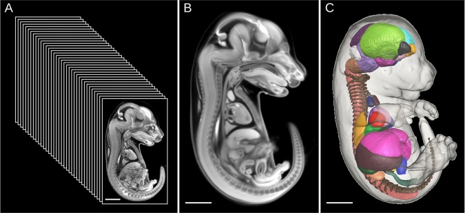
Fig. 1.

Generation of the 3D segmented mouse embryo atlas. (A-C) Thirty-five C57Bl/6 E15.5 mouse embryo micro-CT images (A) were morphed into a high quality representative population average atlas (B) through image registration. Forty-eight organ structures were manually painted through the 3D population average atlas. The resultant 3D mouse embryo segmented atlas (C) can be used to automatically measure organ volumes of new individual embryo images. Figure is adapted from our previous publication in Development (Wong et al., 2012).
