

Abstract
The title compound, C17H18N2S2, exhibits a trans–trans–trans–gauche+ (tttg +) conformation with regard to the NH–CH2–CH2–CH2–NH bond sequence. In the crystal, molecules are connected by N—H⋯S=C and C—H⋯S=C hydrogen bonds, forming a herringbone arrangement along the c-axis direction. The two thioamide groups make dihedral angles of 43.0 (2) and 33.1 (2)° with the adjacent phenyl rings.
Related literature
For the crystal structures and conformations of related compounds, see: for example, Palmer & Brisse (1980 ▶); Brisson & Brisse (1986 ▶); Deguire & Brisse (1988 ▶); Nagasawa et al. (2014 ▶). For the synthesis, see: Hart & Brewbaker (1969 ▶); Cave & Levinson (1985 ▶).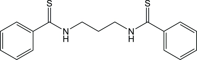
Experimental
Crystal data
C17H18N2S2
M r = 314.45
Orthorhombic,

a = 8.36521 (9) Å
b = 14.13395 (14) Å
c = 26.9223 (3) Å
V = 3183.12 (6) Å3
Z = 8
Cu Kα radiation
μ = 2.97 mm−1
T = 223 K
0.30 × 0.20 × 0.10 mm
Data collection
Bruker APEXII Ultra CCD area-detector diffractometer
Absorption correction: multi-scan (SADABS; Bruker, 2001 ▶) T min = 0.47, T max = 0.76
12276 measured reflections
2882 independent reflections
2736 reflections with I > 2σ(I)
R int = 0.018
Refinement
R[F 2 > 2σ(F 2)] = 0.032
wR(F 2) = 0.085
S = 1.05
2882 reflections
190 parameters
H-atom parameters constrained
Δρmax = 0.26 e Å−3
Δρmin = −0.31 e Å−3
Data collection: APEX2 (Bruker, 2007 ▶); cell refinement: SAINT (Bruker, 2007 ▶); data reduction: SAINT; program(s) used to solve structure: SHELXS2013 (Sheldrick, 2008 ▶); program(s) used to refine structure: SHELXL2013 (Sheldrick, 2008 ▶); molecular graphics: Mercury (Macrae et al., 2006 ▶); software used to prepare material for publication: SHELXL2013.
Supplementary Material
Crystal structure: contains datablock(s) global, I. DOI: 10.1107/S160053681400974X/bv2233sup1.cif
Structure factors: contains datablock(s) I. DOI: 10.1107/S160053681400974X/bv2233Isup2.hkl
Supporting information file. DOI: 10.1107/S160053681400974X/bv2233Isup3.cml
CCDC reference: 1000286
Additional supporting information: crystallographic information; 3D view; checkCIF report
Table 1. Hydrogen-bond geometry (Å, °).
| D—H⋯A | D—H | H⋯A | D⋯A | D—H⋯A |
|---|---|---|---|---|
| N1—H1⋯S2i | 0.87 | 2.59 | 3.4412 (14) | 168 |
| N2—H2⋯S1ii | 0.87 | 2.69 | 3.5135 (12) | 157 |
| C17—H17⋯S1iii | 0.94 | 2.85 | 3.7665 (17) | 166 |
Symmetry codes: (i)  ; (ii)
  ; (iii)
  .
.
supplementary crystallographic information
1. Comment
In a previous paper (Nagasawa et al., 2014), we reported the crystal structure of N, N'-(ethane-1,2-diyl)dibenzothioamide. In this study, we have determined the crystal structure of its homologue, N,N'-(propane-1,3-diyl)dibenzothioamide (referred to here as PDBTA), a monomeric model compound of poly(trimethylene terephthalthioamide), [–(C=S)–C6H4–(C=S)–NH–(CH2)3–NH–]n. Figure 1 shows the molecular structure of PDBTA, whose NH–CH2–CH2–CH2–NH bond sequence adopts the tttg+ conformation. On the other hand, our molecular orbital (MO) calculations at the B3LYP/6–311+G(2 d,p)//B3LYP/6–311+G(2 d,p) level including the solvent effect of dimethyl sulfoxide have predicted that the all-trans form is the most stable of its possible conformations; the crystal conformation, tttg+, was suggested to have a free energy higher than tttt by as much as 1.19 kcal mol-1.
Figure 2 shows the molecular packing of the PDBTA crystal, in which two kinds of intermolecular hydrogen bonds, C=S···H–N and C=S···H–C, seem to be formed (not shown in the figure). For details, see Table 1. The intermolecular attractions may fully compensate the cost of conformational energy of 1.19 kcal mol-1. The PDBTA molecules form a herringbone arrangement along the c-axis direction.
Brisson & Brisse (1986) determined the crystal structure of N,N'-(propane-1,3-diyl)dibenzamide (PDBA), a model compound of poly(trimethylene terephthalamide). The crystalline PDBA molecule lies in the ttg+g+ conformation and forms intermolecular C=O···H—N hydrogen bonds with the neighbors. According to our MO calculations, its most stable conformer is tttg+ (–0.54 kcal mol-1 relative to that of the all-trans state), and the crystal conformation, ttg+g+, has a somewhat higher free energy (+0.32 kcal mol-1).
2. Experimental
Benzoyl chloride (12.0 ml, 103 mmol) was added dropwise to an aqueous solution of 1,3-diaminopropane (3.4 ml, 41 mmol) and sodium hydroxide (61.5 ml, 0.100 mol l-1) stirred by a mechanical stirrer in a three-necked flask equipped with a dropping funnel and a calcium-chloride drying tube, with the flask being bathed in water kept at 10 °C. The mixture was stirred at 10 °C for 1 h, diluted with water (100 ml), and stirred again at room temperature overnight to yield white precipitate. The precipitate was collected by suction filtration, washed with water, and dried. The crude product was recrystallized from a mixture of ethanol and toluene (1:1 in volume) and dried at 40 °C under reduced pressure to yield PDBA (yield 28%). In principle, this synthesis is based on the procedure of Hart & Brewbaker (1969).
PDBA (1.0 g, 3.6 mmol) and Lawesson's reagent (1.7 g, 4.2 mmol) (Cave & Levinson, 1985) were dissolved in toluene (20 ml) stirred in a three-necked flask equipped with a reflux condenser connected to a calcium-chloride drying tube. The mixture was refluxed under dry nitrogen at ca 110 °C for 8 h, and the completion of the reaction was confirmed by thin-layer chromatography. After toluene was removed under reduced pressure, the residual was subjected to column chromatography on silica gel with chloroform, and yellowish solution (retention factor Rf = 0.3) was collected and condensed to yield yellow slurry, which was recrystallized twice from a mixture of ethyl acetate and diethyl ether (1:1 in volume) and dried under reduced pressure to yield PDBTA (yield 48%).
A small quantity of PDBTA was dissolved in chloroform in a glass tube, whose top was sealed with a thin Teflon film. The tube was placed in a vial container including a small amount of n-hexane, and the container was capped and left to stand still in a dark place. After a day, crystals were found to be formed suitable for X-ray diffraction.
3. Refinement
All C—H hydrogen atoms were geometrically positioned with C—H = 0.95 and 0.99 Å for the aromatic and methylene groups respectively, and refined as riding by Uiso(H) = 1.2 Ueq(C). The N—H hydrogen atoms were located and fixed with N—H = 0.87 Å.
Figures
Fig. 1.

Molecular structure of N, N'-(propane-1,2-diyl)dibenzothioamide (PDBTA). Displacement ellipsoids are drawn at the 50% probability level. Isotropic H-atom thermal parameters are represented by spheres of arbitrary size. The labels of hydrogen atoms are omitted for clarity.
Fig. 2.

Packing diagram of PDBTA, viewed down the (a) a, (b) b, and (c) c axes.
Crystal data
| C17H18N2S2 | F(000) = 1328 |
| Mr = 314.45 | Dx = 1.312 Mg m−3 |
| Orthorhombic, Pbca | Cu Kα radiation, λ = 1.54178 Å |
| a = 8.36521 (9) Å | µ = 2.97 mm−1 |
| b = 14.13395 (14) Å | T = 223 K |
| c = 26.9223 (3) Å | Prismatic, yellow |
| V = 3183.12 (6) Å3 | 0.30 × 0.20 × 0.10 mm |
| Z = 8 |
Data collection
| Bruker APEXII Ultra CCD area-detector diffractometer | 2882 independent reflections |
| Radiation source: Bruker TXS fine-focus rotating anode | 2736 reflections with I > 2σ(I) |
| Bruker Helios multilayer mirror monochromator | Rint = 0.018 |
| Phi and ω scans | θmax = 68.3°, θmin = 3.3° |
| Absorption correction: multi-scan (SADABS; Bruker, 2001) | h = −9→10 |
| Tmin = 0.47, Tmax = 0.76 | k = −16→17 |
| 12276 measured reflections | l = −32→32 |
Refinement
| Refinement on F2 | Primary atom site location: structure-invariant direct methods |
| Least-squares matrix: full | Secondary atom site location: difference Fourier map |
| R[F2 > 2σ(F2)] = 0.032 | Hydrogen site location: inferred from neighbouring sites |
| wR(F2) = 0.085 | H-atom parameters constrained |
| S = 1.05 | w = 1/[σ2(Fo2) + (0.0498P)2 + 1.0464P] where P = (Fo2 + 2Fc2)/3 |
| 2882 reflections | (Δ/σ)max = 0.001 |
| 190 parameters | Δρmax = 0.26 e Å−3 |
| 0 restraints | Δρmin = −0.31 e Å−3 |
Special details
| Experimental. SADABS (Sheldrick 1996) |
| Geometry. All e.s.d.'s (except the e.s.d. in the dihedral angle between two l.s. planes) are estimated using the full covariance matrix. The cell e.s.d.'s are taken into account individually in the estimation of e.s.d.'s in distances, angles and torsion angles; correlations between e.s.d.'s in cell parameters are only used when they are defined by crystal symmetry. An approximate (isotropic) treatment of cell e.s.d.'s is used for estimating e.s.d.'s involving l.s. planes. |
| Refinement. Refinement of F2 was performed with all reflections. The weighted R-factor (wR) and goodness of fit (S) are based on F2, while the R-factor on F. The threshold expression of F2 > 2.0 σ(F2) was used only for calculating R-factor. |
Fractional atomic coordinates and isotropic or equivalent isotropic displacement parameters (Å2)
| x | y | z | Uiso*/Ueq | ||
| C1 | 0.04995 (15) | 0.30766 (10) | 0.42684 (5) | 0.0271 (3) | |
| C2 | 0.04043 (16) | 0.33224 (9) | 0.37326 (5) | 0.0261 (3) | |
| C3 | 0.15288 (18) | 0.39267 (10) | 0.35235 (5) | 0.0328 (3) | |
| H3 | 0.236 | 0.4175 | 0.3719 | 0.039* | |
| C4 | 0.1418 (2) | 0.41607 (11) | 0.30247 (6) | 0.0408 (4) | |
| H4 | 0.2193 | 0.4555 | 0.288 | 0.049* | |
| C5 | 0.0177 (2) | 0.38189 (12) | 0.27379 (6) | 0.0435 (4) | |
| H5 | 0.0109 | 0.3981 | 0.24 | 0.052* | |
| C6 | −0.0964 (2) | 0.32377 (11) | 0.29487 (6) | 0.0400 (4) | |
| H6 | −0.1823 | 0.3017 | 0.2756 | 0.048* | |
| C7 | −0.08486 (17) | 0.29786 (10) | 0.34432 (5) | 0.0318 (3) | |
| H7 | −0.1614 | 0.2572 | 0.3584 | 0.038* | |
| C8 | −0.00172 (17) | 0.17921 (12) | 0.48748 (6) | 0.0380 (4) | |
| H8A | −0.092 | 0.1351 | 0.487 | 0.046* | |
| H8B | −0.0292 | 0.231 | 0.5101 | 0.046* | |
| C9 | 0.14371 (17) | 0.12807 (10) | 0.50784 (5) | 0.0340 (3) | |
| H9A | 0.2339 | 0.172 | 0.5108 | 0.041* | |
| H9B | 0.1747 | 0.0765 | 0.4855 | 0.041* | |
| C10 | 0.09938 (18) | 0.08896 (11) | 0.55852 (6) | 0.0364 (3) | |
| H10A | 0.0758 | 0.1419 | 0.5809 | 0.044* | |
| H10B | 0.0019 | 0.051 | 0.5553 | 0.044* | |
| C11 | 0.19903 (16) | −0.02401 (9) | 0.61993 (5) | 0.0268 (3) | |
| C12 | 0.33873 (16) | −0.07725 (9) | 0.63952 (5) | 0.0270 (3) | |
| C13 | 0.34922 (18) | −0.09590 (10) | 0.69027 (5) | 0.0330 (3) | |
| H13 | 0.2681 | −0.0749 | 0.7118 | 0.04* | |
| C14 | 0.4785 (2) | −0.14514 (12) | 0.70912 (6) | 0.0417 (4) | |
| H14 | 0.4858 | −0.1563 | 0.7435 | 0.05* | |
| C15 | 0.5967 (2) | −0.17792 (12) | 0.67788 (7) | 0.0478 (4) | |
| H15 | 0.6842 | −0.2117 | 0.6908 | 0.057* | |
| C16 | 0.5859 (2) | −0.16086 (13) | 0.62749 (7) | 0.0480 (4) | |
| H16 | 0.6657 | −0.1839 | 0.6061 | 0.058* | |
| C17 | 0.45871 (19) | −0.11019 (11) | 0.60823 (6) | 0.0368 (3) | |
| H17 | 0.4534 | −0.098 | 0.5739 | 0.044* | |
| N1 | 0.01881 (14) | 0.21803 (9) | 0.43765 (5) | 0.0324 (3) | |
| H1 | 0.0098 | 0.1789 | 0.4129 | 0.039* | |
| N2 | 0.22457 (14) | 0.03070 (8) | 0.58067 (4) | 0.0301 (3) | |
| H2 | 0.3197 | 0.0319 | 0.5676 | 0.036* | |
| S1 | 0.08969 (5) | 0.39123 (3) | 0.46914 (2) | 0.03743 (13) | |
| S2 | 0.01763 (4) | −0.03524 (3) | 0.64602 (2) | 0.03725 (13) |
Atomic displacement parameters (Å2)
| U11 | U22 | U33 | U12 | U13 | U23 | |
| C1 | 0.0195 (6) | 0.0338 (7) | 0.0279 (7) | 0.0044 (5) | −0.0002 (5) | 0.0037 (5) |
| C2 | 0.0273 (6) | 0.0256 (6) | 0.0253 (6) | 0.0051 (5) | 0.0003 (5) | 0.0011 (5) |
| C3 | 0.0312 (7) | 0.0339 (7) | 0.0333 (8) | 0.0012 (6) | 0.0037 (6) | 0.0018 (5) |
| C4 | 0.0464 (9) | 0.0401 (8) | 0.0360 (8) | 0.0058 (7) | 0.0134 (7) | 0.0093 (6) |
| C5 | 0.0620 (11) | 0.0451 (9) | 0.0235 (7) | 0.0163 (8) | 0.0029 (7) | 0.0045 (6) |
| C6 | 0.0516 (9) | 0.0382 (8) | 0.0301 (7) | 0.0063 (7) | −0.0113 (6) | −0.0039 (6) |
| C7 | 0.0360 (8) | 0.0273 (6) | 0.0320 (7) | 0.0012 (5) | −0.0039 (6) | 0.0008 (5) |
| C8 | 0.0310 (7) | 0.0457 (9) | 0.0372 (8) | 0.0039 (6) | 0.0028 (6) | 0.0188 (7) |
| C9 | 0.0303 (7) | 0.0361 (7) | 0.0356 (8) | 0.0006 (6) | −0.0008 (6) | 0.0106 (6) |
| C10 | 0.0330 (8) | 0.0413 (8) | 0.0349 (8) | 0.0058 (6) | −0.0007 (6) | 0.0122 (6) |
| C11 | 0.0315 (7) | 0.0231 (6) | 0.0259 (6) | −0.0012 (5) | −0.0023 (5) | −0.0014 (5) |
| C12 | 0.0296 (7) | 0.0233 (6) | 0.0281 (6) | −0.0012 (5) | −0.0016 (5) | 0.0010 (5) |
| C13 | 0.0391 (8) | 0.0325 (7) | 0.0275 (7) | 0.0016 (6) | 0.0002 (6) | 0.0016 (5) |
| C14 | 0.0506 (9) | 0.0427 (8) | 0.0317 (8) | 0.0055 (7) | −0.0086 (7) | 0.0083 (7) |
| C15 | 0.0453 (9) | 0.0478 (9) | 0.0502 (10) | 0.0152 (7) | −0.0079 (7) | 0.0108 (8) |
| C16 | 0.0438 (9) | 0.0545 (10) | 0.0456 (9) | 0.0193 (8) | 0.0080 (7) | 0.0056 (8) |
| C17 | 0.0412 (8) | 0.0414 (8) | 0.0279 (7) | 0.0074 (7) | 0.0028 (6) | 0.0045 (6) |
| N1 | 0.0338 (6) | 0.0338 (6) | 0.0298 (6) | 0.0025 (5) | −0.0009 (5) | 0.0071 (5) |
| N2 | 0.0266 (6) | 0.0323 (6) | 0.0315 (6) | 0.0001 (4) | −0.0015 (5) | 0.0075 (5) |
| S1 | 0.0403 (2) | 0.0455 (2) | 0.0265 (2) | 0.00085 (15) | −0.00578 (14) | −0.00393 (14) |
| S2 | 0.0315 (2) | 0.0413 (2) | 0.0390 (2) | 0.00656 (14) | 0.00747 (14) | 0.00916 (15) |
Geometric parameters (Å, º)
| C1—N1 | 1.3256 (18) | C9—H9B | 0.98 |
| C1—C2 | 1.4860 (17) | C10—N2 | 1.4596 (18) |
| C1—S1 | 1.6741 (14) | C10—H10A | 0.98 |
| C2—C3 | 1.390 (2) | C10—H10B | 0.98 |
| C2—C7 | 1.393 (2) | C11—N2 | 1.3270 (17) |
| C3—C4 | 1.386 (2) | C11—C12 | 1.4867 (18) |
| C3—H3 | 0.94 | C11—S2 | 1.6795 (14) |
| C4—C5 | 1.381 (3) | C12—C17 | 1.391 (2) |
| C4—H4 | 0.94 | C12—C13 | 1.3942 (19) |
| C5—C6 | 1.381 (2) | C13—C14 | 1.382 (2) |
| C5—H5 | 0.94 | C13—H13 | 0.94 |
| C6—C7 | 1.384 (2) | C14—C15 | 1.379 (2) |
| C6—H6 | 0.94 | C14—H14 | 0.94 |
| C7—H7 | 0.94 | C15—C16 | 1.381 (2) |
| C8—N1 | 1.4595 (18) | C15—H15 | 0.94 |
| C8—C9 | 1.5175 (19) | C16—C17 | 1.383 (2) |
| C8—H8A | 0.98 | C16—H16 | 0.94 |
| C8—H8B | 0.98 | C17—H17 | 0.94 |
| C9—C10 | 1.518 (2) | N1—H1 | 0.87 |
| C9—H9A | 0.98 | N2—H2 | 0.87 |
| N1—C1—C2 | 115.21 (12) | N2—C10—C9 | 113.41 (12) |
| N1—C1—S1 | 124.33 (11) | N2—C10—H10A | 108.9 |
| C2—C1—S1 | 120.40 (10) | C9—C10—H10A | 108.9 |
| C3—C2—C7 | 119.80 (13) | N2—C10—H10B | 108.9 |
| C3—C2—C1 | 120.04 (12) | C9—C10—H10B | 108.9 |
| C7—C2—C1 | 120.11 (12) | H10A—C10—H10B | 107.7 |
| C4—C3—C2 | 119.58 (14) | N2—C11—C12 | 116.80 (12) |
| C4—C3—H3 | 120.2 | N2—C11—S2 | 122.25 (10) |
| C2—C3—H3 | 120.2 | C12—C11—S2 | 120.93 (10) |
| C5—C4—C3 | 120.58 (15) | C17—C12—C13 | 119.01 (13) |
| C5—C4—H4 | 119.7 | C17—C12—C11 | 121.46 (12) |
| C3—C4—H4 | 119.7 | C13—C12—C11 | 119.52 (12) |
| C4—C5—C6 | 119.84 (14) | C14—C13—C12 | 120.27 (14) |
| C4—C5—H5 | 120.1 | C14—C13—H13 | 119.9 |
| C6—C5—H5 | 120.1 | C12—C13—H13 | 119.9 |
| C5—C6—C7 | 120.29 (14) | C15—C14—C13 | 120.44 (14) |
| C5—C6—H6 | 119.9 | C15—C14—H14 | 119.8 |
| C7—C6—H6 | 119.9 | C13—C14—H14 | 119.8 |
| C6—C7—C2 | 119.87 (14) | C14—C15—C16 | 119.57 (14) |
| C6—C7—H7 | 120.1 | C14—C15—H15 | 120.2 |
| C2—C7—H7 | 120.1 | C16—C15—H15 | 120.2 |
| N1—C8—C9 | 114.63 (12) | C15—C16—C17 | 120.60 (15) |
| N1—C8—H8A | 108.6 | C15—C16—H16 | 119.7 |
| C9—C8—H8A | 108.6 | C17—C16—H16 | 119.7 |
| N1—C8—H8B | 108.6 | C16—C17—C12 | 120.09 (14) |
| C9—C8—H8B | 108.6 | C16—C17—H17 | 120.0 |
| H8A—C8—H8B | 107.6 | C12—C17—H17 | 120.0 |
| C8—C9—C10 | 107.59 (12) | C1—N1—C8 | 125.74 (13) |
| C8—C9—H9A | 110.2 | C1—N1—H1 | 117.1 |
| C10—C9—H9A | 110.2 | C8—N1—H1 | 117.1 |
| C8—C9—H9B | 110.2 | C11—N2—C10 | 122.59 (12) |
| C10—C9—H9B | 110.2 | C11—N2—H2 | 118.7 |
| H9A—C9—H9B | 108.5 | C10—N2—H2 | 118.7 |
Hydrogen-bond geometry (Å, º)
| D—H···A | D—H | H···A | D···A | D—H···A |
| N1—H1···S2i | 0.87 | 2.59 | 3.4412 (14) | 168 |
| N2—H2···S1ii | 0.87 | 2.69 | 3.5135 (12) | 157 |
| C17—H17···S1iii | 0.94 | 2.85 | 3.7665 (17) | 166 |
Symmetry codes: (i) −x, −y, −z+1; (ii) x+1/2, −y+1/2, −z+1; (iii) −x+1/2, y−1/2, z.
Footnotes
Supporting information for this paper is available from the IUCr electronic archives (Reference: BV2233).
References
- Brisson, J. & Brisse, F. (1986). Macromolecules, 19, 2632–2639.
- Bruker (2001). SADABS Bruker AXS Inc., Madison, Wisconsin, USA.
- Bruker (2007). APEX2 and SAINT Bruker AXS Inc., Madison, Wisconsin, USA.
- Cave, M. P. & Levinson, M. I. (1985). Tetrahedron, 41, 5061–5087.
- Deguire, S. & Brisse, F. (1988). Can. J. Chem. 66, 341–347.
- Hart, H. & Brewbaker, J. L. (1969). J. Am. Chem. Soc. 91, 706–711.
- Macrae, C. F., Edgington, P. R., McCabe, P., Pidcock, E., Shields, G. P., Taylor, R., Towler, M. & van de Streek, J. (2006). J. Appl. Cryst. 39, 453–457.
- Nagasawa, M., Sasanuma, Y. & Masu, H. (2014). Acta Cryst. E70, o586. [DOI] [PMC free article] [PubMed]
- Palmer, A. & Brisse, F. (1980). Acta Cryst. B36, 1447–1452.
- Sheldrick, G. M. (2008). Acta Cryst. A64, 112–122. [DOI] [PubMed]
Associated Data
This section collects any data citations, data availability statements, or supplementary materials included in this article.
Supplementary Materials
Crystal structure: contains datablock(s) global, I. DOI: 10.1107/S160053681400974X/bv2233sup1.cif
Structure factors: contains datablock(s) I. DOI: 10.1107/S160053681400974X/bv2233Isup2.hkl
Supporting information file. DOI: 10.1107/S160053681400974X/bv2233Isup3.cml
CCDC reference: 1000286
Additional supporting information: crystallographic information; 3D view; checkCIF report