Summary
Telomeres are specialized structures at the ends of eukaryotic chromosomes that are important for maintaining genome stability and integrity. Telomere dysfunction has been linked to aging and cancer development. In mammalian cells, extensive studies have been carried out to illustrate how core telomeric proteins assemble on telomeres to recruit the telomerase and additional factors for telomere maintenance and protection. In comparison, how changes in growth signaling pathways impact telomeres and telomere-binding proteins remains largely unexplored. The phosphatidylinositol 3-kinase (PI3-K)/Akt (also known as PKB) pathway, one of the best characterized growth signaling cascades, regulates a variety of cellular function including cell proliferation, survival, metabolism, and DNA repair, and dysregulation of PI3-K/Akt signaling has been linked to aging and diseases such as cancer and diabetes. In this study, we provide evidence that the Akt signaling pathway plays an important role in telomere protection. Akt inhibition either by chemical inhibitors or small interfering RNAs induced telomere dysfunction. Furthermore, we found that TPP1 could homodimerize through its OB fold, a process that was dependent on the Akt kinase. Telomere damage and reduced TPP1 dimerization as a result of Akt inhibition was also accompanied by diminished recruitment of TPP1 and POT1 to the telomeres. Our findings highlight a previously unknown link between Akt signaling and telomere protection.
Keywords: TPP1, Akt, telomere protection
Introduction
Chromosomal ends or telomeres are specialized protein-DNA complexes that ensure chromosome stability and integrity (O'Sullivan and Karlseder, 2010; Palm and de Lange, 2008; Xin et al., 2008). In mammalian cells, the six-protein telosome/shelterin complex (TRF1, TRF2, RAP1, TIN2, TPP1, and POT1) assembles on telomeres and recruits the telomerase as well as other factors from diverse pathways (e.g., DNA damage response) for telomere maintenance and protection (de Lange, 2005; Nandakumar and Cech, 2013; O'Connor et al., 2006). Telomere dysregulation can lead to loss of genetic information and genomic instability, cellular senescence, abnormal cell growth and proliferation, pre-mature aging, and cancer (Armanios and Blackburn, 2012; Deng et al., 2008; Donate and Blasco, 2011; Frias et al., 2012). For instance, progressive telomere shortening is directly implicated in replicative senescence, and reactivation of the telomerase represents one of the hallmarks of cancer cells (Shay and Wright, 2011). In addition, mutation and dysfunction of telomerase complex components (e.g., dyskerin) and telosome/shelterin subunits (e.g., TIN2) have been identified in diseases with premature aging phenotypes and predisposition to cancer (Heiss et al., 1998; Sasa et al., 2012; Savage et al., 2008; Vulliamy et al., 2008; Walne et al., 2008; Zhong et al., 2011).
The protein kinase Akt/PKB has also been intimately linked to cancer and aging. Functioning downstream of phosphatidylinositol 3-kinase (PI3-kinase), Akt is frequently activated in human cancers and a prime target for pharmacological intervention (Carnero, 2010; Kloet and Burgering, 2011; Manning and Cantley, 2007; Tzivion and Hay, 2011). Akt regulates a wide array of signaling pathways, including cell growth, survival, proliferation, metabolism, and migration. Its well-studied role in the insulin/IGF-1 pathway among others also connects Akt to aging and longevity (Kloet and Burgering, 2011). Akt has also been shown to suppress DNA damage processing and checkpoint activation in late G2, and promote DNA double-strand break (DSB) repair (Deng et al., 2011; Xu et al., 2010). In addition, several studies point to links between Akt and telomere regulators. For example, Akt was shown to interact with and phosphorylate hTERT and TRF1. Phosphorylation of the hTERT nuclear localization signal appeared critical for hTERT nuclear targeting (Chung et al., 2012; Haendeler et al., 2003; Kang et al., 1999), and Akt may modulate TRF1 levels and telomere length through its interaction with TRF1 (Chen et al., 2009). These studies suggest crosstalk between Akt and telomere regulatory pathways, and implicate Akt in communicating growth and proliferative signals to the telomeres.
To further study how growth signals in the cytosol are transmitted to the nucleus for the maintenance of telomeres, we investigated telomere status in response to nutritional stress and inhibition of Akt activity. Our data indicate telomere damage as well as disrupted TPP1 and POT1 recruitment under these conditions. Interestingly, we also found that TPP1 could homodimerize through its OB-fold, a process sensitive to starvation and regulated by Akt. These findings suggest that Akt can protect telomeres by regulating TPP1 homodimerization, likely enhancing the association of TPP1 and TPP1-POT1 heterodimer with the telomeres.
Results
Akt activity is important for telomere end protection
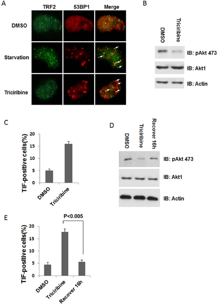
It is plausible that proliferative stimuli may activate protective mechanisms for telomeres to ensure prolonged growth. Conversely, nutritional stress may prove deleterious for telomeres. Indeed, when human HTC75 cells were grown under starvation conditions (0.01% serum), we noticed increased 53BP1 foci at telomeres that accompanied growth inhibition in these cells (Figure 1A). These observations suggest that telomeres may be sensitive to changes in nutrients, and that cell growth signaling may play a positive role in telomere maintenance. Given the importance of Akt in mediating cellular growth responses, we first determined its role in telomere end protection under growth conditions. We treated cells with triciribine, an Akt specific inhibitor, and analyzed telomere dysfunction induced foci (TIF) in these cells. As shown in Figure 1B, treatment with triciribine led to reduced Akt phosphorylation, indicating inhibition of Akt activity and its signaling. Concurrently, we found a ~3 fold increase in the percentage of TIF-positive cells in triciribine-treated cells (Figure 1C), supporting the idea that Akt activity is important for telomere protection.
Figure 1. Akt activity is important for telomere end protection.
(A) TIF analysis of HTC75 cells serum starved (0.01% FBS) for 16 h or treated with triciribine (1µM) for 3 hours. Cells were immunostained with anti-53BP1 (red) and TRF2 (green) antibodies. DMSO-treated cells were used as controls. Arrows indicate overlapping foci. (B) Western blot analysis was carried out using HTC75 cells treated with DMSO or triciribine (1µM) for 3 hours) with the indicated antibodies. (C) Cells from (B) were immunostained as in (A) and the percentage of TIF-positive cells was quantified. Only cells with >4 co-localized foci were scored. Error bars indicate s.e.m. (n=3). (D) HTC75 cells were treated with triciribine for 3 hours, and then maintained for another 16 hours in the absence of triciribine. Western blot analysis was then carried out using the indicated antibodies. (E) Cells from (D) were examined by immunostaining as described in (A) and quantitated. Error bars indicate s.e.m. (n=3). P-value was determined by the Student t-test.
Removing triciribine can reverse its inhibition of Akt and allow cells to resume growth and proliferation. We next examined whether telomere damage as a result of Akt inhibition would persist in cells after the inhibitor was removed. As expected, Akt activity recovered (as indicated by Akt phosphorylation) following removal of triciribine (Figure 1D). This recovery coincided with the decrease in TIF formation in these cells (from 17.8% to 5.6%) (Figure 1E). In fact, we were able to observe fewer telomeric DNA damage foci as early as 3 hours after inhibitor removal (data not shown). These results support the notion that Akt plays a role in protecting telomeres, and that telomere dysfunction as a result of Akt inhibition may be a reversible process.
Akt activity is important for TPP1 and POT1 recruitment to the telomeres
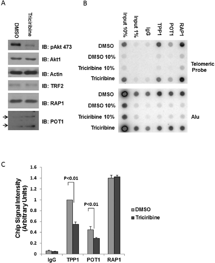
Akt may exert its protective activity through up-regulating total amounts of telomeric proteins, consequently Akt inhibition may down-regulate telomere protein levels. However, we did not observe any drastic changes after triciribine treatment in the levels of core telomeric proteins that we examined (Figure 2A). We therefore speculated that Akt might protect telomeres by promoting the telomeric recruitment of the telosome/shelterin complex. When Akt is inhibited, reduced association of telomeric proteins would lead to exposed telomere ends. Loss of telomere end protection as a result of compromised telomeric protein targeting has been well established. For example, we have shown previously that loss of TPP1 can decrease POT1 telomere recruitment and increase telomere damage (Liu et al., 2004). To test our hypothesis, we performed chromatin immunoprecipitation (ChIP) assays to examine association of core telomere proteins with telomeres in cells treated with triciribine. Of the six core telomeric proteins, TPP1 and POT1 were the only ones that consistently exhibited diminished telomere binding with drug treatment (Figure 2B,C), lending support to the notion that Akt may positively regulate the concentration of TPP1 and POT1 at telomeres.
Figure 2. Telomeric recruitment of TPP1 and POT1 requires Akt activity.
(A) HTC75 cells treated with triciribine or mock treated with DMSO for 3 hours were analyzed by western blotting using the indicated antibodies. Actin was used as loading control. Arrows indicate the two POT1 isoforms. (B) Cells from (A) were examined by telomere chromatin immunoprecipitation (ChIP) using antibodies against TPP1, POT1 and RAP1, followed by dot-blotting with probes against telomere sequences or Alu repeats. Rabbit IgG served as control. (C) Quantification of data from (B) (two independent experiments). ChIP signal intensities were normalized against input DNA. Error bars represent s.d. P-values were determined by the Student t-test.
The Akt1 isoform is the predominant player in telomere protection
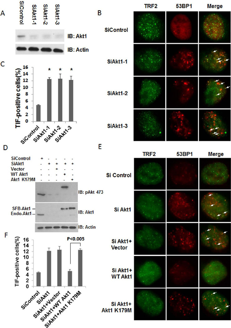
Of the three Akt isoforms (Akt1-3), Akt1 appears the most widely expressed and the best studied to date. In HTC75 cells, Akt1 is highly expressed (Kim et al., 2001). When we transiently transfected three different siRNAs specific for Akt1 into these cells, all three oligos achieved >80% knockdown efficiency (Figure 3A). Importantly, depletion of Akt1 by all three siRNAs elevated TIF formation (Figure 3B,C). In comparison, knocking down Akt2 and Akt3 isoforms had little effect on TIF formation in these cells (suppl. Figure S1), suggesting that Akt1 is the key Akt isoform that mediates telomere end protection.
Figure 3. Akt1 participates in telomere protection.
(A) HTC75 cells transiently transfected with three different siRNA oligos against Akt1 were examined by western blotting. Actin was used as loading control. (B) Cells from (A) were examined by immunostaining using anti-53BP1 (red) and TRF2 (green) antibodies. Arrows indicate overlapping foci. (C) Quantification of data from (B). Only cells with >4 co-localized foci were scored. Error bars indicate s.e.m. (n=3). The symbol * P-values (P <0.005) were determined by the Student t-test. (D) HTC75 cells were transfected with a siRNA oligo against Akt1 (siAkt1-1) in combination with siRNA-resistant SFB-tagged wild-type (WT) Akt1 or kinase-dead Akt1 (Akt1 K179M), and analyzed by western blotting using the indicated antibodies. Actin was used as loading control. Bands that correspond to exogenous and endogenous (Endo) Akt were indicated. (E) Cells from (D) were examined by immunostaining using anti-53BP1 (red) and TRF2 (green) antibodies. Arrows indicate overlapping foci. (F) Quantification of data from (E). Only cells with >4 co-localized foci were scored. Error bars indicate s.e.m. (n=3). P-values were determined by the Student t-test.
To further explore the role of Akt1 activity in telomere protection, we ectopically expressed RNAi-resistant wild-type Akt1 as well as the kinase-dead Akt1 mutant (Akt1 K179M) in Akt1 knockdown cells. Both proteins were expressed at levels comparable to endogenous Akt (Figure 3D). Furthermore, only endogenous and wild-type Akt could be blotted with the anti-phosphoAkt (residue S473) antibody, indicating that Akt1-K179M was indeed inactive. As shown in Figure 3E and F, expression of wild-type Akt1, but not kinase-dead Akt1, could rescue the TIF phenotype of the Akt1 knockdown cells, further supporting the notion that Akt1 is critical for telomere protection and underscoring the importance of its kinase activity in this process.
TPP1 can homodimerize in vivo
Extensive studies have demonstrated the importance and function of multiple pair-wise interactions within the telosome/shelterin complex. By binding to multiple telosome subunits, TIN2 acts as the linchpin for telosome/shelterin assembly, bridging double and single-stranded DNA binding activities (O'Connor et al., 2006; Takai et al., 2011). TIN2-TPP1 interaction ensures TPP1 targeting to the telomeres, and the heterodimer of TPP1-POT1 in turn helps recruit the telomerase and regulate its activity and processivity (Liu et al., 2004; Nandakumar et al., 2012; Wang et al., 2007; Xin et al., 2007; Zhang et al., 2013; Zhong et al., 2012). The disruption of such interactions can compromise telomere length control and end protection.
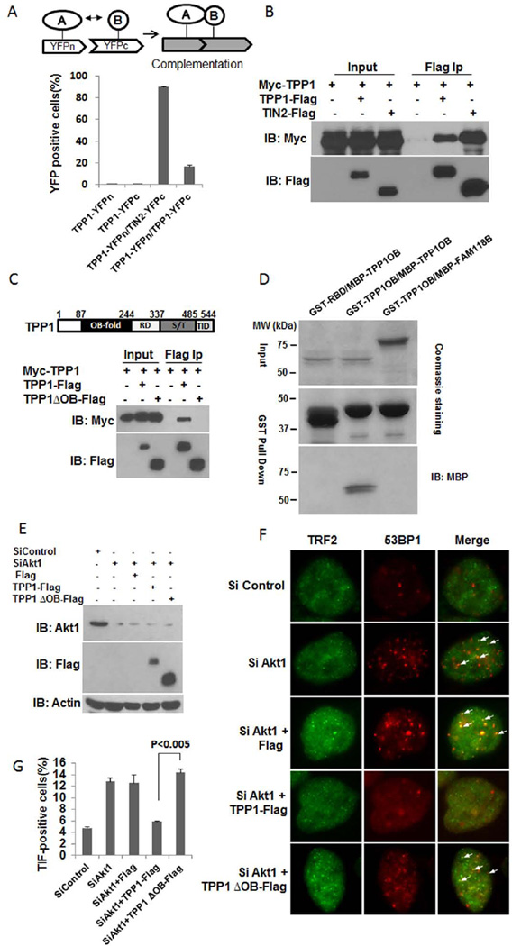
Interactions between telomere proteins can be visualized in live cells using the Bi-molecular Fluorescence Complementation (BiFC/PCA) assay (Kim et al., 2009; Lee et al., 2011). In an YFP-based BiFC assay (Figure 4A), the interaction between two proteins that are tagged respectively with split YFP fragments brings the YFP fragments to close proximity for co-folding and fluorescence complementation (Hu et al., 2002; Wilson et al., 2004). As expected, cells co-expressing the TPP1-TIN2 pair displayed strong YFP fluorescence complementation (Figure 4A). Interestingly, we were also able to detect fluorescence in cells stably expressing the TPP1-TPP1 pair, suggesting homodimerization of the TPP1 protein. To confirm TPP1 dimer formation, we also carried out immunoprecipitation experiments using cells co-expressing Myc- and Flag-tagged TPP1. As shown in Figure 4B, anti-Flag IP could bring down Flag-TPP1 as well as Myc-TPP1, providing further evidence for the ability of TPP1 to homodimerize in vivo.
Figure 4. TPP1 can homodimerize through its OB fold.
(A) HTC75 cells co-expressing YFPn-tagged TPP1 with YFPc-tagged TPP1 or TIN2 were examined by BiFC assays. The percentage of cells displaying fluorescence complementation was quantitated by flow cytometry. Error bars indicate standard error (n=3). (B) 293T cells transiently co-expressing Myc-tagged TPP1 with Flag-tagged TPP1 or TIN2 were immunoprecipitated with anti-Flag antibodies. The immunoprecipitates were western blotted as indicated. (C) 293T cells transiently co-expressing Myc-tagged TPP1 with Flag-tagged TPP1 or TPP1 OB-fold deletion mutant (TPP1ΔOB) were immunoprecipitated with anti-Flag antibodies. The immunoprecipitates were western blotted as indicated. (D) Bacterially purified GST-tagged TPP1 OB fold only mutant (TPP1 OB) was incubated with MBP-tagged TPP1 OB for GST pull-down assays. The precipitates were resolved by SDS-PAGE and visualized by Coomassie staining or western blotting. GST-tagged Raf-1 Ras-binding domain (RBD) and MBP-tagged FAM118B were used as controls. (E) HTC75 cells were transfected with siRNA oligos against Akt1 (siAKT1-1) in combination with Flag-tagged wildtype TPP1 or TPP1 OB fold deletion mutant (TPP1ΔOB), and then analyzed by Western blotting using the indicated antibodies. Actin was used as loading control. (F) Cells from (E) were examined by immunostaining using anti-53BP1 (red) and TRF2 (green) antibodies. Arrows indicate overlapping foci. (G) Quantification of data from (F). Only cells with >4 co-localized foci were scored. Error bars indicate s.e.m. (n=3). P-values were determined by the Student t-test.
The TPP1 OB fold can mediate TPP1 homodimerization
Previous studies have demonstrated that TPP1 utilizes multiple domains for its interaction with other proteins. When we tested the N-terminal OB fold deletion mutant of TPP1 (TPP1-ΔOB), we found that it lost the ability to homodimerize (Figure 4C), indicating that the requirement of the OB fold for TPP1-TPP1 interaction.
To further confirm the role of OB fold in mediating TPP1 homodimerization, we carried out in vitro GST pull-down assays using bacterially expressed GST or MBP-tagged TPP1 OB fold proteins. As controls, MBP-tagged FAM118B (a membrane protein) and GST-tagged Ras-binding domain (RBD) of Raf-1 were also included. As shown in Figure 4D, GST-TPP1 OB was able to precipitate MBP-tagged TPP1 OB, indicating that the OB fold is sufficient for mediating TPP1 homodimerization.
We reasoned that Akt might regulate telomeres through core telomere-binding proteins such as TPP1. To test this idea, we ectopically expressed full-length and OB-fold deletion mutant of TPP1 in Akt1 knockdown cells (Figure 4E) and assessed TIF formation. As shown in Figure 4F and G, expression of wild-type TPP1 in Akt1-depleted cells led to a reduction of TIFs to levels comparable to control cells, supporting the idea that Akt1 can function through TPP1 to regulate telomeres. In addition, deletion of the OB fold abolished the rescue ability of TPP1, again confirming the importance of OB fold and suggesting that OB-fold dependent dimerization of TPP1 might be important for telomere protection.
TPP1 homodimerization depends on Akt1 activity
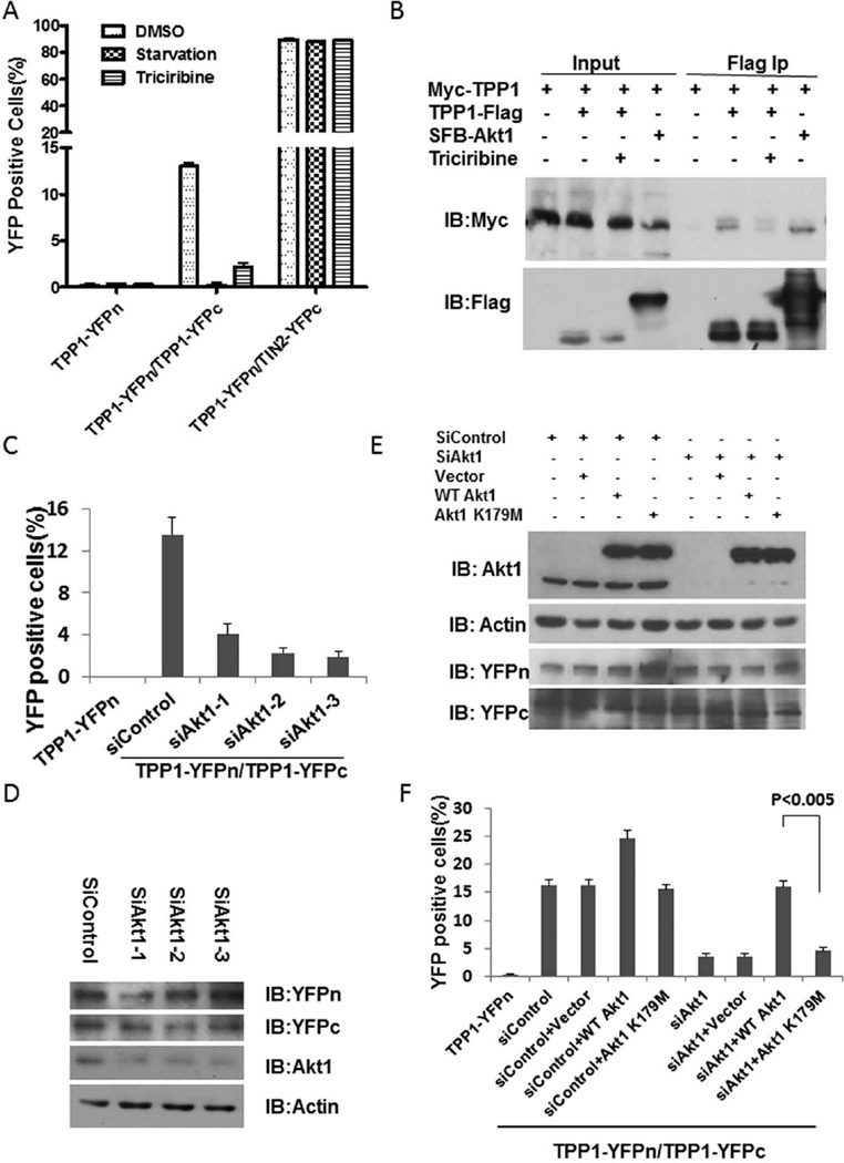
Growth arrest signals often set off cascades of cellular events that lead from inhibition of PI 3-kinase/Akt to dysfunction at the telomeres, perhaps by altering the concentration (or composition) of telomere-bound proteins and protein complexes. Such alterations can be revealed using the BiFC assay, offering clues to the dynamics of the telosome/shelterin complex. To this end, we compared YFP fluorescence complementation signals in HTC75 cells co-expressing YFPn-tagged TPP1 with YFPc-tagged TPP1 or TIN2, under serum starvation conditions or with triciribine. As shown in Figure 4A, the percentage of YFP+ TPP1-TPP1 co-expressing cells was considerably lower than TPP1-TIN2 co-expressing cells. However, only the interaction between TPP1 and TPP1, not TPP1 and TIN2, was sensitive to serum starvation or triciribine treatment (Figure 5A). In support of this observation, co-immunoprecipitation of Myc-TPP1 and TPP1-Flag was also sensitive to triciribine (Figure 5B), suggesting that Akt is important for regulating TPP1 homodimerization. In fact, we found Akt1 could interact with TPP1. As shown in Figure 5B, Myc-TPP1 could co-immunoprecipitate with SFB-Akt1. When Akt1 knockdown cells were examined, TPP1-TPP1 BiFC complementation was abolished with all three siRNAs (Figure 5C,D).
Figure 5. Akt1 can regulate TPP1 homodimerization.
(A) HTC75 cells co-expressing YFPn-tagged TPP1 with YFPc-tagged TPP1 or TIN2 were either serum starved (0.01% FBS) or treated with triciribine, and then examined by BiFC assays. Error bars indicate standard error (n=3). (B) 293T cells transiently co-expressing TPP1-Flag with Myc-TPP1 were treated with DMSO or triciribine. Cells co-expressing SFB-Akt1 and Myc-TPP1 were also examined. Cell lysates were immunoprecipitated with anti-Flag antibodies and blotted as indicated. (C) HTC75 cells co-expressing YFPn-TPP1 with YFPc-TPP1 were transiently transfected with siRNA oligos against Akt1 and analyzed in BiFC assays. Error bars indicate standard errors (n=3). (D) Cells from (C) were examined by western blotting for Akt1 knockdown efficiency. Actin was used as loading control. (E) HTC75 cells co-expressing YFPn-TPP1 and YFPc-TPP1 were transiently co-transfected with control oligos or siRNA oligos against Akt1 in combination with siRNA-resistant SFB-tagged wildtype (WT) Akt1 or kinase-dead Akt1 (Akt1 K179M). The cells were then examined by western blotting using the indicated antibodies. Actin was used as a loading control. (F) Cells from (E) were analyzed in BiFC assays. P-values were determined by the Student t-test.
To further investigate the requirement of Akt1 kinase activity in regulating TPP1 dimer formation, we transiently introduced SFB-tagged wild-type and kinase-dead Akt1 into cells already co-expressing differently tagged TPP1 proteins and carried out BiFC assays (Figure 5E). Consistent with the observation that Akt inhibition could block the formation of TPP1 dimers, rescue expression of wild-type Akt1 was able to restore TPP1 homodimerization to levels similar to control cells (Figure 5F). In fact, in control knockdown cells, ectopic expression of Akt1 enhanced TPP1 dimer formation as well. These data support the notion that Akt1 may participate in telomere protection through regulating TPP1 homodimerization. Importantly, an active Akt1 kinase is required in this process, because the kinase-dead mutant Akt1 K179M failed to rescue TPP1-TPP1 interaction in the BiFC assay (Figure 5F). Collectively, these results suggest that TPP1 homodimerization may promote telosome/shelterin integrity and increase TPP1 concentration at telomeres, a process regulated by cell growth signaling pathways via Akt activation. As a result, perturbations in Akt signaling pathway may impact telomere protection through changes in TPP1 recruitment at the telomeres.
Discussion
In this study, we linked growth signaling pathways mediated by Akt to telomere maintenance. We provide evidence that Akt may directly regulate telomere protection by promoting homodimerization of TPP1. This is the first study to report TPP1 dimer formation. It is likely that increased TPP1 homodimerization would increase the amount of TPP1 at telomeres, which in turn would also enhance POT1 binding on single-stranded telomere DNA, augmenting telomere protection and maintenance. Indeed, when Akt signaling was inhibited, we observed reduced targeting of both TPP1 and POT1 to telomeres, which was accompanied by increased TIF formation.
Compared to its other cellular functions, the precise role of Akt in telomere protection is less well understood. Here we provide additional evidence that indicates a direct role of Akt in protecting chromosomal ends -- positively regulating the concentration of TPP1 and POT1 at the telomeres and promoting telosome/shelterin assembly. In addition to TPP1, Akt may also regulate the assembly and function of the telosome/shelterin through other telosome/shelterin subunits, e.g., TRF1 (Chen et al., 2009). Moreover, using epitope-tagged hTERT, it was shown that Akt phosphorylation of the nuclear localization signal of hTERT was critical for hTERT nuclear targeting (Chung et al., 2012; Kang et al., 1999). These findings suggest that alternative pathways are utilized to integrate signaling cascades that can connect extracellular stimuli to nuclear events, for telomere maintenance in response to cell growth and proliferation.
The three isoforms of Akt (Akt1-3) are encoded by separate genes and share extensive amino acid sequence identity (especially in the kinase domain), with virtually indistinguishable in vitro substrate specificities (Walker et al., 1998). Growing evidence points to distinct function of the three Akt isoforms, likely due to their cell/tissue-specific expression and subcellular localization patterns. This possibility is also supported by our findings that inhibition of Akt2 and Akt3 did not appear to affect telomere protection. It is also possible that due to its high expression in the cells we tested, Akt1 may be the predominant isoform for many cellular processes including telomere protection.
Our study revealed a novel link between telomere maintenance and the PI3-K/Akt pathway. Under normal growth conditions, the activity of Akt ensures telomere integrity and protection that support cell growth and prevent cellular senescence. Conversely, unstable telomeres as a result of attenuated Akt activity from nutritional stress may facilitate growth arrest and checkpoint response. Understanding how such checks and balances may be compromised in human cells will facilitate our quest for uncovering mechanisms of oncogenesis and aging and identifying new treatment and therapeutics.
Experimental Procedures
Vectors, siRNAs, and Bi-molecular Fluorescence Complementation (BiFC) Assay
cDNAs encoding various human wild-type or mutant Akt1, TPP1, and TIN2 were cloned into pBabe- or pCL-based retroviral vectors. The TPP1 OB fold deletion mutant was generated by deleting amino acids 1–240. TPP1 OB contains residues 87–240. TPP1 OB fold alone (residues 87 to 251) were tagged with either GST or MBP for bacterial expression. Human Raf-1 Ras-binding domain (RBD, residues 1– 149) and full-length FAM118B were respectively tagged with GST and maltose-binding protein (MBP) for bacterial expression.
Cells expressing protein pairs respectively tagged with YFPc (residues 156–239 of YFP) and YFPn (residues 1–155 of Venus YFP) were analyzed by flow cytometry as previously described (Lee et al., 2011). 293T cells were used for transient expression and retroviral packaging. HTC75 cells were used for stable protein expression.
siRNAs against Akt1, Akt2, Akt3 were purchased from Sigma:
siAkt1-1: SASI_Hs01_00105952; siAkt1-2 SASI_Hs01_00105953; and siAkt1-3 SASI_Hs01_00105954.
siAkt2-1: SASI_Hs01_00035055; siAkt2-2 SASI_Hs01_00035057.
siAkt3-1: SASI_Hs01_00122808; siAkt3-2: SASI_Hs01_00122809; siAkt3-3: SASI_Hs01_00122810.
For RNAi-resistant expression of Akt1 constructs, we used siAkt1-1 that targets the 3’UTR of Akt1 mRNA to knockdown Akt1.
RT-qPCR
Total RNAs were isolated using the RNeasy mini kit (Qiagen). Reverse transcription was performed using iScript Select cDNA synthesis kit (Bio-Rad). Real-time PCR was performed using an ABI StepOnePlus real-time PCR system and SYBR Green Master Mix (Invitrogen).
Telomere dysfunction-induced foci (TIF) assays
TIF assays were performed essentially as described previously (Kim et al., 2009). Briefly, cells grown overnight on cover slips were first permeabilized with 0.5% TritonX-100 in 1× phosphate-buffered saline (PBS), fixed with 4% paraformaldehyde, and then permeabilized again with 0.5% TritonX-100 plus 300mM sucrose. The cells were subsequently co-immunostained with anti-53BP1 (Novus, NB100-304) and anti-TRF2 (Calbiochem, OP129) antibodies, and visualized under a Nikon TE200 fluorescence microscope.
Co-immunoprecipitation and Western blotting
To prepare cell lysates for co-immunoprecipitation, cells were lysed in NETN (20 mM Tris (pH8.0), 1 mM EDTA, 100 mM NaCl, 0.5% NP-40) buffer supplemented with protease inhibitor cocktails (Sigma) and subjected to centrifuge at 14,000 g for 30 min. After centrifugation, the supernatant was used for immunoprecipitation with anti-Flag M2 affinity resins (Sigma) on ice for 1h. After four washes with NETN buffer, the proteins were eluted with Flag peptide (200 µg/ml), resolved by SDS-PAGE, and western blotted.
The antibodies used are: polyclonal anti-Flag (Sigma, F7425), anti-Flag M2 (Sigma, A8592), polyclonal anti-GFP (Abcam, ab290), monoclonal anti-GFP (N-term) (EPITOMICS, 1533-1), anti-hTRF2 (CalBiochem, OP129), anti-Akt (Cell Signaling Technology, 40D4), anti-phospho-Akt (Ser473) (Cell Signaling Technology, 9271), polyclonal anti-RAP1 (Bethyl Laboratories), anti-POT1 (Novus, NB500), anti-MBP (Millipore, 05-499), anti-Myc (Santa Cruz Biotechnology, sc-40), and anti-actin (Sigma, A3853).
ChIP
Chromatin immunoprecipitation (ChIP) analysis was performed as described previously (Liang et al., 2008). Briefly, cells were fixed in 1% formaldehyde and lysed in 1% Triton X-100, 150 mM NaCl, and 2 mM EDTA, and 50 mM HEPES (pH7.5). Sonicated lysates were incubated with the appropriate antibodies (3µg), and the DNA was eluted in 0.5%SDS and 0.1 M NaHCO3 and purified with the QIAquick PCR purification kit (Qiagen). The DNA was slot-blotted onto a Hybond N+ membrane and hybridized with a telomeric G-rich probe (5'-TTAGGGTTAGGGTTAGGGT-3') or Alu probe (5'-GGCCGGGCGCGGTGGCTCACGCCTGTAATCCCAGCA-3'). The signals were quantitated using a PhosphorImager. The antibodies used for ChIP are: rabbit polyclonal POT1 (N-terminal) (O'Connor et al., 2006), rabbit polyclonal TPP1 (467) (Liu et al., 2004), polyclonal anti-RAP1 (Bethyl Laboratories) and rabbit IgG (Santa Cruz, SC2027).
In vitro binding assays
GST-tagged TPP1 OB fold and Raf-1 Ras-binding domain (RBD) proteins, and maltose-binding protein (MBP)-tagged TPP1 OB fold and FAM118B proteins were purified from Escherichia coli BL21 cells. GST-TPP1 OB was first immobilized on glutathione-sepharose 4B beads and then incubated with MBP-TPP1 OB for 2 h at 4°C. The beads were subsequently washed and used for SDS-PAGE followed by Coomassie staining and western blotting analysis.
Supplementary Material
Supplementary Figure S1. Depletion of Akt2 or Akt3 did not affect telomere protection. (A) HTC75 cells transiently transfected with two different siRNA oligos against Akt2 were examined by RT-qPCR. Error bars indicate standard error (n=3). (B) Cells from (A) were examined by immunostaining using anti-53BP1 (red) and TRF2 (green) antibodies. (C) Quantification of data from (B). Only cells with >4 co-localized foci were scored. Error bars indicate s.e.m. (n=3). (D) HTC75 cells transiently transfected with three different siRNA oligos against Akt3 were examined by RT-qPCR. Error bars indicate standard error (n=3). (E) Cells from (D) were examined by immunostaining using anti-53BP1 (red) and TRF2 (green) antibodies. (F) Quantification of data from (E). Only cells with >4 co-localized foci were scored. Error bars indicate s.e.m. (n=3).
Acknowledgements
We thank Drs. Hyeung Kim, Jun Xu, and Ka-wing Fong for technical help. This study was supported by the National Basic Research Program (973 Program) (2010CB945401 and 2012CB911201), National Natural Science Foundation (91019020 and 91213302), Specialized Research Fund for the Doctoral Program of Higher Education (20100171110028), Introduced Innovative R&D Team of Guangdong Province (201001Y0104687244), and Changjiang Scholars Programme of China. We would also like to acknowledge the support of NCI CA133249, NIGMS GM095599, the Welch Foundation Q-1673, and the GRSA Shared Resource at the Dan L. Duncan Cancer Center (P30CA125123). The project described was also supported in part by BCM IDDRC (5P30HD024064) from the Eunice Kennedy Shriver National Institute of Child Health & Human Development. The content is solely the responsibility of the authors and does not necessarily represent the official views of the Eunice Kennedy Shriver National Institute of Child Health & Human Development or the National Institutes of Health.
Footnotes
Author Contributions
D.L., J.C., and Z.S. designed the experiments. X.H. performed the experiments. Y.Z. and Y.L. prepared the samples for GST in vitro pull down assays. W.L. prepared samples for RT-qPCR. X.H., D.L., and Z.S. wrote the manuscript.
Authors’ Conflict of Interest Statement
The authors declare no conflict of interest.
References
- Armanios M, Blackburn EH. The telomere syndromes. Nat Rev Genet. 2012;13:693–704. doi: 10.1038/nrg3246. [DOI] [PMC free article] [PubMed] [Google Scholar]
- Carnero A. The PKB/AKT pathway in cancer. Curr Pharm Des. 2010;16:34–44. doi: 10.2174/138161210789941865. [DOI] [PubMed] [Google Scholar]
- Chen YC, Teng SC, Wu KJ. Phosphorylation of telomeric repeat binding factor 1 (TRF1) by Akt causes telomere shortening. Cancer Invest. 2009;27:24–28. doi: 10.1080/07357900802027081. [DOI] [PubMed] [Google Scholar]
- Chung J, Khadka P, Chung IK. Nuclear import of hTERT requires a bipartite nuclear localization signal and Akt-mediated phosphorylation. J Cell Sci. 2012;125:2684–2697. doi: 10.1242/jcs.099267. [DOI] [PubMed] [Google Scholar]
- de Lange T. Shelterin: the protein complex that shapes and safeguards human telomeres. Genes Dev. 2005;19:2100–2110. doi: 10.1101/gad.1346005. [DOI] [PubMed] [Google Scholar]
- Deng R, Tang J, Ma JG, Chen SP, Xia LP, Zhou WJ, Li DD, Feng GK, Zeng YX, Zhu XF. PKB/Akt promotes DSB repair in cancer cells through upregulating Mre11 expression following ionizing radiation. Oncogene. 2011;30:944–955. doi: 10.1038/onc.2010.467. [DOI] [PubMed] [Google Scholar]
- Deng Y, Chan SS, Chang S. Telomere dysfunction and tumour suppression: the senescence connection. Nat Rev Cancer. 2008;8:450–458. doi: 10.1038/nrc2393. [DOI] [PMC free article] [PubMed] [Google Scholar]
- Donate LE, Blasco MA. Telomeres in cancer and ageing. Philos Trans R Soc Lond B Biol Sci. 2011;366:76–84. doi: 10.1098/rstb.2010.0291. [DOI] [PMC free article] [PubMed] [Google Scholar]
- Frias C, Pampalona J, Genesca A, Tusell L. Telomere dysfunction and genome instability. Front Biosci. 2012;17:2181–2196. doi: 10.2741/4044. [DOI] [PubMed] [Google Scholar]
- Haendeler J, Hoffmann J, Rahman S, Zeiher AM, Dimmeler S. Regulation of telomerase activity and anti-apoptotic function by protein-protein interaction and phosphorylation. FEBS Lett. 2003;536:180–186. doi: 10.1016/s0014-5793(03)00058-9. [DOI] [PubMed] [Google Scholar]
- Heiss NS, Knight SW, Vulliamy TJ, Klauck SM, Wiemann S, Mason PJ, Poustka A, Dokal I. X-linked dyskeratosis congenita is caused by mutations in a highly conserved gene with putative nucleolar functions. Nat Genet. 1998;19:32–38. doi: 10.1038/ng0598-32. [DOI] [PubMed] [Google Scholar]
- Hu CD, Chinenov Y, Kerppola TK. Visualization of interactions among bZIP and Rel family proteins in living cells using bimolecular fluorescence complementation. Mol Cell. 2002;9:789–798. doi: 10.1016/s1097-2765(02)00496-3. [DOI] [PubMed] [Google Scholar]
- Kang SS, Kwon T, Kwon DY, Do SI. Akt protein kinase enhances human telomerase activity through phosphorylation of telomerase reverse transcriptase subunit. J Biol Chem. 1999;274:13085–13090. doi: 10.1074/jbc.274.19.13085. [DOI] [PubMed] [Google Scholar]
- Kim D, Kim S, Koh H, Yoon SO, Chung AS, Cho KS, Chung J. Akt/PKB promotes cancer cell invasion via increased motility and metalloproteinase production. FASEB J. 2001;15:1953–1962. doi: 10.1096/fj.01-0198com. [DOI] [PubMed] [Google Scholar]
- Kim H, Lee OH, Xin H, Chen LY, Qin J, Chae HK, Lin SY, Safari A, Liu D, Songyang Z. TRF2 functions as a protein hub and regulates telomere maintenance by recognizing specific peptide motifs. Nat Struct Mol Biol. 2009;16:372–379. doi: 10.1038/nsmb.1575. [DOI] [PubMed] [Google Scholar]
- Kloet DE, Burgering BM. The PKB/FOXO switch in aging and cancer. Biochim Biophys Acta. 2011;1813:1926–1937. doi: 10.1016/j.bbamcr.2011.04.003. [DOI] [PubMed] [Google Scholar]
- Lee OH, Kim H, He Q, Baek HJ, Yang D, Chen LY, Liang J, Chae HK, Safari A, Liu D, et al. Genome-wide YFP fluorescence complementation screen identifies new regulators for telomere signaling in human cells. Mol Cell Proteomics. 2011;10 doi: 10.1074/mcp.M110.001628. M110 001628. [DOI] [PMC free article] [PubMed] [Google Scholar]
- Liang J, Wan M, Zhang Y, Gu P, Xin H, Jung SY, Qin J, Wong J, Cooney AJ, Liu D, et al. Nanog and Oct4 associate with unique transcriptional repression complexes in embryonic stem cells. Nat Cell Biol. 2008;10:731–739. doi: 10.1038/ncb1736. [DOI] [PubMed] [Google Scholar]
- Liu D, Safari A, O'Connor MS, Chan DW, Laegeler A, Qin J, Songyang Z. PTOP interacts with POT1 and regulates its localization to telomeres. Nat Cell Biol. 2004;6:673–680. doi: 10.1038/ncb1142. [DOI] [PubMed] [Google Scholar]
- Manning BD, Cantley LC. AKT/PKB signaling: navigating downstream. Cell. 2007;129:1261–1274. doi: 10.1016/j.cell.2007.06.009. [DOI] [PMC free article] [PubMed] [Google Scholar]
- Nandakumar J, Bell CF, Weidenfeld I, Zaug AJ, Leinwand LA, Cech TR. The TEL patch of telomere protein TPP1 mediates telomerase recruitment and processivity. Nature. 2012;492:285–289. doi: 10.1038/nature11648. [DOI] [PMC free article] [PubMed] [Google Scholar]
- Nandakumar J, Cech TR. Finding the end: recruitment of telomerase to telomeres. Nat Rev Mol Cell Biol. 2013;14:69–82. doi: 10.1038/nrm3505. [DOI] [PMC free article] [PubMed] [Google Scholar]
- O'Connor MS, Safari A, Xin H, Liu D, Songyang Z. A critical role for TPP1 and TIN2 interaction in high-order telomeric complex assembly. Proc Natl Acad Sci U S A. 2006;103:11874–11879. doi: 10.1073/pnas.0605303103. [DOI] [PMC free article] [PubMed] [Google Scholar]
- O'Sullivan RJ, Karlseder J. Telomeres: protecting chromosomes against genome instability. Nat Rev Mol Cell Biol. 2010;11:171–181. doi: 10.1038/nrm2848. [DOI] [PMC free article] [PubMed] [Google Scholar]
- Palm W, de Lange T. How shelterin protects mammalian telomeres. Annu Rev Genet. 2008;42:301–334. doi: 10.1146/annurev.genet.41.110306.130350. [DOI] [PubMed] [Google Scholar]
- Sasa GS, Ribes-Zamora A, Nelson ND, Bertuch AA. Three novel truncating TINF2 mutations causing severe dyskeratosis congenita in early childhood. Clin Genet. 2012;81:470–478. doi: 10.1111/j.1399-0004.2011.01658.x. [DOI] [PMC free article] [PubMed] [Google Scholar]
- Savage SA, Giri N, Baerlocher GM, Orr N, Lansdorp PM, Alter BP. TINF2, a component of the shelterin telomere protection complex, is mutated in dyskeratosis congenita. Am J Hum Genet. 2008;82:501–509. doi: 10.1016/j.ajhg.2007.10.004. [DOI] [PMC free article] [PubMed] [Google Scholar]
- Shay JW, Wright WE. Role of telomeres and telomerase in cancer. Semin Cancer Biol. 2011;21:349–353. doi: 10.1016/j.semcancer.2011.10.001. [DOI] [PMC free article] [PubMed] [Google Scholar]
- Takai KK, Kibe T, Donigian JR, Frescas D, de Lange T. Telomere protection by TPP1/POT1 requires tethering to TIN2. Mol Cell. 2011;44:647–659. doi: 10.1016/j.molcel.2011.08.043. [DOI] [PMC free article] [PubMed] [Google Scholar]
- Tzivion G, Hay N. PI3K-AKT-FoxO axis in cancer and aging. Biochim Biophys Acta. 2011;1813:1925. doi: 10.1016/j.bbamcr.2011.08.014. [DOI] [PubMed] [Google Scholar]
- Vulliamy T, Beswick R, Kirwan M, Marrone A, Digweed M, Walne A, Dokal I. Mutations in the telomerase component NHP2 cause the premature ageing syndrome dyskeratosis congenita. Proc Natl Acad Sci U S A. 2008;105:8073–8078. doi: 10.1073/pnas.0800042105. [DOI] [PMC free article] [PubMed] [Google Scholar]
- Walker KS, Deak M, Paterson A, Hudson K, Cohen P, Alessi DR. Activation of protein kinase B beta and gamma isoforms by insulin in vivo and by 3-phosphoinositide-dependent protein kinase-1 in vitro: comparison with protein kinase B alpha. Biochem J. 1998;331(Pt 1):299–308. doi: 10.1042/bj3310299. [DOI] [PMC free article] [PubMed] [Google Scholar]
- Walne AJ, Vulliamy T, Beswick R, Kirwan M, Dokal I. TINF2 mutations result in very short telomeres: analysis of a large cohort of patients with dyskeratosis congenita and related bone marrow failure syndromes. Blood. 2008;112:3594–3600. doi: 10.1182/blood-2008-05-153445. [DOI] [PMC free article] [PubMed] [Google Scholar]
- Wang F, Podell ER, Zaug AJ, Yang Y, Baciu P, Cech TR, Lei M. The POT1-TPP1 telomere complex is a telomerase processivity factor. Nature. 2007;445:506–510. doi: 10.1038/nature05454. [DOI] [PubMed] [Google Scholar]
- Wilson CG, Magliery TJ, Regan L. Detecting protein-protein interactions with GFP-fragment reassembly. Nat Methods. 2004;1:255–262. doi: 10.1038/nmeth1204-255. [DOI] [PubMed] [Google Scholar]
- Xin H, Liu D, Songyang Z. The telosome/shelterin complex and its functions. Genome Biol. 2008;9:232. doi: 10.1186/gb-2008-9-9-232. [DOI] [PMC free article] [PubMed] [Google Scholar]
- Xin H, Liu D, Wan M, Safari A, Kim H, Sun W, O'Connor MS, Songyang Z. TPP1 is a homologue of ciliate TEBP-beta and interacts with POT1 to recruit telomerase. Nature. 2007;445:559–562. doi: 10.1038/nature05469. [DOI] [PubMed] [Google Scholar]
- Xu N, Hegarat N, Black EJ, Scott MT, Hochegger H, Gillespie DA. Akt/PKB suppresses DNA damage processing and checkpoint activation in late G2. J Cell Biol. 2010;190:297–305. doi: 10.1083/jcb.201003004. [DOI] [PMC free article] [PubMed] [Google Scholar]
- Zhang Y, Chen LY, Han X, Xie W, Kim H, Yang D, Liu D, Songyang Z. Phosphorylation of TPP1 regulates cell cycle-dependent telomerase recruitment. Proc Natl Acad Sci U S A. 2013;110:5457–5462. doi: 10.1073/pnas.1217733110. [DOI] [PMC free article] [PubMed] [Google Scholar]
- Zhong F, Savage SA, Shkreli M, Giri N, Jessop L, Myers T, Chen R, Alter BP, Artandi SE. Disruption of telomerase trafficking by TCAB1 mutation causes dyskeratosis congenita. Genes Dev. 2011;25:11–16. doi: 10.1101/gad.2006411. [DOI] [PMC free article] [PubMed] [Google Scholar]
- Zhong FL, Batista LF, Freund A, Pech MF, Venteicher AS, Artandi SE. TPP1 OB-fold domain controls telomere maintenance by recruiting telomerase to chromosome ends. Cell. 2012;150:481–494. doi: 10.1016/j.cell.2012.07.012. [DOI] [PMC free article] [PubMed] [Google Scholar]
Associated Data
This section collects any data citations, data availability statements, or supplementary materials included in this article.
Supplementary Materials
Supplementary Figure S1. Depletion of Akt2 or Akt3 did not affect telomere protection. (A) HTC75 cells transiently transfected with two different siRNA oligos against Akt2 were examined by RT-qPCR. Error bars indicate standard error (n=3). (B) Cells from (A) were examined by immunostaining using anti-53BP1 (red) and TRF2 (green) antibodies. (C) Quantification of data from (B). Only cells with >4 co-localized foci were scored. Error bars indicate s.e.m. (n=3). (D) HTC75 cells transiently transfected with three different siRNA oligos against Akt3 were examined by RT-qPCR. Error bars indicate standard error (n=3). (E) Cells from (D) were examined by immunostaining using anti-53BP1 (red) and TRF2 (green) antibodies. (F) Quantification of data from (E). Only cells with >4 co-localized foci were scored. Error bars indicate s.e.m. (n=3).


