Abstract
We have shown that intranasal coapplication of Bacillus anthracis protective Ag (PA) together with a B. anthracis edema factor (EF) mutant having reduced adenylate cyclase activity (i.e., EF-S414N) enhances anti-PAAb responses, but also acts as a mucosal adjuvant for coadministered unrelated Ags. To elucidate the role of edema toxin (EdTx) components in its adjuvanticity, we examined how a PA mutant lacking the ability to bind EF (PA-U7) or another mutant that allows the cellular uptake of EF, but fails to efficiently mediate its translocation into the cytosol (PA-dFF), would affect EdTx-induced adaptive immunity. Native EdTx promotes costimulatory molecule expression by macrophages and B lymphocytes, and a broad spectrum of cytokine responses by cervical lymph node cells in vitro. These effects were reduced or abrogated when cells were treated with EF plus PA-dFF, or PA-U7 instead of PA. We also intranasally immunized groups of mice with a recombinant fusion protein of Yersinia pestis F1 and LcrVAgs (F1-V) together with EdTx variants consisting of wild-type or mutants PA and EF. Analysis of serum and mucosal Ab responses against F1-V or EdTx components (i.e., PA and EF) revealed no adjuvant activity in mice that received PA-U7 instead of PA. In contrast, coimmunization with PA-dFF enhanced serum Ab responses. Finally, immunization with native PA and an EF mutant lacking adenylate cyclase activity (EF-K346R) failed to enhance Ab responses. In summary, a fully functional PA and a minimum of adenylate cyclase activity are needed for EdTx to act as a mucosal adjuvant.
Edema toxin (EdTx) is one of the exotoxins produced by the Gram-positive, spore-forming rod Bacillus anthracis (1). EdTx is composed of two subunits. The binding subunit, or protective Ag (PA), allows the binding of these toxins to the anthrax toxin receptors that are expressed by most cells. The enzymatic subunit, or edema factor (EF), is a calmodulin- and calcium-dependent adenylate cyclase that catalyzes the conversion of ATP to cAMP (1, 2). We recently showed that intranasal coapplication of PA together with an EF mutant with reduced adenylate cyclase activity (i.e., EF-S414N) enhanced anti-PA Ab responses above levels achieved by intranasal immunization with PA alone (3). The EdTx S414N also was found to be an adjuvant that promotes mucosal and systemic immunity to intranasally coadministered unrelated vaccine Ags (3). However, the exact contribution of each subunit of EdTx to its mucosal adjuvanticity remained unclear.
Most information regarding the mechanisms employed by bacterial toxins to regulate mucosal immunity comes from studies with cholera toxin (CT) and the related heat-labile enterotoxin I of Escherichia coli (LT-I or LT). Like EdTx, these molecules are AB-type toxins. Both toxins carry an enzymatic A subunit capable of increasing cellular cAMP concentrations (4). In contrast, they target distinct cellular receptors, such as CT binding to GM1 gangliosides (5), whereas LT-I is more promiscuous and binds other gangliosides besides GM1, including asialo GM1 and GM2 (6–8). Studies have shown that the ADP-ribosyl transferase activity of enterotoxins is not required for the intranasal adjuvanticity of LT-I (9, 10) and CT (11, 12). These findings led to the concept that the ability to increase cAMP was not absolutely required for bacterial toxins to display adjuvant activity, at least by the intranasal route. How enterotoxin mutants without enzymatic activity retain adjuvant activity remains unclear, but it is believed that the presence of an A subunit improves the stability of the molecule. In this regard, the binding B subunit of CT is not an adjuvant for Ags coadministered to mice by the intranasal route. In contrast, the more promiscuous LT-B, which in addition to GM1 binds other gangliosides, was reported to exert adjuvant activity (13). The manner in which the enzymatic A subunit of enterotoxins is targeted to cells also has important implications for their adjuvant activity. For example, the CTA-1DD molecule, which targets an enzymatically active CT-A subunit to B cells, also is an adjuvant capable of promoting mucosal and systemic immunity to intranasally coadministered Ags (14). It generally is accepted that subunits of enterotoxins play a role in the polarization of immune responses toward a Th1 or a Th2 type. Studies have compared immune responses induced by the chimera of CT-A/LT-B and LT-A/CT-B as nasal adjuvants (15, 16).
This study employed mutants of PA and EF and in vitro and in vivo approaches to establish how the subcellular delivery of EF in immune cells and its adenylate cyclase activity affect the mucosal adjuvant activity of nasally administered EdTx.
Materials and Methods
Mice
Female C57BL/6 mice, 6–7 wk of age, were obtained from Charles River Laboratories (Wilmington, MA) or The Jackson Laboratory (Bar Harbor, ME) and were 9–12 wk of age when used. All studies were performed in accordance with both National Institutes of Health and Ohio State University Institutional Animal Care and Use Committee guidelines to avoid pain and distress.
Cell lines and toxins
The macrophage cell lines J774A.1 and RAW264.7, which are both derived from BALB/c mice (I-Ad), were obtained from American Type Culture Collection (Manassas, VA). Cells were maintained in RPMI 1640 medium containing 10 mM HEPES, 2 mM l-glutamine, 100 U/ml penicillin, 100 µg/ml streptomycin, 2 × 10⁻⁵ M 2-ME, 1 mM sodium pyruvate, and 10% FCS. Anthrax EdTx components and derivatives that were used are listed in Table I. The PA, PA-U7, and PA-dFF proteins were purified from cultures of a recombinant strain of B. anthracis, as previously described (17). The EF-S414NS was obtained from List Biological Laboratories (Campbell, CA; product 173) and was produced in a recombinant strain of B. anthracis, as previously noted (3). This protein was previously denoted EF-S447N (3), based on numbering that included the signal sequence. This EF-S414N mutant protein has ~20-fold less activity on cells than the native EF (18). EF wild type (EFwt) and EF-K346R were produced in E. coli, as previously described (19, 20). CT was obtained from List Biological Laboratories.
Table I.
PA and EF derivatives used in these studies
| Molecules | Characteristics | References |
|---|---|---|
| PA | wt (or native) PA | |
| PA-dFF (E308D) | Mutant with deletion of F314 and F315; internalizes EF, LF to endosomes, but does not translocate them efficiently to the cytosol (E308D has no known functional effect) | 43 |
| PA-U7 | Mutant lacking the RKKR furin cleavage site, and therefore unable to bind and internalize EF, LF | 44 |
| EF | wt (or native) EF | |
| EF-S414N | Mutant with reduced ability to increase cAMP levels in cells | 18 |
| EF-K346R | EF mutant devoid of adenylate cyclase catalytic activity due to a point mutation in the ATP binding site | 19 |
Effect of EdTx derivatives on Ag uptake and costimulatory molecule expression in vitro
Mouse macrophage cell lines RAW264.7 and J774A.1 (American Type Culture Collection) were used to address the effect of EdTx on APC functions in vitro, including Ag uptake and expression of costimulatory molecules. The cells were plated on a 24-well plate at 106 cells/ml/well and treated for 24 h with the following combination of EdTx subunits (2 µg/ml): 1) PAwt; 2) EF-S414N; 3) EFwt; 4) PAwt + EF-S414N; 5) PAwt + EFwt; 6) PA-U7 + EF-S414N; and 7) PA-dFF + EF-S414N. Medium was removed and cells were incubated for 15 min with OVA-Alexa Fluor 488 (5 µg/ml; Molecular Probes, Eugene, OR). The expression of costimulatory molecules and the uptake of OVA were assessed by flow cytometry. In separate experiments, mouse cervical lymph node and spleen cells were incubated for 6, 24, or 48 h with these toxin derivatives. Cytokine responses were evaluated by real-time RT-PCR, and the expression of costimulatory molecules was assessed by flow cytometry.
Immunostaining for flow cytometry
Cells were washed and resuspended in staining buffer (PBS, 1% BSA, 0.01% NaN3). For flow cytometry analysis, cells were incubated for 30 min at 4°C with one or a combination of the following fluorescent anti-mouse Abs: anti-CD40, anti-CD80, anti-CD86, anti–I-Ab (MHC class II), anti-CD11b, and anti-B220 (CD45; BD Biosciences, San Jose, CA). Cells were washed twice in staining buffer and analyzed by flow cytometry (BD FACSCalibur, BD Biosciences; C6 flow cytometer, Accuri Cytometers, Ann Arbor, MI). In selected experiments, cells were fixed in 1% paraformaldehyde and kept at 4°C before analysis.
Quantification of cytokine mRNA by real-time RT-PCR
Cytokine mRNA responses to EdTx components and derivatives were evaluated, as previously described (21). Cells were collected and washed in RPMI 1640 without FCS, and RNA was isolated using STAT-60 (Tel-Test, Friendswood, TX), according to the manufacturer’s instructions. Reverse transcription was performed with Superscript II reverse transcriptase, dNTPs, and oligo(dT). Real-time PCR (Stratagene, La Jolla, CA) was performed with primers generated with Oligo software (Plymouth, MN) and the SYBR green detection system, according to the manufacturer. Results are expressed as threshold cycle (Ct), defined as the cycle at which the fluorescence rises appreciably above the background fluorescence as determined by the second derivative maximum method. The following formula was used to calculate the logarithm of the relative mRNA levels of a given cytokine: 20 – (meanCtcytokine – meanCtβ-actin). This formula allows the normalization of all results against β-actin levels to correct for differences in cDNA concentration between the starting templates. In order to better visualize the effect of treatments, some mRNA responses were expressed as treatment-induced relative mRNA levels (TmRNA = 2−[ΔΔCt treated cells − ΔΔCt of nontreated cells])
Immunization
A recombinant fusion protein of Yersinia pestis F1 and LcrV Ags (F1-V; Biodefense and Emerging Infections Research Resources Repository, Manassas, VA) was used as Ag to examine the role of EdTx components in its adjuvant activity for unrelated intranasal vaccines. Briefly, mice were immunized intranasally three times at weekly intervals with 20 µg F1-V alone, or F1-V plus 2 µg EdTx consisting of PA and EFwt, or F1-V plus 2 µg PA-U7 and 2 µg EF, or F1-V plus 2 µg PA-dFF and 2 µg EF. The PA-U7 and PA-dFF (or PA-dFF E308D) molecules are mutants of PA, the first one lacking the furin site and the latter being unable to efficiently translocate EF to the cytosol. To examine the role of the adenylate cyclase activity of EF in the adjuvanticity of EdTx, mice were immunized intranasally with F1-V (20 µg), F1-V plus wt EdTx (PA plus EF; 2 µg), or F1-V plus PA plus EF-K346R. We also examined the contribution of these toxin components on the anti-PA responses. For these studies, mice were intranasally immunized with 2 µg PA alone; 2 µg PA and 2 µg EF; 2 µg PA-U7 and 2 µg EF; PA-dFF and 2 µg EF; or 2 µg PA and 2 µg EF-K346R. Mice were lightly anesthetized with a solution of ketamine/xylazine and given 10 µl vaccine per nostril. Blood and external secretions (fecal extracts, vaginal washes, and saliva) were collected, as previously described (16, 22).
Evaluation of F1-V– and PA-specific Ab isotypes and IgG subclass responses
ELISA was used to assess Ag-specific Ab levels in plasma and external secretions, as described previously (16, 22). Briefly, microtiter plates were coated with F1-V (5 µg/ml), PA (5 µg/ml), or EF (5 µg/ml) for analysis of F1-V–, PA-, and EF-specific Ab responses, respectively. The IgM, IgG, or IgA Abs were detected with HRP-conjugated goat anti-mouse µ-, γ-, or α-H chain-specific antisera (Southern Biotechnology Associates, Birmingham, AL). Biotin-conjugated rat anti-mouse IgG1 (clone A85–1), IgG2a (clone R19–15), IgG2b (clone R12–3), or IgG3 (clone R40–82) mAbs and HRP-conjugated streptavidin (BD Biosciences) were used to measure IgG subclass Ab responses. The color was developed with the addition of ABTS substrate (Sigma-Aldrich, St. Louis, MO), and the absorbance was measured at 415 nm. End-point titers were expressed as the log2 dilution giving an OD415 of ≥0.1 above those obtained with nonimmunized control mouse samples.
Macrophage toxicity assay to assess anti-PA neutralizing Abs
The protective effects of PA-specific Abs were determined, as previously described (3). Briefly, J774 macrophages (5 × 10⁴ cells/well) were added to 96-well, flat-bottom plates. After 12 h of incubation, plasma or external secretion samples were added together with lethal toxin (1 µg/ml PA plus 1 µg/ml lethal factor [LF]) and incubated for an additional 12 h, as described (3, 23). Viable macrophages were evaluated after addition of MTT (Sigma-Aldrich) (23).
Statistics
Results are expressed as the mean ± 1 SD. Unless otherwise indicated, statistical significance was determined by Student t test or by ANOVA, followed by the Fisher least significant difference test. For statistical analysis, Ab levels below the detection limit were recorded as 2 log2 below the detection limit. For analysis of mRNA responses, one-way ANOVA followed by a Duncan’s multiple range test were used to compare 20-ΔCt values among the groups (i.e., no treatment, PA, PA + EF, and mutants of PA + EF). All tests were considered significant at a probability of p ≤ 0.05. Statistical analyses were performed with the Statistica 9.0 software package (StatSoft, Tulsa, OK) or the Statview software for MacIntosh computers.
Results
Intracellular delivery of EF influences Ag uptake by macrophages
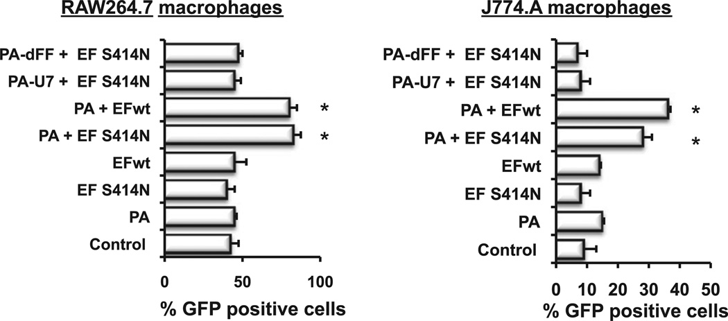
The abilities of APCs to take up Ag and express costimulatory molecules are crucial for the development of adaptive immunity after immunization. In order to elucidate the relative contribution of the cell-binding component (i.e., PA) and the enzymatic component responsible for the adjuvant activity of the B. anthracis EdTx, we first examined the uptake of a soluble Ag (i.e., OVA-Alexa Fluor 488) by macrophages exposed to mutants of PA and EF (Table I). One of the PA mutants (PA-U7) lacks the furin site, and thus cannot be proteolytically activated so as to bind EF. The other mutant (PA-dFF E308D) has amino acid changes (deletion of F314 and F315) that greatly decrease its ability to translocate EF to the cytosol (whereas the E308D substitution has no known effect). We also examined the contribution of the enzymatic subunit by using a mutant of EF (EF-S414N) that has a greatly reduced ability to increase cellular cAMP concentrations (Table I). Exposure of macrophage cell lines to PA alone, or EF alone, did not affect their ability to uptake Ag. Macrophages exposed to PA plus EF-S414N or EFwt exhibited significantly higher uptake of the soluble Ag than control-untreated cells (Fig. 1). In contrast, pretreatment of macrophages with EF-S414N and PA-dFF or PA-U7 failed to enhance uptake of the soluble Ag (Fig. 1).
FIGURE 1.
OVA uptake by macrophage cell lines treated with EdTx derivatives. Mouse macrophage cell lines RAW264.7 and J774A.1 were plated on a 24-well plate at 106 cells/ml and treated for 24 h with the following combination of EdTx subunits (2 µg/ml): 1) PAwt (PA) alone; 2) EF-S414N alone; 3) EF; 4) PA + EF-S414N; 5) PA + EF; 6) PA-U7 + EF-S414N; 7) PA-dFF + EF-S414N. Medium was removed and cells were incubated for 15 min with OVA-Alexa Fluor 488 (5 µg/ml; Molecular Probes). The uptake of OVA was assessed by flow cytometry and expressed as percentage of GFP-positive cells (n = 3 separate experiments). Results are expressed as mean ± 1 SD. Statistical significance: *p ≤ 0.05 when compared with control-untreated cells.
Intracellular delivery of EF influences costimulatory molecule expression
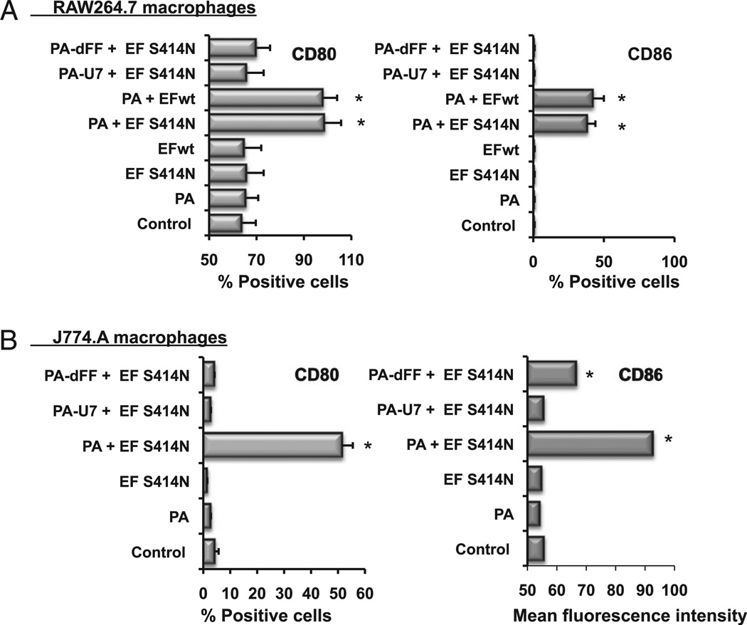
We next examined the role of EdTx components in the expression of CD80 and CD86 by macrophage cell lines (i.e., RAW264.7 and J774A.1). As expected, treatment of macrophage cultures with PA alone, or EF alone, did not enhance their expression of these costimulatory molecules (Fig. 2). Consistent with our previous report (3), CD80 and CD86 expression were up-regulated in macrophages exposed to PA plus EF-S414N. Interestingly, the same profiles of CD80 and CD86 expression were seen after exposure of macrophages to PA plus the fully functional EFwt. The levels of CD80 and CD86 expression were not increased in macrophages cultured in the presence of EF-S414N and PA-U7, where the same levels of costimulatory molecule expression were found as in control cells cultured without effectors or in the presence of PA alone (Fig. 2). The presence of EF-S414N and PA-dFF induced a slight, but no significant increase in CD80 expression by RAW264.7 cells. However, EF-S414N and PA-dFF significantly increased the mean fluorescence intensity of CD86 expression by J774A.1 cells (Fig. 2).
FIGURE 2.
Costimulatory molecule expression in macrophage cell lines treated with EdTx derivatives. Mouse macrophage cell lines RAW264.7 (A) and J774A.1 (B) were plated on a 24-well plate at 106 cells/ml and treated for 24 h with the following combination of EdTx subunits (2 µg/ml): 1) PAwt (PA) alone; 2) EF-S414N alone; 3) EF; 4) PA + EF-S414N; 5) PA + EF; 6) PA-U7 + EF-S414N; 7) PA-dFF + EF-S414N. The expression of costimulatory molecules CD80 and CD86 was assessed by flow cytometry. Results are expressed as percentage of positive cells or mean fluorescence intensity and are from four separate experiments. Results are expressed as mean ± 1 SD. Statistical significance: *p ≤ 0.05 when compared with control untreated cells.
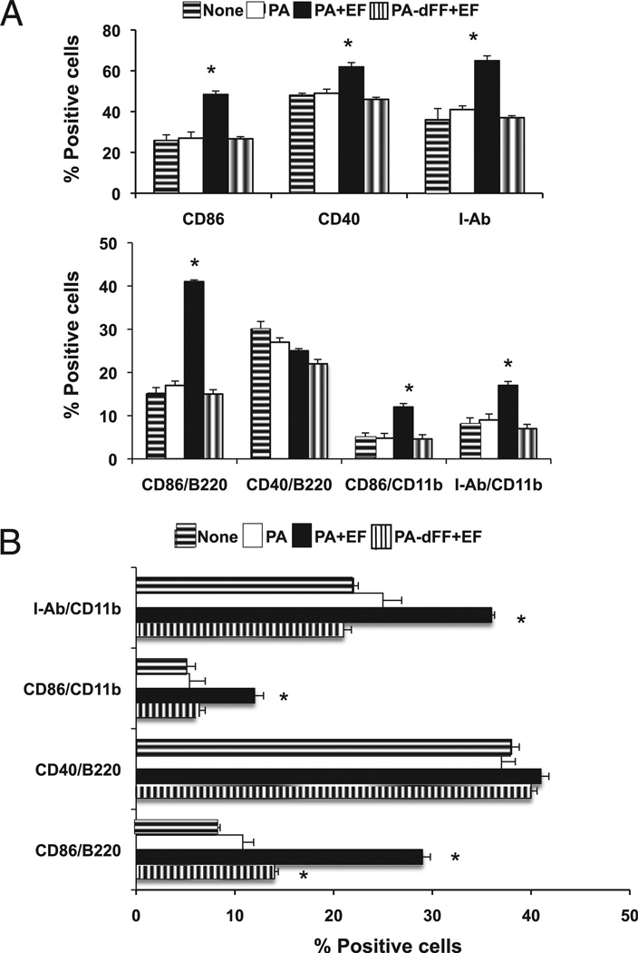
It is clear that macrophage cell lines, and even bone marrow-derived APCs (i.e., macrophages and dendritic cells), may not recapitulate the behavior of cells in the complex environment of lymphoid tissues. Thus, we next examined the responses of cervical lymph node cells to in vitro treatment with EdTx components. Neither PA alone nor PA-dFF plus EF regulated expression of CD86, CD40, or I-Ab (MHC class II) by cervical lymph node cells. As seen with macrophage cell lines, a combination of PA plus EF enhanced CD86 expression in cervical lymph node cells, and this effect extended to both B220+ (B lymphocytes) and CD11b+ (macrophages) cells (Fig. 3A). This treatment also increased the expression of I-Ab, mostly on macrophages (Fig. 3A). CD40 expression was also increased by PA plus EF, but this effect seemed to be restricted to cells other than B220+ cells (B lymphocytes) (Fig. 3A). Treatment of spleen cells with EdTx components showed the same regulatory effect of PA plus EF on CD86, CD40, and I-Ab expression (Fig. 3B). However, PA-dFF plus EF was able to enhance CD86 expression by splenic B cells (B220+) (Fig. 3B). Neither EF alone nor EF plus PA-U7 affected costimulatory molecule expression by cervical lymph node or spleen cells (data not shown).
FIGURE 3.
Costimulatory molecule and MHC class II expression cervical lymph node and spleen cells treated with EdTx derivatives. Mouse cervical lymph node cells (A) and spleen cells (B) were plated on a 24-well plate at 106 cells/ml and incubated for 24 h with medium (None) or the following combination of EdTx subunits: 1) PAwt (PA) alone (2 µg/ml); 2) PA + E F (2 µg/ml); 3) PA-dFF + EF (2 µg/ml). The expression of costimulatory molecules (i.e., CD86 and CD40) and MHC class II (I-Ab) by B cells (B220) and macrophages (CD11b) was assessed by flow cytometry. Results are expressed as percentage of positive cells and are from four separate experiments. Results are expressed as mean ± 1 SD. Statistical significance: *p ≤ 0.05 when compared with control-untreated cells (None).
Intracellular delivery of EF influences cytokine responses
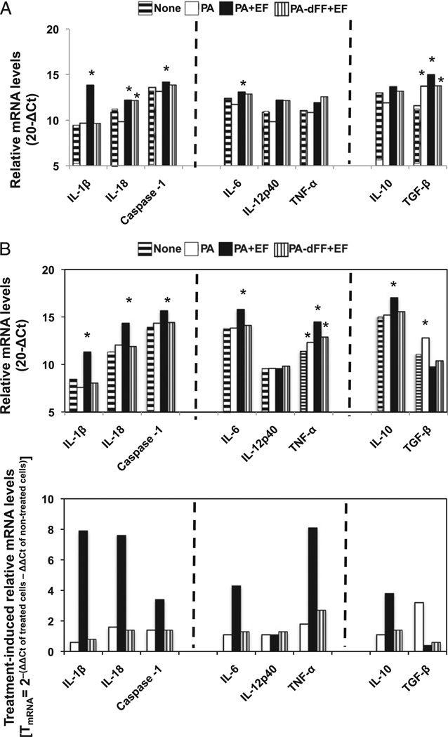
Cytokines secreted by APCs in response to adjuvant exposure play a central role in the induction of adaptive immunity. The mucosal adjuvant CT induces IL-6 (24) and IL-1 (25) secretion by APCs, and we (26) and others (27) have shown that these cytokines promote adaptive immune responses. We found that a 6-h exposure of cervical lymph node cells to PA plus EF induced IL-1β, IL-18, caspase-1, IL-6, and TGF-β mRNA responses (Fig. 4A). The PA-dFF plus EF treatment was also able to enhance IL-18 and TGF-β mRNA, but not any of the other cytokines tested (Fig. 4A). Interestingly, cervical lymph node cells exposed to PA plus EF for 24 h exhibited a different profile of cytokine response, which consisted of high IL-1β, IL-18, caspase-1, IL-6, TNF-α, and IL-10 mRNA responses (Fig. 4B, upper panel). Also interesting was our finding that, at this 24-h time point, the PA-dFF plus EF treatment only induced TNF-α mRNA (Fig. 4B, upper panel). Neither EF alone nor EF plus PA-U7 induced significant cytokine mRNA responses (data not shown). To facilitate visualization of the effect of the treatments, mRNA responses were also expressed as treatment-induced relative mRNA levels (Fig. 4B, lower panel).
FIGURE 4.
Cytokine mRNA responses to EdTx components and derivatives. Relative cytokine mRNA expression in cervical lymph node cells after 6 h (A) or 24 h (B) of culture in the presence of EdTx components and derivatives. Relative mRNA levels were analyzed by real-time RT-PCR. All the results were normalized against β-actin expression to correct for differences in cDNA concentration of the starting template and are expressed as 20 – (meanCtcytokine – meanCtβ-actin). One-way ANOVA, followed by Duncan’s multiple range test, was used to compare 20-ΔCt values of treated groups (i.e., PA, PA + EF, and mutants of PA + EF) with control untreated. Differences were considered significant at a probability of *p ≤ 0.05.
Intracellular delivery of EF enhances systemic and mucosal immunity against PA
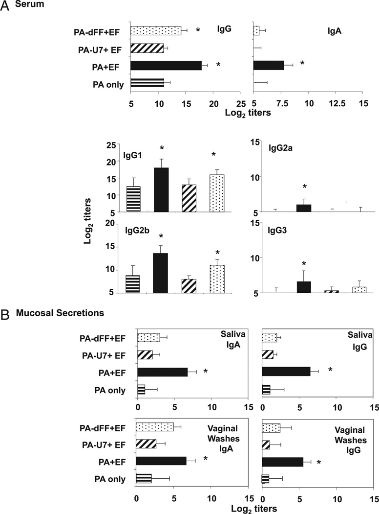
We previously showed that intranasal immunization with PA plus EF-S414N promotes anti-PA Ab responses in both the peripheral blood and mucosal secretions (3). There also was a report by others suggesting that, when used in higher concentrations, EF (28), LF, or fusion proteins containing the N-terminal portion of LF can enter cells in the absence of PA (29, 30). Therefore, we examined how the cellular site of EF delivery would affect EdTx-induced immunity to PA. When compared with mice intranasally immunized with PA plus EF-S414N, mice that received EF-S414N plus the PA-U7 (devoid of the furin cleavage site) exhibited significantly reduced PA-specific Abs in the serum (Fig. 5A). In fact, PA-specific Ab levels in these mice were not different from those measured in mice given PA alone (Fig. 5A). We also found that mice that received EF-S414N plus PA-dFF were able to mount high levels of PA-specific serum IgG1 and IgG2b Abs, although these levels were below those achieved by immunization with the native PA and EF-S414N. PA-specific serum and mucosal IgA Abs were significantly reduced in mice given EF-S414N plus the PA-U7, but these responses were less affected in samples from mice given EF-S414N plus PA-dFF (Fig. 5B). As expected, no anti-PA Abs were detected in control mice given FIV alone (Fig. 5).
FIGURE 5.
PA-specific Abs after intranasal immunization with PA mutants and EFwt. A, Serum PA-specific Ab isotype and IgG subclass responses after intranasal administration of EdTx derivatives. Mice were immunized three times at weekly intervals with 2 µg PA only (horizontal-striped bar); 2 µg PA plus 2 µg EF (filled bar); 2 µg PA-U7 plus 2 µg EF (diagnol-striped bar); or 2 µg PA-dFF plus 2 µg EF (stippled bar). The PA-specific IgA, IgG, and IgG subclass responses were determined 1 wk after the last immunization. B, PA-specific Ab responses in saliva and vaginal secretions were determined 2 wk after the last immunization. The results are expressed as the reciprocal log2 titers ± 1 SD and are from three experiments and five mice/group. Statistical significance: *p ≤ 0.05 when compared with mice immunized with PA only.
It also was important to establish the biological significance of anti-PA Abs induced by intranasal immunization with EdTx derivatives consisting of EF-S414N plus PA-U7 or PA-dFF. The in vitro macrophage toxicity assay revealed low levels of anti-PA neutralizing Abs in the serum of mice given PA alone and significantly higher titers in the serum of mice immunized with PA plus EF-S414N (Table II). Coadministration of EF-S414N plus PA-U7 generated low levels of anti-PA neutralizing Abs, which were of the same magnitude as those measured in the serum of mice given PA alone. Interestingly, coimmunization of EF-S414N plus PA-dFF also enhanced serum levels of neutralizing anti-PA Abs above the levels seen after intranasal immunization with PA alone (Table II). Finally, neutralizing anti-PA Abs were detected in the saliva, but only in mice immunized with PA plus EF-S414N and, to a lesser extent, in saliva of mice that received EF-S414N plus PA-dFF (Table II). No other mucosal secretion was found to contain significant levels of neutralizing anti-PA Abs.
Table II.
Neutralizing anti-PA Abs produced in response to intranasal administration of EdTx derivatives
| Neutralizing Ab Titers
(1/Dilution) |
|||
|---|---|---|---|
| Immunization | Plasma | Saliva | Vaginal Washes |
| PA only | 426 ± 13 | BD | BD |
| PA plus EF-S414N | 7103 ± 82* | 10 ± 3* | 3 ± 2 |
| PA-U7 plus EF-S414N | 391 ± 23 | BD | BD |
| PA-dFF plus EF-S414N | 2109 ± 12* | BD | BD |
Serial dilutions of each sample were added to J774 macrophage cultures in the presence of lethal toxin. The neutralizing titers were determined as the last dilution yielding an MTT OD equal to twice the background value. Results are shown as neutralizing Ab titers and are expressed as the reciprocal dilution ± 1 SE of three separate experiments, and five mice per group per experiment.
p < 0.05 when compared with control samples from mice given PA alone. BD, below detection level.
The adjuvant activity of B. anthracis EdTx for coadministered vaccine Ags is influenced by the subcellular site of EF delivery
We next examined to what extent the subcellular site of EF delivery would affect the adjuvant activity of EdTx for a coadministered vaccine Ag and subsequent establishment of systemic and mucosal immune responses. For this purpose, groups of mice were immunized three times at weekly intervals with the following: 1) 20 µg F1-V alone; 2) F1-V plus 2 µg PA and 2 µg EF; 3) F1-V plus 2 µg PA-U7 and 2 µg EF; and 4) F1-V plus 2 µg PA-dFF and 2 µg EF. The EFwt was used in this series of experiments because our previous studies showed no difference between the adjuvant activity of EFwt and EF-S414N.
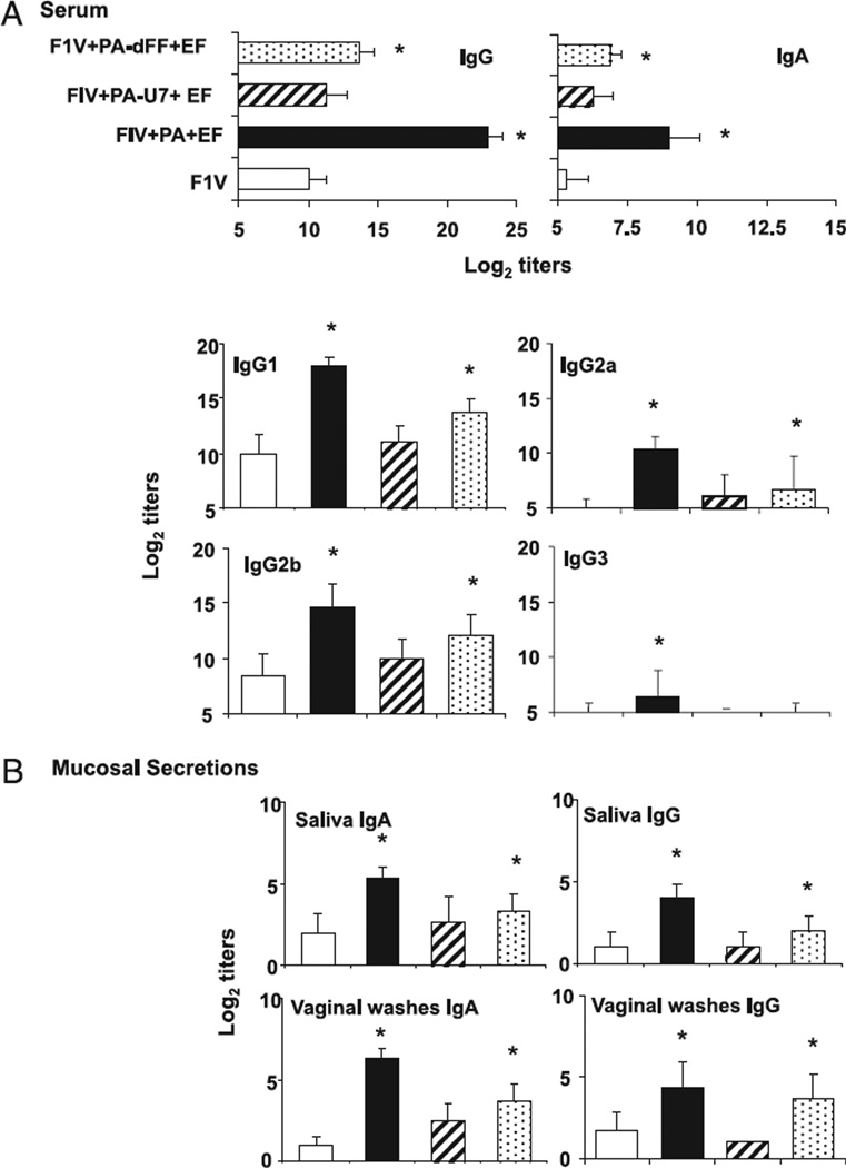
High levels of F1-V–specific IgA and IgG Abs were detected in the serum of mice given PA plus EF as adjuvant (Fig. 6). As described above for PA-specific Ab responses, intranasal coapplication of F1-V together with EF plus PA-dFF, which allows the entry of EF to endosomes, but not an efficient translocation of EF into the cytosol, also gave rise to significant levels of F1-V– specific IgG Abs in the plasma (Fig. 6). The profile of IgG subclasses, and more specifically the ratio of IgG1/IgG2a Abs, is an indirect indication of the Th cytokines supporting the Ab responses (31). In this regard, mice immunized with EF and PA-dFF exhibited the same profile of IgG subclasses of mice given PA plus EF with IgG1 > IgG2b > IgG2a > IgG3 (Fig. 6). However, the magnitude of these anti-F1V responses was lower than those induced by native EdTx (i.e., PA plus EF) as adjuvant. Furthermore, immunization in the presence of EF and PA-dFF also induced F1-V–specific IgG, but not IgA, in vaginal secretions and saliva (Fig. 6). In contrast with PA-dFF, PA-U7, which is devoid of the furin cleavage site and, thus, does not mediate the entry of EF into the cells, failed to enhance F1-V–specific Ab responses in the blood or in mucosal secretions (Fig. 6).
FIGURE 6.
F1-V–specific Abs after intranasal immunization with PA mutants and EFwt. A, Serum F1-V–specific isotype and IgG subclass responses after intranasal administration of EdTx derivatives. Mice were immunized three times at weekly intervals with 20 µg F1-V only (open bar); F1-V plus 2 µg PA plus 2 µg EF (filled bar); F1-V plus 2 µg PA-U7 plus 2 µg EF (diagnol-striped bar); or F1-V plus 2 µg PA-dFF plus 2 µg EF (stippled bar). F1-V–specific IgA, IgG, and IgG subclass Ab responses were determined 1 wk after the last immunization. B, Ag-specific Ab levels in mucosal secretions (saliva and vaginal washes) were determined 2 wk after the last immunization. The results are expressed as the reciprocal log2 titers ± 1 SD and are from three experiments and five mice/group. Statistical significance: *p ≤ 0.05 when compared with mice immunized with F1-V only.
EdTx requires an enzymatically active EF subunit to enhance immunity to PA and coadministered unrelated vaccine Ag
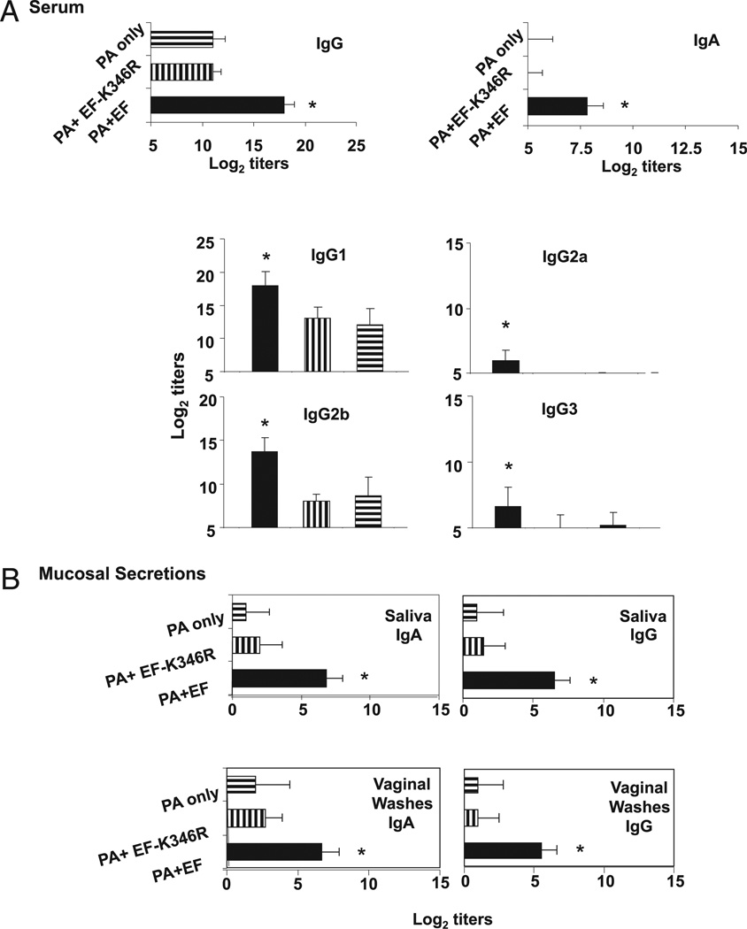
Finally, we asked whether an EdTx derivative consisting of native PA and a mutant EF devoid of adenylate cyclase activity would retain the adjuvant activity of native EdTx when used for intranasal immunizations. To address the role of the adenylate cyclase activity in the adjuvant activity for PA, mice were immunized three times at weekly intervals with the following: 1) 2 µg PA alone; 2) 2 µg PA plus 2 µg EF (EdTx); or 3) 2 µg PA plus 2 µg EF-K346R (EdTx derivative). As expected, mice immunized with PA plus EF exhibited enhanced anti-PA Ab responses compared with mice immunized with PA only. Intranasal immunization with PA plus EF-K346R failed to enhance PA-specific Ab responses above levels achieved in the serum (Fig. 7A) or in mucosal secretions (Fig. 7B) after immunization with PA alone.
FIGURE 7.
PA-specific Abs after intranasal immunization with wt PA and EF mutants. A, PA-specific serum Ab isotype and IgG subclass responses after intranasal administration of EdTx derivatives. Mice were immunized three times at weekly intervals with 2 µg PA alone; 2 µg PA plus 2 µg EF; or 2 µg PA-U7 plus 2 µg EF-K346R. The IgA and IgG subclass (IgG1 and IgG2a) Ab responses were determined 1 wk after the last immunization. B, PA-specific Ab levels in mucosal secretions (saliva and vaginal washes) were assessed 2 wk after the last immunization. The results are expressed as the reciprocal log2 titers ± 1 SD and are from three experiments and five mice/group. Statistical significance: *p ≤ 0.05 when compared with mice immunized with PA only.
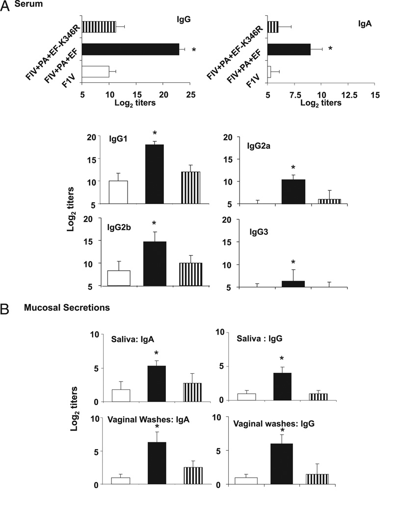
Several reports have documented that ganglioside-binding enterotoxins lacking enzymatic activity can retain their adjuvant activity and enhance mucosal and systemic immunity to intranasally coadministered vaccine Ags (9–12). Therefore, we tested whether intranasal application of PA plus EF-K346R could enhance immune responses to coadministered F1-V. Mice intranasally immunized with 20 µg F1-V plus EdTx (2 µg PA plus 2 µg EF) exhibited elevated anti–F1-V Ab levels in both the serum and mucosal secretions (Fig. 8). In contrast, the magnitude of the F1-V–specific Ab responses in the blood and mucosal secretions was not significantly different between groups of mice given F1-V alone and F1-V together with PA plus EF-K346R (Fig. 8). Taken together, these results clearly indicate that the EF-K346R molecule lacking enzymatic activity is not an adjuvant, and that some adenylate cyclase activity is needed for EdTx to act as an adjuvant for intranasal vaccines.
FIGURE 8.
F1-V–specific Abs after intranasal immunization with wt PA and EF mutants. A, F1-V–specific serum Ab isotype and IgG subclass responses after intranasal administration of EdTx derivatives. Mice were immunized three times at weekly intervals with 20 µg F1-V only; F1-V plus 2 µg PA plus 2 µg EF; or F1-V plus 2 µg PA-U7 plus 2 µg EF-K346R. The F1-V–specific IgA, IgG, and IgG subclass Ab responses were determined 1 wk after the last immunization. B, F1-V–specific Ab levels in mucosal secretions (saliva and vaginal washes) were assessed 2 wk after the last immunization. The results are expressed as the reciprocal log2 titers ± 1 SD and are from three experiments and five mice/group. Statistical significance: *p ≤ 0.05 when compared with mice immunized with F1-V only.
Discussion
A better understanding of the relative contribution of the binding/targeting and enzymatic subunits of enterotoxins and other bacterial toxin derivatives to the mucosal adjuvant activity of these molecules is crucial for the design of more effective immunization strategies. The results summarized in this manuscript clearly indicate key requirements for B. anthracis EdTx to act as an adjuvant and promote mucosal and systemic immunity. Thus, the enzymatic subunit EF needs to be delivered into the cytosol of host cells by functional PA, and a minimum of adenylate cyclase activity is required for EF to stimulate APCs and enhance vaccine-specific Ab responses.
One of the major findings of this work is the fact that the adjuvant activity of EdTx is completely abrogated when the enzymatic subunit EF is devoid of adenylate cyclase activity. As mentioned earlier, a number of studies have shown that the ADP-ribosyl transferase activity is not required for nasal adjuvanticity of LT-I (9, 10) and CT (11, 12). In contrast, our results clearly indicate that a minimum of enzymatic activity is needed for EF to enhance immunity. This apparent discrepancy could be explained by the difference in mechanisms involved in the ability of EdTx and the enterotoxins CT and LT to increase cAMP. For example, CT and LT are ADP ribosyl transferases (5) that activate the endogenous adenylate cyclase of cells, whereas EdTx is itself an adenylate cyclase (2, 32). It also is possible that the multimeric binding B subunits of CT and LT provide cells with addition signals by cross-linking their receptor molecules. In this regard, although CT and LT are closely related enterotoxins, CT as a mucosal adjuvant promotes strong Th2 responses, whereas LT supports mixed Th1/Th2 responses. The difference in the profile of Th cytokine responses supported by these adjuvants has been attributed to their targeting of different cell surface molecules.
The expression of costimulatory molecules by APCs and the ability to take up Ags are critical steps for the development of adaptive immunity. Our in vitro studies showed that binding of PA to the cell surface anthrax toxin receptor is not sufficient for triggering stimulatory signals, which in turn would up-regulate the expression of costimulatory molecules and promote Ag uptake. More specifically, we found that correct subcellular delivery of EF by a functional PA was a prerequisite for EdTx to exert its adjuvant activity. Our finding is consistent with previous reports that B. anthracis PA is a sophisticated system for delivery of exogenous molecules in mammalian cells. The combination of anthrax PA plus the 1–254 N-terminal fragment of LF (LFn1–254) or EF (EFn1–254) has been used as a molecular syringe to introduce foreign Ags into target cells. Mechanistic studies with PA mutants attenuated in self-assembly or translocation (33, 34) or cells with disrupted furin genes (35) showed that delivery of Ag involves the same toxin self-assembly and translocation steps that occur during intoxication. This delivery system primed CTL responses in both BALB/c (H-2d) and C57BL/6 (H-2b) mice (36). In addition, injection of mice with dendritic cells treated in vitro with a LFn1–254 fusion protein and PA effectively primed CTL responses (37).
Because several studies suggested that EF (or LF) could enter mammalian cells in the absence of PA, at least when added at higher concentrations, one might expect to measure some signs of stimulation on macrophages exposed to EF alone or EF plus PA-U7. In this regard, studies with the proteasome inhibitor lactacystin have shown that the LFn1–254- and PA-mediated delivery of proteins switches their processing from the cytosolic degradative pathway into the classical processing pathway, for presentation by MHC class I molecules (29, 33). In addition, whereas the cellular delivery of LFn-coupled Ag optimally occurred in the presence of a functional PA, LFn fusion proteins were able to enter the classical MHC class I degradation pathway and prime CTL responses in the absence of PA (29, 30). This PA-independent LFn delivery of Ags could be abrogated in the presence of a phagocytosis inhibitor (cytochalasin B) or an inhibitor of proteins exocytosis (brefeldin) (29). Finally, the Ags delivered in the absence of PA seemed to colocalize with proteasomes, which degrade proteins into peptides capable of entering the classical MHC class I pathway (30). In conclusion, it is very likely that the lack of regulatory activity of EF added alone to cells or added in the presence of PA-U7 is due to its degradation within the proteasome.
Another striking observation from this study is the ability of EF to retain some stimulatory effects on APC functions and Ab responses, when delivered into cells by PA-dFF. This limited, but constitutive regulatory effect suggests that cAMP is not exclusively produced by EF in the cytosol and that a limited level of cAMP can be induced by EF in the endosomes. Alternatively, the channel produced by the PA dFF protein may retain a limited ability to translocate EF. In this regard, even rather small increases in cAMP may be able to fully activate protein kinase A and the downstream pathways. Real-time imaging of cells expressing cAMP biosensors have shown that EF enters the cytosol from late endosomes and remains associated with these compartments (38). Anthrax toxins are known to alter immune cell functions (39). However, anthrax EdTx was also reported to induce the maturation of dendritic cells (40), an effect that certainly contributes to its adjuvanticity. In this study, we show that EdTx (i.e., PA plus EF), and to some extent EF given together with PA-dFF, stimulates expression of costimulatory and production of cytokines, including IL-1, known to mimic most of the adjuvant activity of CT for IgG Ab responses (27). Cytokine mRNA responses in our studies are consistent with a previous report that i.v. administration of EdTx increased several cytokines, including IL-1, IL-6, and IL-10 (41). The role of each individual cytokine tested in the adjuvant activity of EdTx derivatives remains to be elucidates. Nonetheless, our studies clearly demonstrate that mutation of EdTx components that alter these cytokine responses also affects the adjuvant activity of EdTx derivatives for both systemic IgG and mucosal IgA responses. Our in vitro studies with macrophages, cervical lymph node, and spleen cells also highlight the point that different cells display heterogeneous levels of susceptibility to EdTx, which in part may be due to differences in the expression of cellular receptors TEM8 and CMG2 (1, 42). Finally, the fact that EF given together with PA-dFF retains some of the adjuvant activity of EdTx for IgG, but not IgA, provides us with a potentially useful system for dissecting mechanisms underlying the ability of vaccine adjuvants to promote secretory IgA Ab responses.
Acknowledgments
This work was supported by National Institutes of Health Grant AI 43197.
We thank Kathleen Hayes-Ozello for editorial assistance.
Abbreviations used in this paper
- BD
below detection level
- Ct
threshold cycle
- CT
cholera toxin
- EdTx
edema toxin
- EF
edema factor
- LF
lethal factor
- PA
protective Ag
- wt
wild type
Footnotes
Disclosures
The authors have no financial conflicts of interest.
References
- 1.Young JA, Collier RJ. Anthrax toxin: receptor binding, internalization, pore formation, and translocation. Annu. Rev. Biochem. 2007;76:243–265. doi: 10.1146/annurev.biochem.75.103004.142728. [DOI] [PubMed] [Google Scholar]
- 2.Tang WJ, Guo Q. The adenylyl cyclase activity of anthrax edema factor. Mol. Aspects Med. 2009;30:423–430. doi: 10.1016/j.mam.2009.06.001. [DOI] [PMC free article] [PubMed] [Google Scholar]
- 3.Duverger A, Jackson RJ, van Ginkel FW, Fischer R, Tafaro A, Leppla SH, Fujihashi K, Kiyono H, McGhee JR, Boyaka PN. Bacillus anthracis edema toxin acts as an adjuvant for mucosal immune responses to nasally administered vaccine antigens. J. Immunol. 2006;176:1776–1783. doi: 10.4049/jimmunol.176.3.1776. [DOI] [PubMed] [Google Scholar]
- 4.Gill DM, King CA. The mechanism of action of cholera toxin in pigeon erythrocyte lysates. J. Biol. Chem. 1975;250:6424–6432. [PubMed] [Google Scholar]
- 5.vanHeyningen S. Cholera toxin: interaction of subunits with ganglioside GM1. Science. 1974;183:656–657. doi: 10.1126/science.183.4125.656. [DOI] [PubMed] [Google Scholar]
- 6.Holmgren J, Lindblad M, Fredman P, Svennerholm L, Myrvold H. Comparison of receptors for cholera and Escherichia coli enterotoxins in human intestine. Gastroenterology. 1985;89:27–35. doi: 10.1016/0016-5085(85)90741-3. [DOI] [PubMed] [Google Scholar]
- 7.Griffiths SL, Finkelstein RA, Critchley DR. Characterization of the receptor for cholera toxin and Escherichia coli heat-labile toxin in rabbit intestinal brush borders. Biochem. J. 1986;238:313–322. doi: 10.1042/bj2380313. [DOI] [PMC free article] [PubMed] [Google Scholar]
- 8.Fukuta S, Magnani JL, Twiddy EM, Holmes RK, Ginsburg V. Comparison of the carbohydrate-binding specificities of cholera toxin and Escherichia coli heat-labile enterotoxins LTh-I, LT-IIa, and LT-IIb. Infect. Immun. 1988;56:1748–1753. doi: 10.1128/iai.56.7.1748-1753.1988. [DOI] [PMC free article] [PubMed] [Google Scholar]
- 9.Douce G, Turcotte C, Cropley I, Roberts M, Pizza M, Domenghini M, Rappuoli R, Dougan G. Mutants of Escherichia coli heat-labile toxin lacking ADP-ribosyltransferase activity act as nontoxic, mucosal adjuvants. Proc. Natl. Acad. Sci. USA. 1995;92:1644–1648. doi: 10.1073/pnas.92.5.1644. [DOI] [PMC free article] [PubMed] [Google Scholar]
- 10.Dickinson BL, Clements JD. Dissociation of Escherichia coli heat-labile enterotoxin adjuvanticity from ADP-ribosyltransferase activity. Infec. Immun. 1995;63:1617–1623. doi: 10.1128/iai.63.5.1617-1623.1995. [DOI] [PMC free article] [PubMed] [Google Scholar]
- 11.Yamamoto S, Kiyono H, Yamamoto M, Imaoka K, Fujihashi K, Van Ginkel FW, Noda M, Takeda Y, McGhee JR. A nontoxic mutant of cholera toxin elicits Th2-type responses for enhanced mucosal immunity. Proc. Natl. Acad. Sci. USA. 1997;94:5267–5272. doi: 10.1073/pnas.94.10.5267. [DOI] [PMC free article] [PubMed] [Google Scholar]
- 12.Hagiwara Y, Kawamura YI, Kataoka K, Rahima B, Jackson RJ, Komase K, Dohi T, Boyaka PN, Takeda Y, Kiyono H, et al. A second generation of double mutant cholera toxin adjuvants: enhanced immunity without intracellular trafficking. J. Immunol. 2006;177:3045–3054. doi: 10.4049/jimmunol.177.5.3045. [DOI] [PMC free article] [PubMed] [Google Scholar]
- 13.Williams NA. Immune modulation by the cholera-like enterotoxin B-subunits: from adjuvant to immunotherapeutic. Int. J. Med. Microbiol. 2000;290:447–453. doi: 10.1016/S1438-4221(00)80062-4. [DOI] [PubMed] [Google Scholar]
- 14.Agren L, Lo¨wenadler B, Lycke N. A novel concept in mucosal adjuvanticity: the CTA1-DD adjuvant is a B cell-targeted fusion protein that incorporates the enzymatically active cholera toxin A1 subunit. Immunol. Cell Biol. 1998;76:280–287. doi: 10.1046/j.1440-1711.1998.00750.x. [DOI] [PubMed] [Google Scholar]
- 15.Bowman CC, Clements JD. Differential biological and adjuvant activities of cholera toxin and Escherichia coli heat-labile enterotoxin hybrids. Infect. Immun. 2001;69:1528–1535. doi: 10.1128/IAI.69.3.1528-1535.2001. [DOI] [PMC free article] [PubMed] [Google Scholar]
- 16.Boyaka PN, Ohmura M, Fujihashi K, Koga T, Yamamoto M, Kweon MN, Takeda Y, Jackson RJ, Kiyono H, Yuki Y, McGhee JR. Chimeras of labile toxin one and cholera toxin retain mucosal adjuvanticity and direct Th cell subsets via their B subunit. J. Immunol. 2003;170:454–462. doi: 10.4049/jimmunol.170.1.454. [DOI] [PubMed] [Google Scholar]
- 17.Ramirez DM, Leppla SH, Schneerson R, Shiloach J. Production, recovery and immunogenicity of the protective antigen from a recombinant strain of Bacillus anthracis . J. Ind. Microbiol. Biotechnol. 2002;28:232–238. doi: 10.1038/sj/jim/7000239. [DOI] [PubMed] [Google Scholar]
- 18.Cooksey BA, Sampey GC, Pierre JL, Zhang X, Karwoski JD, Choi GH, Laird MW. Production of biologically active Bacillus anthracis edema factor in Escherichia coli . Biotechnol. Prog. 2004;20:1651–1659. doi: 10.1021/bp049725n. [DOI] [PubMed] [Google Scholar]
- 19.Drum CL, Yan SZ, Bard J, Shen YQ, Lu D, Soelaiman S, Grabarek Z, Bohm A, Tang WJ. Structural basis for the activation of anthrax adenylyl cyclase exotoxin by calmodulin. Nature. 2002;415:396–402. doi: 10.1038/415396a. [DOI] [PubMed] [Google Scholar]
- 20.Soelaiman S, Wei BQ, Bergson P, Lee YS, Shen Y, Mrksich M, Shoichet BK, Tang WJ. Structure-based inhibitor discovery against adenylyl cyclase toxins from pathogenic bacteria that cause anthrax and whooping cough. J. Biol. Chem. 2003;278:25990–25997. doi: 10.1074/jbc.M301232200. [DOI] [PubMed] [Google Scholar]
- 21.Fischer R, McGhee JR, Vu HL, Atkinson TP, Jackson RJ, Tomé D, Boyaka PN. Oral and nasal sensitization promote distinct immune responses and lung reactivity in a mouse model of peanut allergy. Am. J. Pathol. 2005;167:1621–1630. doi: 10.1016/S0002-9440(10)61246-1. [DOI] [PMC free article] [PubMed] [Google Scholar]
- 22.Boyaka PN, Tafaro A, Fischer R, Leppla SH, Fujihashi K, McGhee JR. Effective mucosal immunity to anthrax: neutralizing antibodies and Th cell responses following nasal immunization with protective antigen. J. Immunol. 2003;170:5636–5643. doi: 10.4049/jimmunol.170.11.5636. [DOI] [PubMed] [Google Scholar]
- 23.Mosmann T. Rapid colorimetric assay for cellular growth and survival: application to proliferation and cytotoxicity assays. J. Immunol. Methods. 1983;65:55–63. doi: 10.1016/0022-1759(83)90303-4. [DOI] [PubMed] [Google Scholar]
- 24.Bromander AK, Kjerrulf M, Holmgren J, Lycke N. Cholera toxin enhances alloantigen presentation by cultured intestinal epithelial cells. Scand. J. Immunol. 1993;37:452–458. doi: 10.1111/j.1365-3083.1993.tb03318.x. [DOI] [PubMed] [Google Scholar]
- 25.Lycke N, Bromander AK, Ekman L, Karlsson U, Holmgren J. Cellular basis of immunomodulation by cholera toxin in vitro with possible association to the adjuvant function in vivo. J. Immunol. 1989;142:20–27. [PubMed] [Google Scholar]
- 26.Boyaka PN, Marinaro M, Jackson RJ, Menon S, Kiyono H, Jirillo E, McGhee JR. IL-12 is an effective adjuvant for induction of mucosal immunity. J. Immunol. 1999;162:122–128. [PubMed] [Google Scholar]
- 27.Staats HF, Ennis FA., Jr IL-1 is an effective adjuvant for mucosal and systemic immune responses when coadministered with protein immunogens. J. Immunol. 1999;162:6141–6147. [PubMed] [Google Scholar]
- 28.Hong J, Beeler J, Zhukovskaya NL, He W, Tang WJ, Rosner MR. Anthrax edema factor potency depends on mode of cell entry. Biochem. Biophys. Res. Commun. 2005;335:850–857. doi: 10.1016/j.bbrc.2005.07.132. [DOI] [PubMed] [Google Scholar]
- 29.Cao H, Agrawal D, Kushner N, Touzjian N, Essex M, Lu Y. Delivery of exogenous protein antigens to major histocompatibility complex class I pathway in cytosol. J. Infect. Dis. 2002;185:244–251. doi: 10.1086/338448. [DOI] [PubMed] [Google Scholar]
- 30.Kushner N, Zhang D, Touzjian N, Essex M, Lieberman J, Lu Y. A fragment of anthrax lethal factor delivers proteins to the cytosol without requiring protective antigen. Proc. Natl. Acad. Sci. USA. 2003;100:6652–6657. doi: 10.1073/pnas.1131930100. [DOI] [PMC free article] [PubMed] [Google Scholar]
- 31.Finkelman FD, Holmes J, Katona IM, Urban JF, Jr, Beckmann MP, Park LS, Schooley KA, Coffman RL, Mosmann TR, Paul WE. Lymphokine control of in vivo immunoglobulin isotype selection. Annu. Rev. Immunol. 1990;8:303–333. doi: 10.1146/annurev.iy.08.040190.001511. [DOI] [PubMed] [Google Scholar]
- 32.Mock M, Fouet A. Anthrax. Annu. Rev. Microbiol. 2001;55:647–671. doi: 10.1146/annurev.micro.55.1.647. [DOI] [PubMed] [Google Scholar]
- 33.Goletz TJ, Klimpel KR, Arora N, Leppla SH, Keith JM, Berzofsky JA. Targeting HIV proteins to the major histocompatibility complex class I processing pathway with a novel gp120-anthrax toxin fusion protein. Proc. Natl. Acad. Sci. USA. 1997;94:12059–12064. doi: 10.1073/pnas.94.22.12059. [DOI] [PMC free article] [PubMed] [Google Scholar]
- 34.Ballard JD, Collier RJ, Starnbach MN. Anthrax toxin-mediated delivery of a cytotoxic T-cell epitope in vivo. Proc. Natl. Acad. Sci. USA. 1996;93:12531–12534. doi: 10.1073/pnas.93.22.12531. [DOI] [PMC free article] [PubMed] [Google Scholar]
- 35.Zhang Y, Kida Y, Kuwano K, Misumi Y, Ikehara Y, Arai S. Role of furin in delivery of a CTL epitope of an anthrax toxin-fusion protein. Microbiol. Immunol. 2001;45:119–125. doi: 10.1111/j.1348-0421.2001.tb01279.x. [DOI] [PubMed] [Google Scholar]
- 36.Doling AM, Ballard JD, Shen H, Krishna KM, Ahmed R, Collier RJ, Starnbach MN. Cytotoxic T-lymphocyte epitopes fused to anthrax toxin induce protective antiviral immunity. Infect. Immun. 1999;67:3290–3296. doi: 10.1128/iai.67.7.3290-3296.1999. [DOI] [PMC free article] [PubMed] [Google Scholar]
- 37.Moriya O, Matsui M, Osorio M, Miyazawa H, Rice CM, Feinstone SM, Leppla SH, Keith JM, Akatsuka T. Induction of hepatitis C virus-specific cytotoxic T lymphocytes in mice by immunization with dendritic cells treated with an anthrax toxin fusion protein. Vaccine. 2001;20:789–796. doi: 10.1016/s0264-410x(01)00407-8. [DOI] [PubMed] [Google Scholar]
- 38.Dal Molin F, Tonello F, Ladant D, Zornetta I, Zamparo I, Di Benedetto G, Zaccolo M, Montecucco C. Cell entry and cAMP imaging of anthrax edema toxin. EMBO J. 2006;25:5405–5413. doi: 10.1038/sj.emboj.7601408. [DOI] [PMC free article] [PubMed] [Google Scholar]
- 39.Tournier JN, Rossi Paccani S, Quesnel-Hellmann A, Baldari CT. Anthrax toxins: a weapon to systematically dismantle the host immune defenses. Mol. Aspects Med. 2009;30:456–466. doi: 10.1016/j.mam.2009.06.002. [DOI] [PubMed] [Google Scholar]
- 40.Maldonado-Arocho FJ, Bradley KA. Anthrax edema toxin induces maturation of dendritic cells and enhances chemotaxis towards macrophage inflammatory protein 3beta. Infect. Immun. 2009;77:2036–2042. doi: 10.1128/IAI.01329-08. [DOI] [PMC free article] [PubMed] [Google Scholar]
- 41.Firoved AM, Miller GF, Moayeri M, Kakkar R, Shen Y, Wiggins JF, McNally EM, Tang WJ, Leppla SH. Bacillus anthracis edema toxin causes extensive tissue lesions and rapid lethality in mice. Am. J. Pathol. 2005;167:1309–1320. doi: 10.1016/S0002-9440(10)61218-7. [DOI] [PMC free article] [PubMed] [Google Scholar]
- 42.Chen KH, Liu S, Bankston LA, Liddington RC, Leppla SH. Selection of anthrax toxin protective antigen variants that discriminate between the cellular receptors TEM8 and CMG2 and achieve targeting of tumor cells. J. Biol. Chem. 2007;282:9834–9845. doi: 10.1074/jbc.M611142200. [DOI] [PMC free article] [PubMed] [Google Scholar]
- 43.Singh Y, Klimpel KR, Arora N, Sharma M, Leppla SH. The chymotrypsin-sensitive site, FFD315, in anthrax toxin protective antigen is required for translocation of lethal factor. J. Biol. Chem. 1994;269:29039–29046. [PubMed] [Google Scholar]
- 44.Liu S, Bugge TH, Leppla SH. Targeting of tumor cells by cell surface urokinase plasminogen activator-dependent anthrax toxin. J. Biol. Chem. 2001;276:17976–17984. doi: 10.1074/jbc.M011085200. [DOI] [PubMed] [Google Scholar]


