Abstract
Objectives:
The objective of this study was to evaluate stress distribution in the dentin and alveolar bone created by load application on simulated endodontically treated teeth with two different esthetic posts.
Materials and Methods:
A finite element model was made and elastic moduli and poissons ratio of all the materials fed to the software. For both the models, a 100N force was applied on the lingual surface of the tooth at an angle of 45°. Stress concentration and distribution were evaluated and noted down for both the posts.
Results:
Finite element method revealed that Glass fibre post had homogenous distribution of stress whereas in zirconia post the stress was concentrated in the post.
Conclusion:
The present findings suggest that glass fibre post should be used in well-conserved radicular tooth structure and Zirconia post is indicated in weakened and grossly destructed tooth structure.
Keywords: Esthetic post, finite element analysis, glass fiber post, stress analysis, stress distribution, zirconia post
INTRODUCTION
The aim of prosthodontics should be an attempt to converge to the dictum of DeVan “Perpetual preservation of what remains rather than meticulous restoration of what is lost.” Contemporary restorative dentistry has the main purpose of restoring function and esthetic of teeth, which had their structure severely degraded due to caries or fracture.[1]
Restoration of endodontically treated teeth is a complicated procedure because of the various factors that need to be considered.[2] The primary function of coronoradicular post is to provide retention for the core and to reinforce and to replace the remaining coronal tooth structure.[3,4] Materials used for post and core can be metal or non-metal. Besides causing greyish discolouration of all ceramic crowns and gingiva, metal posts can cause corrosion reaction, metallic taste, oral burning, oral pain, sensitization allergic reaction and also resisted lateral forces without distortion and this resulted in stress transfer to dentin causing potential root cracking and fracture.[5] Hence, from the point of view of health, strength and esthetics, a wide range of esthetic post has become commercially available. They provide enhanced esthetics and improved material strength. The finite element method is a highly approved method to simulate biophysical phenomena in computerized models of teeth and their periodontium.[6] The finite element method is considered to be an extremely useful tool to simulate the mechanical effects of chewing forces acting on the periodontal ligament and on the dental hard tissues.[6] The finite element method is based on a mathematical model, which approximates the geometry, loading and constraint conditions of a structure to be analyzed. Deformations and stresses at any point within the model can be evaluated and highly stressed regions can be analyzed.[7] The elastic modulus and the poisons ratio for the modeled materials are specified for each element. A system of simultaneous equations is generated and solved to yield predictable stress distribution in each throughout a structure.[8] However, the result of the finite element method depends on its modeling methods and the values assigned to the material properties. The present study applies the finite element method to determine the stress distribution in maxillary central incisor, restored with two different commercially available prefabricated esthetic posts.
METHODOLOGY
Three-dimensional models of maxillary central incisors with 21 mm length and 7 mm diameter at the level of crown margin were created by 3D geometry creations in ANSYS 12.0 (Canonsburg, PA, USA) software using extrusion and Boolean operations using wheelers data. These models were fabricated including supporting structures, namely periodontal ligament (0.175 mm), cortical bone (0.5 mm) and spongy bone. Geometric measurement of the tooth and surrounding structure was according to measurements in wheeler. The average thickness of root dentin was taken as 1 mm to mimic a weakened root structure. The model included enamel, dentin, post, core, periodontal ligament, cortical bone, cancellous bone, crown and Gutta-percha. Cement layer between the post and dentin was not modeled and was treated as part of dentin because of its thinness in young's modulus of elasticity of dentin and cement. Cementum was also not modeled.
The Model I represent a pulpless tooth, with 4 mm of remaining Gutta-percha apical seal, restored with a glass fiber post (Contec Blanco, Hahnenkratt GmbH, Kφnigsbach-Stein, Germany), 10 mm long and 2 mm wide centered in the root cavity and filled with the composite resin. They have the advantage of being biocompatible and elastic modulus similar to that of dentin.
The model II represented the same tooth structure, but with zirconia post (CosmoPost, Ivoclar Vivadent, Schaan, Liechtenstein) according to the indirect technique. They have high rigidity and excellent esthetics. Fractures originate at microscopic manufacturing defects, which can cause crack nucleation and propagation.
Material properties
All materials included in the finite element used in the simulation were assumed to be homogeneous, isotropic and linear elastic. The elastic modulus and the Poisson's ratio of the materials involved in the finite element analysis are described in Table 1.[1]
Table 1.
Elastic modulus and the Poisson's ratio of the materials involved in the finite element analysis
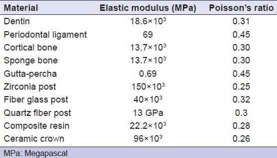
The key point plot followed by line plot, area plot and volume generation was done. Geometric build-up of the surrounding structure was done. Meshing was done with 4 noded tetra elements solid 45 software. All the three models had 320488 quadratic elements and 54733 nodes. To simulate masticatory loading, a 100 N oblique force at an angle of 45° is applied on the lingual face of the central incisor. The outer cortical bone surfaces are face constrained in all directions to prevent movement of the body.
RESULT
The results were captured for shear stress and Von Mises stress in the dentin, bone (cortical and cancellous), post, Gutta-percha, composite, periodontal ligament and All-ceramic crown. All the stresses were expressed in Megapascals.
The finite element analysis is summarized in Table 2.
Table 2.
Summary of the results of finite element analysis

INFERENCE
The restoration geometry and the elastic moduli of the materials involved in the procedure can influence the stress distribution pattern developed in the restored tooth under occlusal load. With both the posts, alveolar bone had more stress concentrated at the apex followed by the labial plate. The highest magnitude of stress concentration in alveolar bone was noticed glass fiber post, mainly in the cervical and middle third of the tooth [Figure 1]. It can be concluded that in a fiber post there is a homogenous distribution of stress. As a result of this, there is stress concentration in the cervical region of the tooth [Figure 2]. Hence, fiber posts are better suited for well-conserved radicular structure. The ceramic post restored incisor presented under loading, shear stress concentration in the middle of the post close to the tooth cervical region [Figure 3]. Small areas of concentrated shear stress were also observed at the apex of the post. Von Mises stress indicates that the highest level of this stress under loading was achieved in the inner post within the middle third of the tooth [Figure 4]. Hence, ceramic posts are indicated in case of weakened tooth roots.
Figure 1.
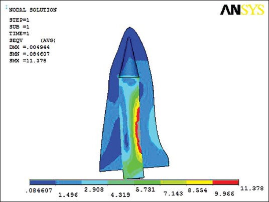
Stress in post dentine interface in glass fiber post
Figure 2.
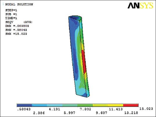
Von mises stress showing less concentration of stress in glass fiber post
Figure 3.
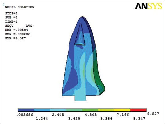
Stress in post dentine interface in zirconia post
Figure 4.
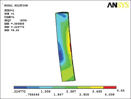
Von mises stress showing increased concentration of stress in zirconia post
DISCUSSION
Careful post-endodontic restoration is required as endodontically treated teeth are mainly lost due to restorative difficulties and failure of the root canal treatment. The demand for conservation of tooth resulted in frequent use of endodontics with post-core-crown restoration. Finite element analysis result revealed that in glass fiber post the stress concentration in bone was in the labial plate near the cervical region of the tooth. In dentin, the stress was concentrated in the middle third of the tooth near the cervical region. Stress was uniformly distributed to all the structures of the tooth. Zirconia post showed stress concentration in bone in the labial plate near the cervical region of the tooth. In dentin, the stress was concentrated in the middle third of the tooth. However, highest stress concentration was observed in the post proper in the middle third of the post. Our study revealed that the restoration geometry and the elastic moduli of the materials involved influence the stress distribution pattern on load. This result was in accordance to the result obtained by Martha et al.
From our study result, we can conclude that fiber posts are advocated in well-conserved radicular structure, as there is a homogenous distribution of stress in tooth, bone, periodontal ligament, post core and all-ceramic crown. This result is supported by Simone Grandini, Nicoletta Chieffi et al. (2008) who conducted a study on GC fiber post, ParaPost Fiber White, FibreKor, Double Taper Light-Post radiopaque, FRC Postec and Luscent Anchor post to evaluate their fatigue resistance and structural integrity. Ceramic posts reduced the stress concentration in the dentin due to high modulus of elasticity of the zirconia post. The force is transmitted directly to the post tooth interface without being absorbed. Consequently, stresses acting on dentin decreases. This result is in agreement with the result of Seo et al.[9] who conducted a finite element study to investigate the effect of rigidity of post-core system on stress distribution. Hence, precise definition of these forces followed by appropriate application of finite element method at critical points within a restored tooth is required for predicting the longevity and success of restoration.
Clinical implication of the study
The growing realization for esthetics has led to an alarming need to know the stress distribution in the esthetic endodontic post for the success of the restoration.
Based on this in vitro study, the following considerations need to be noted by the clinicians. Clinicians must cautiously consider a post-core system as a treatment alternative as they only help to retain the overlying prosthesis and does not improve the fracture resistance of endodontically treated teeth. Glass fiber post and should be considered in cases of well-conserved radicular structure. Zirconia posts are indicated in cases of weakened tooth structure. They are indicated in cases of grossly destructed teeth, high lip line and thin gingiva for better esthetics.
CONCLUSION
Successful rehabilitation of an endodontically treated tooth is the greatest challenge for the prosthodontist. This dispute becomes graver with the restoration of an anterior tooth. Within the limitations of this study, it is concluded that knowledge on the stress distribution in esthetic post is important for the selection of an appropriate post for a particular situation. Glass fiber posts are indicated in a well-conserved radicular structure and zirconia post are indicated in grossly destructed tooth, areas with heavy forces and in high lip line and thin gingival tissue for better esthetics. A careful consideration must be given to the treatment options in patients who require such restorative care.
Footnotes
Source of Support: Nil.
Conflict of Interest: None declared
REFERENCES
- 1.Vasconcellos AM, Pereira SV, Darwish IA, Camarao AF. Virtual analysis of stresses in human teeth restored with esthetic posts. Mater Res. 2008;11:459–63. [Google Scholar]
- 2.Papadogiannis D, Lakes RS, Palaghias G, Papadogiannis Y. Creep and dynamic viscoelastic behavior of endodontic fiber-reinforced composite posts. J Prosthodont Res. 2009;53:185–92. doi: 10.1016/j.jpor.2009.07.001. [DOI] [PubMed] [Google Scholar]
- 3.Hegde M, Sureshchandra B. Esthetic posts-An update. Endodontology. 2010;22:100–7. [Google Scholar]
- 4.Robbins JW. Restoration of the endodontically treated tooth. Dent Clin North Am. 2002;46:367–84. doi: 10.1016/s0011-8532(01)00006-4. [DOI] [PubMed] [Google Scholar]
- 5.Bateman G, Ricketts DN, Saunders WP. Fibre-based post systems: A review. Br Dent J. 2003;195:43–8. doi: 10.1038/sj.bdj.4810278. [DOI] [PubMed] [Google Scholar]
- 6.Lupke M, Gardemin M, Kopke S, Seifert H, Staszyk C. Finite element analysis of the equine periodontal ligament under masticatory loading. Wien Tierazil Mschr Vet Med Austriaca. 2010;97:101–6. [Google Scholar]
- 7.Li XN, Shi YK, Li ZC, Song CY, Chen XD, Guan ZQ, et al. Three dimensional finite element analysis of a maxillary central incisor restored with different post-core materials. Int Chin J Dent. 2008;8:21–7. [Google Scholar]
- 8.Boschian Pest L, Guidotti S, Pietrabissa R, Gagliani M. Stress distribution in a post-restored tooth using the three-dimensional finite element method. J Oral Rehabil. 2006;33:690–7. doi: 10.1111/j.1365-2842.2006.01538.x. [DOI] [PubMed] [Google Scholar]
- 9.Seo MS, Shon WJ, Lee WC, Yoo HM, Cho BH, Baek SH. Finite element analysis of maxillary central incisors restored with various post-and-core applications. J Korean Acad Cons Dent. 2009;34:324–32. [Google Scholar]


